CHAPTER 3 Tumours of the Stomach - Pathology Outlines
CHAPTER 3 Tumours of the Stomach - Pathology Outlines
CHAPTER 3 Tumours of the Stomach - Pathology Outlines

Create successful ePaper yourself
Turn your PDF publications into a flip-book with our unique Google optimized e-Paper software.
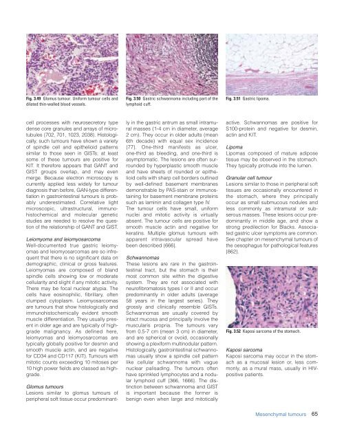
Fig. 3.49 Glomus tumour. Uniform tumour cells and<br />
dilated thin-walled blood vessels.<br />
Fig. 3.50 Gastric schwannoma including part <strong>of</strong> <strong>the</strong><br />
lymphoid cuff.<br />
Fig. 3.51 Gastric lipoma.<br />
cell processes with neurosecretory type<br />
dense core granules and arrays <strong>of</strong> microtubules<br />
{702, 701, 1023, 2038}. Histologically,<br />
such tumours have shown a variety<br />
<strong>of</strong> spindle cell and epi<strong>the</strong>lioid patterns<br />
similar to those seen in GISTs; at least<br />
some <strong>of</strong> <strong>the</strong>se tumours are positive for<br />
KIT. It <strong>the</strong>refore appears that GANT and<br />
GIST groups overlap, and may even<br />
merge. Because electron microscopy is<br />
currently applied less widely for tumour<br />
diagnosis than before, GAN-type differentiation<br />
in gastrointestinal tumours is probably<br />
underestimated. Correlative light<br />
microscopic, ultrastructural, immunohistochemical<br />
and molecular genetic<br />
studies are needed to resolve <strong>the</strong> question<br />
<strong>of</strong> <strong>the</strong> relationship <strong>of</strong> GANT and GIST.<br />
Leiomyoma and leiomyosarcoma<br />
Well-documented true gastric leiomyomas<br />
and leiomyosarcomas are so infrequent<br />
that <strong>the</strong>re is no significant data on<br />
demographic, clinical or gross features.<br />
Leiomyomas are composed <strong>of</strong> bland<br />
spindle cells showing low or moderate<br />
cellularity and slight if any mitotic activity.<br />
There may be focal nuclear atypia. The<br />
cells have eosinophilic, fibrillary, <strong>of</strong>ten<br />
clumped cytoplasm. Leiomyosarcomas<br />
are tumours that show histologically and<br />
immunohistochemically evident smooth<br />
muscle differentiation. They usually present<br />
in older age and are typically <strong>of</strong> highgrade<br />
malignancy. As defined here,<br />
leiomyomas and leiomyosarcomas are<br />
typically globally positive for desmin and<br />
smooth muscle actin, and are negative<br />
for CD34 and CD117 (KIT). <strong>Tumours</strong> with<br />
mitotic counts exceeding 10 mitoses per<br />
10 high power fields are classed as highgrade.<br />
Glomus tumours<br />
Lesions similar to glomus tumours <strong>of</strong><br />
peripheral s<strong>of</strong>t tissue occur predominantly<br />
in <strong>the</strong> gastric antrum as small intramural<br />
masses (1-4 cm in diameter, average<br />
2 cm). They occur in older adults (mean<br />
6th decade) with equal sex incidence<br />
{77}. One-third manifests as ulcer,<br />
one-third as bleeding, and one-third is<br />
asymptomatic. The lesions are <strong>of</strong>ten surrounded<br />
by hyperplastic smooth muscle<br />
and have sheets <strong>of</strong> rounded or epi<strong>the</strong>lioid<br />
cells with sharp cell borders outlined<br />
by well-defined basement membranes<br />
demonstrable by PAS-stain or immunostaining<br />
for basement membrane proteins<br />
such as laminin and collagen type IV.<br />
The tumour cells have small, uniform<br />
nuclei and mitotic activity is virtually<br />
absent. The tumour cells are positive for<br />
smooth muscle actin and negative for<br />
keratins. Multiple glomus tumours with<br />
apparent intravascular spread have<br />
been described {666}.<br />
Schwannomas<br />
These lesions are rare in <strong>the</strong> gastrointestinal<br />
tract, but <strong>the</strong> stomach is <strong>the</strong>ir<br />
most common site within <strong>the</strong> digestive<br />
system. They are not associated with<br />
neur<strong>of</strong>ibromatosis types I or II and occur<br />
predominantly in older adults (average<br />
58 years in <strong>the</strong> largest series). They<br />
grossly and clinically resemble GISTs.<br />
Schwannomas are usually covered by<br />
intact mucosa and principally involve <strong>the</strong><br />
muscularis propria. The tumours vary<br />
from 0.5-7 cm (mean 3 cm) in diameter,<br />
and are spherical or ovoid, occasionally<br />
showing a plexiform multinodular pattern.<br />
Histologically, gastrointestinal schwannomas<br />
usually show a spindle cell pattern<br />
like cellular schwannoma with vague<br />
nuclear palisading. The tumours <strong>of</strong>ten<br />
have sprinkled lymphocytes and a nodular<br />
lymphoid cuff {366, 1666}. The distinction<br />
between schwannoma and GIST<br />
is important because <strong>the</strong> former is<br />
benign even when large and mitotically<br />
active. Schwannomas are positive for<br />
S100-protein and negative for desmin,<br />
actin and KIT.<br />
Lipoma<br />
Lipomas composed <strong>of</strong> mature adipose<br />
tissue may be observed in <strong>the</strong> stomach.<br />
They typically protrude into <strong>the</strong> lumen.<br />
Granular cell tumour<br />
Lesions similar to those in peripheral s<strong>of</strong>t<br />
tissues are occasionally encountered in<br />
<strong>the</strong> stomach, where <strong>the</strong>y principally<br />
occur as small submucous nodules and<br />
less commonly as intramural or subserous<br />
masses. These lesions occur predominantly<br />
in middle age, and show a<br />
strong predilection for Blacks. Associated<br />
gastric ulcer symptoms are common.<br />
See chapter on mesenchymal tumours <strong>of</strong><br />
<strong>the</strong> oesophagus for pathological features<br />
{862}.<br />
Fig. 3.52 Kaposi sarcoma <strong>of</strong> <strong>the</strong> stomach.<br />
Kaposi sarcoma<br />
Kaposi sarcoma may occur in <strong>the</strong> stomach<br />
as a mucosal lesion or, less commonly,<br />
as a mural mass, usually in HIVpositive<br />
patients.<br />
Mesenchymal tumours<br />
65