CHAPTER 3 Tumours of the Stomach - Pathology Outlines
CHAPTER 3 Tumours of the Stomach - Pathology Outlines
CHAPTER 3 Tumours of the Stomach - Pathology Outlines

You also want an ePaper? Increase the reach of your titles
YUMPU automatically turns print PDFs into web optimized ePapers that Google loves.
There are two main types <strong>of</strong> intestinal<br />
metaplasia: ‘complete’ (also designated<br />
as ‘small intestinal type’ or type I), and<br />
‘incomplete’ (types II and III) {843}.<br />
Different mucin expression patterns characterize<br />
<strong>the</strong> metaplasias: complete shows<br />
decreased expression <strong>of</strong> ‘gastric’ (MUC1,<br />
MUC5AC and MUC6) mucins and<br />
expression <strong>of</strong> MUC2, an intestinal mucin.<br />
In incomplete intestinal metaplasia, ‘gastric’<br />
mucins are co-expressed with MUC2<br />
mucin. These findings show that incomplete<br />
intestinal metaplasia has a mixed<br />
gastric and intestinal phenotype reflecting<br />
an aberrant differentiation program<br />
not reproducing any normal adult gastrointestinal<br />
epi<strong>the</strong>lial phenotype {1574}.<br />
Intraepi<strong>the</strong>lial neoplasia<br />
Intraepi<strong>the</strong>lial neoplasia (dysplasia) arises<br />
in ei<strong>the</strong>r <strong>the</strong> native gastric or <strong>of</strong> intestinalized<br />
gastric epi<strong>the</strong>lia. Pyloric gland adenoma<br />
is a form <strong>of</strong> intraepi<strong>the</strong>lial neoplasia<br />
arising in <strong>the</strong> native mucosa {2066, 1885}.<br />
In <strong>the</strong> multi-stage <strong>the</strong>ory <strong>of</strong> gastric oncogenesis,<br />
intraepi<strong>the</strong>lial neoplasia lies<br />
between atrophic metaplastic lesions<br />
and invasive cancer (Table 3.01).<br />
Problems associated with diagnosing<br />
gastric intraepi<strong>the</strong>lial neoplasia include<br />
<strong>the</strong> distinction from reactive or regenerative<br />
changes associated with active<br />
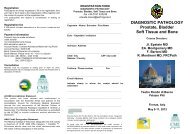
Fig. 3.21 Reactive gastritis with marked foveolar<br />
hyperplasia.<br />
inflammation, and <strong>the</strong> distinction between<br />
intraepi<strong>the</strong>lial and invasive carcinoma<br />
{1683, 1025}. Several proposals have<br />
been made for <strong>the</strong> terminology <strong>of</strong> <strong>the</strong><br />
morphological spectrum <strong>of</strong> lesions that lie<br />
between non-neoplastic changes and<br />
early invasive cancer, including <strong>the</strong><br />
recent international Padova classification<br />
{1636}.<br />
Indefinite for intraepi<strong>the</strong>lial neoplasia<br />
Sometimes, doubts arise as to whe<strong>the</strong>r a<br />
lesion is neoplastic or non-neoplastic (i.e.<br />
reactive or regenerative), particularly in<br />
small biopsies. In such cases, <strong>the</strong> dilemma<br />
is usually solved by cutting deeper<br />
levels <strong>of</strong> <strong>the</strong> block, by obtaining additional<br />
biopsies, or after removing possible<br />
sources <strong>of</strong> cellular hyperproliferation. One<br />
important source <strong>of</strong> a potentially alarming<br />
lesion is <strong>the</strong> regeneration associated with<br />
NSAID-induced injury or superficial erosion/ulceration<br />
caused by gastric acid.<br />
Cases lacking all <strong>the</strong> attributes required<br />
for a definitive diagnosis <strong>of</strong> intraepi<strong>the</strong>lial<br />
neoplasia may be placed into <strong>the</strong> category<br />
‘indefinite for intraepi<strong>the</strong>lial neoplasia’.<br />
In native gastric mucosa, foveolar hyperproliferation<br />
may be indefinite for dysplasia,<br />
showing irregular and tortuous tubular<br />
structures with epi<strong>the</strong>lial mucus depletion,<br />
a high nuclear-cytoplasmic ratio and loss<br />
<strong>of</strong> cellular polarity. Large, oval/round,<br />
hyperchromatic nuclei associate with<br />
prominent mitoses, usually located near<br />
<strong>the</strong> proliferative zone in <strong>the</strong> mucous neck<br />
region.<br />
In intestinal metaplasia, areas indefinite<br />
for intraepi<strong>the</strong>lial neoplasia exhibit a<br />
hyperproliferative metaplastic epi<strong>the</strong>lium.<br />
The glands may appear closely packed,<br />
lined by cells with large, hyperchromatic,<br />
rounded or elongated, basally located<br />
nuclei. Nucleoli are an inconsistent finding.<br />
The cyto-architectural alterations tend<br />
to decrease from <strong>the</strong> base <strong>of</strong> <strong>the</strong> glands to<br />
<strong>the</strong>ir superficial portion.<br />
Intraepi<strong>the</strong>lial neoplasia<br />
It has flat, polypoid, or slightly depressed<br />
growth patterns; <strong>the</strong> flat pattern may lack<br />
any endoscopic changes on conventional<br />
endoscopy, but shows an irregular<br />
appearance on dye endoscopy. In<br />
Western countries, <strong>the</strong> term adenoma is<br />
applied when <strong>the</strong> proliferation produces<br />
a macroscopic, usually discrete, protruding<br />
lesion. However, in Japan, adenomas<br />
include all gross types (i.e. flat, elevated<br />
and depressed). Gastric adenomas are<br />
less common than hyperplastic polyps;<br />
overall, <strong>the</strong>y account for approximately<br />
10% <strong>of</strong> gastric polyps {1843}. They tend<br />
to arise in <strong>the</strong> antrum or mid stomach in<br />
areas <strong>of</strong> intestinal metaplasia.<br />
Morphologically, adenomas can be<br />
described as tubular (<strong>the</strong> most common),<br />
tubulovillous, or villous; <strong>the</strong> latter<br />
two have also been called papillotubular<br />
and papillary. Most have epi<strong>the</strong>lium <strong>of</strong><br />
intestinal type, but some have gastric<br />
foveolar features.<br />
Low-grade intraepi<strong>the</strong>lial neoplasia<br />
This lesion shows a slightly modified<br />
mucosal architecture, including <strong>the</strong> presence<br />
<strong>of</strong> tubular structures with budding<br />
and branching, papillary enfolding, crypt<br />
leng<strong>the</strong>ning with serration, and cystic<br />
changes. Glands are lined by enlarged<br />
columnar cells with minimal or no mucin.<br />
Homogeneously blue vesicular, rounded<br />
or ovoid nuclei are usually pseudostratified<br />
in <strong>the</strong> proliferation zone located at<br />
<strong>the</strong> superficial portion <strong>of</strong> <strong>the</strong> dysplastic<br />
tubules.<br />
High-grade intraepi<strong>the</strong>lial neoplasia<br />
There is increasing architectural distortion<br />
with glandular crowding and prominent<br />
cellular atypia. Tubules can be irregular in<br />
shape, with frequent branching and fold-<br />
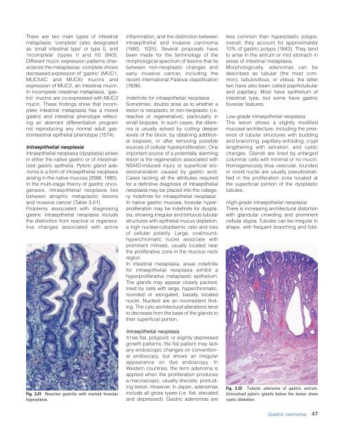
Fig. 3.22 Tubular adenoma <strong>of</strong> gastric antrum.<br />
Uninvolved pyloric glands below <strong>the</strong> lesion show<br />
cystic dilatation.<br />
Gastric carinoma<br />
47