CHAPTER 3 Tumours of the Stomach - Pathology Outlines
CHAPTER 3 Tumours of the Stomach - Pathology Outlines
CHAPTER 3 Tumours of the Stomach - Pathology Outlines

Create successful ePaper yourself
Turn your PDF publications into a flip-book with our unique Google optimized e-Paper software.
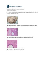
A<br />
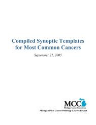
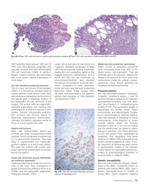
Fig. 3.33 A Type I ECL-cell carcinoid in a patient with pernicious anaemia. B Type II ECL-cell carcinoid in a patient with MEN1 and ZES.<br />
B<br />
Ki67 labelling index (above 1000 per 10<br />
HPF) and more frequent lymphatic and<br />
vascular invasion than well differentiated<br />
ECL-cell carcinoids {1589}. In addition,<br />
deeply invasive tumours are associated<br />
with local and/or distant metastases in<br />
most cases.<br />
EC-cell, serotonin-producing carcinoid<br />
This is a very rare tumour in <strong>the</strong> stomach<br />
{1591}. It is formed by rounded nests <strong>of</strong><br />
closely packed small tumour cells, <strong>of</strong>ten<br />
with peripheral palisading, reminiscent <strong>of</strong><br />
<strong>the</strong> typical type A histologic pattern <strong>of</strong><br />
<strong>the</strong> argentaffin EC-cell carcinoid <strong>of</strong> <strong>the</strong><br />
midgut. The tumour cells are argentaffin,<br />
intensely argyrophilic and reactive with<br />
chromogranin A and anti-serotonin antibodies.<br />
Electron microscopic examination<br />
confirms <strong>the</strong> EC-cell nature by<br />
detecting characteristic pleomorphic,<br />
intensely osmiophilic granules similar to<br />
those <strong>of</strong> normal gastric EC-cells.<br />
Gastrin-cell tumours<br />
Most well differentiated gastrin-cell<br />
tumours are small mucosal-submucosal<br />
nodules, found incidentally at endoscopy<br />
or in a gastrectomy specimen. They may<br />
show a characteristic thin trabeculargyriform<br />
pattern or a solid nest pattern.<br />
The cells are uniform with scanty cytoplasm<br />
and show predominant immunoreactivity<br />
for gastrin.<br />
Small cell carcinoma (poorly differentiated<br />
endocrine neoplasm)<br />
These are identical to small cell carcinomas<br />
<strong>of</strong> <strong>the</strong> lung. They correspond to<br />
grade 3 tumours according to Rindi et al.<br />
{1589}, and are particularly aggressive,<br />
malignant tumours {1591}.<br />
Large cell neuroendocrine carcinoma is a<br />
malignant neoplasm composed <strong>of</strong> large<br />
cells having organoid, nesting, trabecular,<br />
rosette-like and palisading patterns that<br />
suggest endocrine differentiation, and in<br />
which <strong>the</strong> last can be confirmed by<br />
immunohistochemistry and electron<br />
microscopy. In contrast to small cell carcinoma,<br />
cytoplasm is more abundant,<br />
nuclei are more vesicular and nucleoli are<br />
prominent {1954}. These tumours have<br />
not been well described in <strong>the</strong> gastrointestinal<br />
tract because <strong>of</strong> <strong>the</strong>ir apparent<br />
low frequency {1188}.<br />
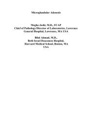
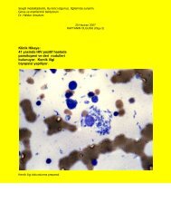
Fig. 3.34 ECL-cell carcinoid showing immunoexpression<br />
<strong>of</strong> chromogranin A.<br />
Mixed exocrine-endocrine carcinomas<br />
These consist <strong>of</strong> neoplastic endocrine<br />
cells composing more than 30% <strong>of</strong> <strong>the</strong><br />
whole tumour cell population. They are<br />
relatively rare in <strong>the</strong> stomach, despite <strong>the</strong><br />
frequent occurrence <strong>of</strong> minor endocrine<br />
components inside <strong>the</strong> ordinary adenocarcinoma.<br />
They should generally be<br />
classified as adenocarcinomas.<br />
Precursor lesions<br />
ECL-cell carcinoids arising in hypergastrinaemic<br />
conditions (types I and II)<br />
develop through a sequence <strong>of</strong> hyperplasia-dysplasia-neoplasia<br />
that has been<br />
well documented in histopathological<br />
studies {1777}. The successive stages <strong>of</strong><br />
hyperplasia are termed simple, linear,<br />
micronodular, and adenomatoid. Dysplasia<br />
is characterized by relatively atypical<br />
cells with features <strong>of</strong> enlarging or fusing<br />
micronodules, micro-invasion or newly<br />
formed stroma. When <strong>the</strong> nodules<br />
increase in size to > 0.5 mm or invade<br />
into <strong>the</strong> submucosa, <strong>the</strong> lesion is classified<br />
as a carcinoid. The entire spectrum<br />
<strong>of</strong> ECL-cell growth, from hyperplasia to<br />
dysplasia and neoplasia has been<br />
observed in MEN-1/ZES and autoimmune<br />
chronic atrophic gastritis (A-CAG). A similar<br />
sequence <strong>of</strong> lesions has been shown<br />
in experimental models <strong>of</strong> <strong>the</strong> disease,<br />
mostly based on hypergastrinaemia secondary<br />
to pharmacological inhibition <strong>of</strong><br />
acid secretion in rodents {1896}.<br />
Genetic susceptibility<br />
ECL-cell carcinoids are integral components<br />
<strong>of</strong> <strong>the</strong> MEN-1 syndrome {1042}. In<br />
patients with familial MEN-1/ZES, type II<br />
gastric carcinoids arise in 13-30% <strong>of</strong><br />
cases {854, 1042}. However, patients<br />
Endocrine tumours<br />
55