CHAPTER 3 Tumours of the Stomach - Pathology Outlines
CHAPTER 3 Tumours of the Stomach - Pathology Outlines
CHAPTER 3 Tumours of the Stomach - Pathology Outlines

You also want an ePaper? Increase the reach of your titles
YUMPU automatically turns print PDFs into web optimized ePapers that Google loves.
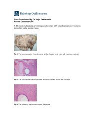
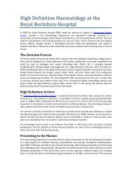
Fig. 3.40 Low-grade B-cell MALT lymphoma. Small<br />
lymphoid cells form a diffuse infiltrate extending<br />
into <strong>the</strong> submucosa.<br />
cells, is due to a mechanism mediated<br />
via T-cells and that this help is contact<br />
dependant. Fur<strong>the</strong>r studies have shown<br />
that <strong>the</strong> T-cells responsible for <strong>the</strong> proliferative<br />
drive are specifically those found<br />
within <strong>the</strong> tumour and <strong>the</strong>ir function cannot<br />
be replaced by T-cells derived from<br />
elsewhere (e.g. <strong>the</strong> spleen) in <strong>the</strong> same<br />
patient {769}.<br />
Histopathology<br />
The organisation <strong>of</strong> <strong>the</strong> lymphoma mimics<br />
that <strong>of</strong> normal MALT and <strong>the</strong> cellular<br />
morphology and immunophenotype is<br />
essentially that <strong>of</strong> <strong>the</strong> marginal zone<br />
B-cell. The neoplastic cells infiltrate<br />
between pre-existing lymphoid follicles,<br />
initially loca-lised outside <strong>the</strong> follicular<br />
mantle zone in a marginal zone pattern.<br />
As <strong>the</strong> lesion progresses, <strong>the</strong> neoplastic<br />
cells erode, colonize and eventually<br />
overrun <strong>the</strong> lymphoid follicles resulting in<br />
a vague nodularity to an o<strong>the</strong>rwise diffuse<br />
lymphomatous infiltrate {800}. The<br />
morphology <strong>of</strong> <strong>the</strong> neoplastic cell can be<br />
variable even within a single case.<br />
Characteristically, <strong>the</strong> cell is <strong>of</strong> intermediate<br />
size with pale cytoplasm and an<br />
irregular nucleus. The resemblance <strong>of</strong><br />
<strong>the</strong>se cells to <strong>the</strong> centrocyte <strong>of</strong> <strong>the</strong> follicle<br />
centre has led to <strong>the</strong> term ‘centrocyte-like<br />
(CCL)’ cell being applied to <strong>the</strong> neoplastic<br />
component <strong>of</strong> MALT lymphomas. In<br />
some cases, <strong>the</strong> CCL cell may be more<br />
reminiscent <strong>of</strong> a mature small B lymphocyte<br />
while in o<strong>the</strong>r cases, <strong>the</strong> cell may<br />
have a monocytoid appearance with<br />
more abundant, pale cytoplasm and a<br />
well defined cell border. Plasma cell differentiation<br />
is typical and may be very<br />
prominent. Dutcher bodies may be identified.<br />
The CCL cells infiltrate and destroy<br />
adjacent gastric glands to form lymphoepi<strong>the</strong>lial<br />
lesions. Lympho-epi<strong>the</strong>lial<br />
lesions typical for MALT lymphoma are<br />
defined as infiltration <strong>of</strong> <strong>the</strong> glandular<br />
epi<strong>the</strong>lium by clusters <strong>of</strong> neoplastic lymphoid<br />
cells with associated destruction<br />
<strong>of</strong> gland architecture and morphological<br />
changes within <strong>the</strong> epi<strong>the</strong>lial cells,<br />
including increased eosinophilia.<br />
Immunohistochemistry<br />
The immunophenotype <strong>of</strong> <strong>the</strong> CCL cell is<br />
similar to that <strong>of</strong> <strong>the</strong> marginal zone B-cell.<br />
There is expression <strong>of</strong> pan-B-cell antigens<br />
such as CD20 and CD79a and <strong>the</strong><br />
more mature B-cell markers CD21 and<br />
CD35. The cells do not express CD10.<br />
They are usually positive for bcl-2 protein<br />
and may express CD43 but do not<br />
express CD5 or CD23. They express surface<br />
and, to a lesser extent, cytoplasmic<br />
immunoglobulin (usually IgM or IgA,<br />
rarely IgG) and show light chain restriction.<br />
Immunostaining with anti-cytokeratin<br />
antibodies is useful in demonstrating<br />
lymphoepi<strong>the</strong>lial lesions. Immunostaining<br />
with antibodies that highlight follicular<br />
dendritic cells (anti-CD21, anti-CD23 or<br />
anti-CD35) help to demonstrate underlying<br />
follicular dendritic cell networks in<br />
those cases in which <strong>the</strong> lymphoid follicles<br />
have been completely overrun by<br />
<strong>the</strong> lymphoma.<br />
Differential diagnosis<br />
The distinction between florid gastritis<br />
and low-grade MALT lymphoma may be<br />
difficult. In such cases it is essential to<br />
have sufficient biopsy material (up to<br />
eight biopsies from endoscopically suspicious<br />
areas) with good preservation <strong>of</strong><br />
morphology and correct orientation <strong>of</strong><br />
<strong>the</strong> biopsy specimen. For <strong>the</strong> distinction<br />
between reactive and neoplastic infiltrates,<br />
histological evaluation remains <strong>the</strong><br />
gold standard, but accessory studies<br />
may be helpful. In both reactive and neoplastic<br />
cases, lymphoid follicles are present<br />
and <strong>the</strong>se may be associated with<br />
active inflammation, crypt abscesses<br />
and reactive epi<strong>the</strong>lial changes. In gastritis,<br />
<strong>the</strong> infiltrate surrounding <strong>the</strong> lymphoid<br />
follicles in <strong>the</strong> lamina propria is<br />
plasma cell predominant while in MALT<br />
lymphoma <strong>the</strong> infiltrate contains a dominant<br />
population <strong>of</strong> lymphocytes with CCL<br />
cell morphology, infiltrating through <strong>the</strong><br />
lamina propria and around glands.<br />
Prominent lymphoepi<strong>the</strong>lial lesions,<br />
Dutcher bodies and moderate cytological<br />
atypia are associated only with lymphoma.<br />
All <strong>of</strong> <strong>the</strong>se features may not be<br />
present in biopsy material from a single<br />
case. In some cases it is justifiable to<br />
make <strong>the</strong> diagnosis <strong>of</strong> low-grade MALT<br />
lymphoma in <strong>the</strong> absence <strong>of</strong> one or more<br />
<strong>of</strong> <strong>the</strong>se features if <strong>the</strong> overall histological<br />
appearances are those <strong>of</strong> lymphoma.<br />
Rare or questionable lymphoepi<strong>the</strong>lial<br />
lesions, dense lymphoid infiltration, mild<br />
cytological atypia and muscularis mucosae<br />
invasion are features more <strong>of</strong>ten<br />
associated with, but not limited to, lymphoma<br />
{2212}.<br />
In some cases it will not be possible to<br />
make a definite distinction between reactive<br />
infiltrates and lymphoma and in<br />
<strong>the</strong>se cases a diagnosis <strong>of</strong> ‘atypical lymphoid<br />
infiltrate <strong>of</strong> uncertain nature’ is<br />
appropriate.<br />
Effect <strong>of</strong> H. pylori eradication<br />
The histological appearances <strong>of</strong> gastric<br />
biopsies from patients showing complete<br />
regression <strong>of</strong> lymphoma after H. pylori<br />
A<br />
B<br />
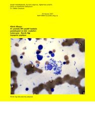
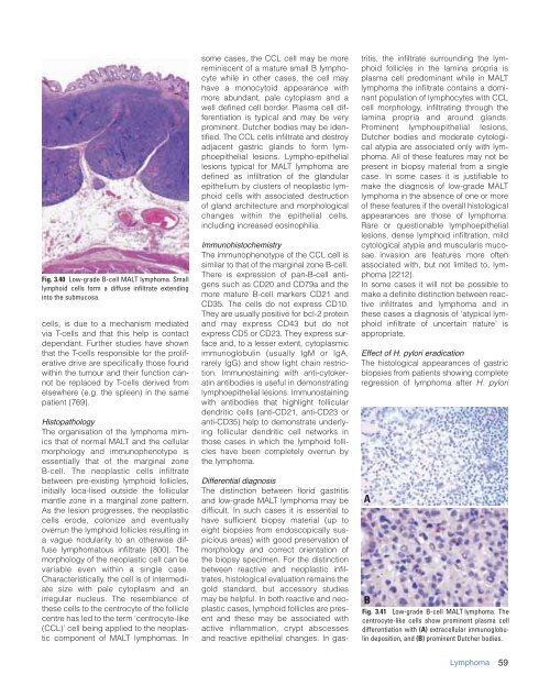
Fig. 3.41 Low-grade B-cell MALT lymphoma. The<br />
centrocyte-like cells show prominent plasma cell<br />
differentiation with (A) extracellular immunoglobulin<br />
deposition, and (B) prominent Dutcher bodies.<br />
Lymphoma<br />
59