CHAPTER 3 Tumours of the Stomach - Pathology Outlines
CHAPTER 3 Tumours of the Stomach - Pathology Outlines
CHAPTER 3 Tumours of the Stomach - Pathology Outlines

You also want an ePaper? Increase the reach of your titles
YUMPU automatically turns print PDFs into web optimized ePapers that Google loves.
A<br />
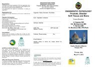
Fig. 3.11 A, B Tubular adenocarcinoma.<br />
A<br />
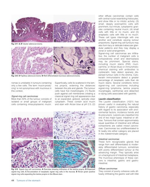
Fig. 3.12 A Papillary adenocarcinoma. B Well differentiated mucinous adenocarcinoma.<br />
nomas is unreliable in tumours containing<br />
only a few cells. The term ‘mucin-producing’<br />
is not synonymous with mucinous in<br />
this context.<br />
Signet-ring cell carcinomas<br />
More than 50% <strong>of</strong> <strong>the</strong> tumour consists <strong>of</strong><br />
isolated or small groups <strong>of</strong> malignant<br />
cells containing intracytoplasmic mucin.<br />
A<br />
C<br />
Superficially, cells lie scattered in <strong>the</strong> lamina<br />
propria, widening <strong>the</strong> distances<br />
between <strong>the</strong> pits and glands. The tumour<br />
cells have five morphologies: (1) Nuclei<br />
push against cell membranes creating a<br />
classical signet ring cell appearance due<br />
to an expanded, globoid, optically clear<br />
cytoplasm. These contain acid mucin<br />
and stain with Alcian blue at pH 2.5; (2)<br />
Fig. 3.13 Signet-ring cell carcinomas. A Overview showing Infiltration <strong>of</strong> <strong>the</strong> lamina propria. B Dispersed<br />
signet-ring cells. C Accumulation <strong>of</strong> neoplastic signet ring cells in <strong>the</strong> mucosa. D Alcian green positive<br />
signet-ring cells expanding <strong>the</strong> lamina propria in this Movat stain.<br />
B<br />
B<br />
B<br />
D<br />
o<strong>the</strong>r diffuse carcinomas contain cells<br />
with central nuclei resembling histiocytes,<br />
and show little or no mitotic activity; (3)<br />
small, deeply eosinophilic cells with<br />
prominent, but minute, cytoplasmic granules<br />
containing neutral mucin; (4) small<br />
cells with little or no mucin, and (5)<br />
anaplastic cells with little or no mucin.<br />
These cell types intermingle with one<br />
ano<strong>the</strong>r and constitute varying tumour<br />
proportions. Signet-ring cell tumours may<br />
also form lacy or delicate trabecular glandular<br />
patterns and <strong>the</strong>y may display a<br />
zonal or solid arrangement.<br />
Signet-ring cell carcinomas are infiltrative;<br />
<strong>the</strong> number <strong>of</strong> malignant cells is<br />
comparatively small and desmoplasia<br />
may be prominent. Special stains,<br />
including mucin stains (PAS, mucicarmine,<br />
or Alcian blue) or immunohistochemical<br />
staining with antibodies to<br />
cytokeratin, help detect sparsely dispersed<br />
tumour cells in <strong>the</strong> stroma. Cytokeratin<br />
immunostains detect a greater<br />
percentage <strong>of</strong> neoplastic cells than do<br />
mucin stains. Several conditions mimic<br />
signet-ring cell carcinoma including<br />
signet-ring lymphoma, lamina propria<br />
muciphages, xanthomas and detached<br />
or dying cells associated with gastritis.<br />
Laurén classification<br />
The Laurén classification {1021} has<br />
proven useful in evaluating <strong>the</strong> natural<br />
history <strong>of</strong> gastric carcinoma, especially<br />
with regard to its association with environmental<br />
factors, incidence trends and<br />
its precursors. Lesions are classified into<br />
one <strong>of</strong> two major types: intestinal or diffuse.<br />
<strong>Tumours</strong> that contain approximately<br />
equal quantities <strong>of</strong> intestinal and diffuse<br />
components are called mixed carcinomas.<br />
Carcinomas too undifferentiated to<br />
fit neatly into ei<strong>the</strong>r category are placed<br />
in <strong>the</strong> indeterminate category.<br />
Intestinal carcinomas<br />
These form recognizable glands that<br />
range from well differentiated to moderately<br />
differentiated tumours, sometimes<br />
with poorly differentiated tumour at <strong>the</strong><br />
advancing margin. They typically arise<br />
on a background <strong>of</strong> intestinal metaplasia.<br />
The mucinous phenotype <strong>of</strong> <strong>the</strong>se cancers<br />
is intestinal, gastric and gastrointestinal.<br />
Diffuse carcinomas<br />
They consist <strong>of</strong> poorly cohesive cells diffusely<br />
infiltrating <strong>the</strong> gastric wall with little<br />
44 <strong>Tumours</strong> <strong>of</strong> <strong>the</strong> stomach