CHAPTER 3 Tumours of the Stomach - Pathology Outlines
CHAPTER 3 Tumours of the Stomach - Pathology Outlines
CHAPTER 3 Tumours of the Stomach - Pathology Outlines

Create successful ePaper yourself
Turn your PDF publications into a flip-book with our unique Google optimized e-Paper software.
tiple and multicentric. Of 152 cases studied<br />
by endoscopy, 57% had more than<br />
two growths {1561}.<br />
Type II ECL-cell carcinoids<br />
Hypertrophic, hypersecretory gastropathy<br />
and high levels <strong>of</strong> circulating gastrin<br />
are critical diagnostic findings. In all<br />
cases, ECL-cell hyperplasia and/or dysplasia<br />
were noted in <strong>the</strong> fundic peritumoural<br />
mucosa {1590}. These gastric<br />
carcinoids are usually multiple and smaller<br />
than 1.5 cm in size in <strong>the</strong> majority <strong>of</strong><br />
cases {1590}.<br />
Type III (sporadic) ECL-cell carcinoids<br />
These lesions are not associated with<br />
hypergastinaemia or A-CAG. They are<br />
generally solitary growths, and arise in <strong>the</strong><br />
setting <strong>of</strong> gastric mucosa devoid <strong>of</strong><br />
ECL-cell hyperplasia/dysplasia and <strong>of</strong><br />
significant pathologic lesions except for<br />
gastritis (o<strong>the</strong>r than A-CAG). Rare multiple<br />
tumours have been observed {1590}.<br />
Clinically, type III tumours present (1) as a<br />
mass lesion with no evidence <strong>of</strong> endocrine<br />
symptoms (nonfunctioning carcinoid)<br />
and with clinical findings similar to<br />
those <strong>of</strong> adenocarcinoma, including gastric<br />
haemorrhage, obstruction and metastasis,<br />
or (2) with endocrine symptoms <strong>of</strong><br />
an ‘atypical carcinoid syndrome’ with red<br />
cutaneous flushing and absence <strong>of</strong> diarrhoea,<br />
usually coupled with liver metastases<br />
and production <strong>of</strong> histamine and<br />
5-hydroxytryptophan {1386, 1598}.<br />
Non ECL-cell gastric carcinoids.<br />
These uncommon tumours may present<br />
with ZES due to <strong>the</strong>ir gastrin production<br />
(which is more frequently found in duodenal<br />
gastrinomas) or with Cushing syndrome<br />
due to secretion <strong>of</strong> adrenocorticotrophic<br />
hormone (ACTH) {711, 1791}.<br />
Macroscopy<br />
Type I ECL-cell carcinoids are multiple in<br />
57% <strong>of</strong> cases {1590}, usually appearing<br />
as small tan nodules or polyps that are<br />
circumscribed in <strong>the</strong> mucosa or, more<br />
<strong>of</strong>ten, to <strong>the</strong> submucosa. Most tumours<br />
(77%) are < 1 cm in maximum diameter<br />
and 97% <strong>of</strong> tumours are < 1.5 cm. The<br />
muscularis propria is involved in only a<br />
minority <strong>of</strong> cases (7%) {1590}.<br />
The stomachs with type II tumours are<br />
enlarged and show a thickened gastric<br />
wall (0.6-4.5 cm) due to severe hypertrophic-hypersecretory<br />
gastropathy and<br />
multiple mucosal-submucosal nodules<br />
which, though larger than those <strong>of</strong> type I,<br />
are generally smaller than 1.5 cm in size<br />
in 75% <strong>of</strong> cases {1590}.<br />
Type III ECL-cell tumours are usually single<br />
and in 33% <strong>of</strong> <strong>the</strong> cases larger than 2<br />
cm in diameter. Infiltration <strong>of</strong> <strong>the</strong> muscularis<br />
propria is found in 76%, and <strong>of</strong> <strong>the</strong><br />
serosa in 53% <strong>of</strong> cases {1590}.<br />
Histopathology<br />
The histopathological categorization <strong>of</strong><br />
endocrine tumours <strong>of</strong> <strong>the</strong> stomach<br />
described here, is a modification <strong>of</strong> <strong>the</strong><br />
WHO classification <strong>of</strong> endocrine tumours<br />
{1784}.<br />
Carcinoid tumour<br />
A carcinoid is defined morphologically<br />
as a well differentiated neoplasm <strong>of</strong> <strong>the</strong><br />
diffuse endocrine system.<br />
ECL-cell carcinoid<br />
The majority <strong>of</strong> type I and type II<br />
ECL-cell carcinoids are characterized<br />
by small, microlobular-trabecular aggregates<br />
formed by regularly distributed,<br />
<strong>of</strong>ten aligned cells (mosaic-like pattern),<br />
with regular, monomorphic nuclei, usually<br />
inapparent nucleoli, ra<strong>the</strong>r abundant,<br />
fairly eosinophilic cytoplasm, almost<br />
absent mitoses, and infrequent angioinvasion.<br />
<strong>Tumours</strong> with <strong>the</strong>se features (grade 1<br />
according to Rindi et al {1589}) are generally<br />
limited to mucosa or submucosa<br />
{1589} and can be considered as<br />
tumours with benign behaviour. The ECL<br />
nature <strong>of</strong> <strong>the</strong> tumours is confirmed by<br />
strong argyrophilia by Grimelius or<br />
Sevier Munger techniques and positive<br />
immunoreactivity for chromogranin A, in<br />
<strong>the</strong> absence <strong>of</strong> reactivity for <strong>the</strong><br />
argentaffin or diazonium tests for serotonin,<br />
and no or only occasional<br />
immunoreactivity for hormonal products<br />
{1591}. Minor cell sub-populations expressing<br />
serotonin, gastrin, somatostatin,<br />
pancreatic polypeptide (PP), or<br />
α-hCG have been detected in a minority<br />
<strong>of</strong> tumours {1591}. A few ECL-cell<br />
tumours produce histamine and<br />
5-hydroxy-tryptophan; <strong>the</strong>se lesions,<br />
when <strong>the</strong>y metastasize, can produce<br />
‘atypical’ carcinoid syndrome {1591}<br />
Vesicular monoamine transporter type 2<br />
(VMAT-2) is a suitable and specific marker<br />
for ECL-cell tumours {1592} while histamine<br />
or histidine decarboxylase immunohistochemical<br />
analysis, although specific,<br />
is less suitable for routinely processed<br />
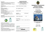
Fig. 3.32 Sporadic (type III) ECL-cell carcinoid <strong>of</strong> <strong>the</strong><br />
gastric body. The surrounding mucosa is normal.<br />
specimens {1865}. The ECL-cell nature <strong>of</strong><br />
argyrophil tumours is ultimately assessed<br />
by demonstrating ECL-type granules by<br />
electron microscopy {232, 1591}.<br />
Sporadic ECL-cell carcinoids are usually<br />
more aggressive than those associated<br />
with A-CAG or MEN-1. Histopathologically,<br />
<strong>the</strong>se tumours show a prevalence<br />
<strong>of</strong> solid cellular aggregates and large trabeculae,<br />
crowding, and irregular distribution<br />
<strong>of</strong> round to spindle and polyhedral<br />
tumour cells, fairly large vesicular nuclei<br />
with prominent eosinophilic nucleoli, or<br />
smaller, hyperchromatic nuclei with irregular<br />
chromatin clumps and small nucleoli,<br />
considerable mitotic activity, sometimes<br />
with atypical mitotic figures and<br />
scarce necrosis.<br />
<strong>Tumours</strong> with <strong>the</strong>se histological features<br />
or grade 2 features {1589} show a higher<br />
mitotic rate (mean <strong>of</strong> 9 per 10 HPF), a frequent<br />
expression <strong>of</strong> p53 (60%), a higher<br />
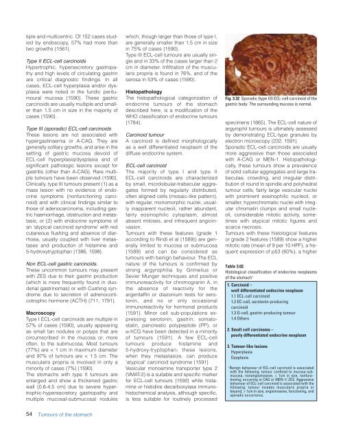
Table 3.02.<br />
Histological classification <strong>of</strong> endocrine neoplasms<br />
<strong>of</strong> <strong>the</strong> stomach 1<br />
1. Carcinoid –<br />
well differentiated endocrine neoplasm<br />
1.1 ECL-cell carcinoid<br />
1.2 EC-cell, serotonin-producing<br />
carcinoid<br />
1.3 G-cell, gastrin-producing tumour<br />
1.4 O<strong>the</strong>rs<br />
2. Small cell carcinoma –<br />
poorly differentiated endocrine neoplasm<br />
3. Tumour-like lesions<br />
Hyperplasia<br />
Dysplasia<br />
1<br />
Benign behaviour <strong>of</strong> ECL-cell carcinoid is associated<br />
with <strong>the</strong> following: tumour confined to mucosa-submucosa,<br />
nonangioinvasive, < 1cm in size, nonfunctioning;<br />
occurring in CAG or MEN-1/ ZES. Aggressive<br />
behaviour <strong>of</strong> ECL-cell carcinoid is associated with <strong>the</strong><br />
following: tumour invades muscularis propria or<br />
beyond, > 1cm in size, angioinvasive, functioning, and<br />
sporadic occurrence.<br />
54 <strong>Tumours</strong> <strong>of</strong> <strong>the</strong> stomach