CHAPTER 3 Tumours of the Stomach - Pathology Outlines
CHAPTER 3 Tumours of the Stomach - Pathology Outlines
CHAPTER 3 Tumours of the Stomach - Pathology Outlines

You also want an ePaper? Increase the reach of your titles
YUMPU automatically turns print PDFs into web optimized ePapers that Google loves.
Polyps<br />
Hyperplastic polyps<br />
Hyperplastic polyps are one <strong>of</strong> <strong>the</strong> commonest<br />
gastric polyps. They are sessile<br />
or pedunculated lesions, usually < 2.0<br />
cm in diameter, typically arising in <strong>the</strong><br />
antrum on a background <strong>of</strong> H. pylori gastritis.<br />
They contain a proliferation <strong>of</strong> surface<br />
foveolar cells lining elongated, distorted<br />
pits extending deep into <strong>the</strong><br />
stroma. They may contain pyloric glands,<br />
chief cells and parietal cells. The surface<br />
<strong>of</strong>ten erodes. In a minority <strong>of</strong> cases, carcinoma<br />
develops within <strong>the</strong> polyps in<br />
areas <strong>of</strong> intestinal metaplasia and dysplasia.<br />
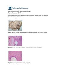
A<br />
B<br />
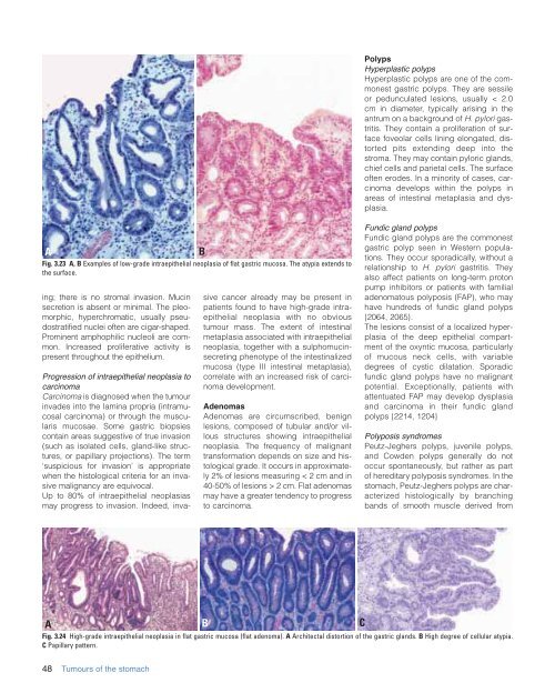
Fig. 3.23 A, B Examples <strong>of</strong> low-grade intraepi<strong>the</strong>lial neoplasia <strong>of</strong> flat gastric mucosa. The atypia extends to<br />
<strong>the</strong> surface.<br />
ing; <strong>the</strong>re is no stromal invasion. Mucin<br />
secretion is absent or minimal. The pleomorphic,<br />
hyperchromatic, usually pseudostratified<br />
nuclei <strong>of</strong>ten are cigar-shaped.<br />
Prominent amphophilic nucleoli are common.<br />
Increased proliferative activity is<br />
present throughout <strong>the</strong> epi<strong>the</strong>lium.<br />
Progression <strong>of</strong> intraepi<strong>the</strong>lial neoplasia to<br />
carcinoma<br />
Carcinoma is diagnosed when <strong>the</strong> tumour<br />
invades into <strong>the</strong> lamina propria (intramucosal<br />
carcinoma) or through <strong>the</strong> muscularis<br />
mucosae. Some gastric biopsies<br />
contain areas suggestive <strong>of</strong> true invasion<br />
(such as isolated cells, gland-like structures,<br />
or papillary projections). The term<br />
‘suspicious for invasion’ is appropriate<br />
when <strong>the</strong> histological criteria for an invasive<br />
malignancy are equivocal.<br />
Up to 80% <strong>of</strong> intraepi<strong>the</strong>lial neoplasias<br />
may progress to invasion. Indeed, invasive<br />
cancer already may be present in<br />
patients found to have high-grade intraepi<strong>the</strong>lial<br />
neoplasia with no obvious<br />
tumour mass. The extent <strong>of</strong> intestinal<br />
metaplasia associated with intraepi<strong>the</strong>lial<br />
neoplasia, toge<strong>the</strong>r with a sulphomucinsecreting<br />
phenotype <strong>of</strong> <strong>the</strong> intestinalized<br />
mucosa (type III intestinal metaplasia),<br />
correlate with an increased risk <strong>of</strong> carcinoma<br />
development.<br />
Adenomas<br />
Adenomas are circumscribed, benign<br />
lesions, composed <strong>of</strong> tubular and/or villous<br />
structures showing intraepi<strong>the</strong>lial<br />
neoplasia. The frequency <strong>of</strong> malignant<br />
transformation depends on size and histological<br />
grade. It occurs in approximately<br />
2% <strong>of</strong> lesions measuring < 2 cm and in<br />
40-50% <strong>of</strong> lesions > 2 cm. Flat adenomas<br />
may have a greater tendency to progress<br />
to carcinoma.<br />
Fundic gland polyps<br />
Fundic gland polyps are <strong>the</strong> commonest<br />
gastric polyp seen in Western populations.<br />
They occur sporadically, without a<br />
relationship to H. pylori gastritis. They<br />
also affect patients on long-term proton<br />
pump inhibitors or patients with familial<br />
adenomatous polyposis (FAP), who may<br />
have hundreds <strong>of</strong> fundic gland polyps<br />
{2064, 2065}.<br />
The lesions consist <strong>of</strong> a localized hyperplasia<br />
<strong>of</strong> <strong>the</strong> deep epi<strong>the</strong>lial compartment<br />
<strong>of</strong> <strong>the</strong> oxyntic mucosa, particularly<br />
<strong>of</strong> mucous neck cells, with variable<br />
degrees <strong>of</strong> cystic dilatation. Sporadic<br />
fundic gland polyps have no malignant<br />
potential. Exceptionally, patients with<br />
attentuated FAP may develop dysplasia<br />
and carcinoma in <strong>the</strong>ir fundic gland<br />
polyps {2214, 1204}<br />
Polyposis syndromes<br />
Peutz-Jeghers polyps, juvenile polyps,<br />
and Cowden polyps generally do not<br />
occur spontaneously, but ra<strong>the</strong>r as part<br />
<strong>of</strong> hereditary polyposis syndromes. In <strong>the</strong><br />
stomach, Peutz-Jeghers polyps are characterized<br />
histologically by branching<br />
bands <strong>of</strong> smooth muscle derived from<br />
A B C<br />
Fig. 3.24 High-grade intraepi<strong>the</strong>lial neoplasia in flat gastric mucosa (flat adenoma). A Architectal distortion <strong>of</strong> <strong>the</strong> gastric glands. B High degree <strong>of</strong> cellular atypia.<br />
C Papillary pattern.<br />
48 <strong>Tumours</strong> <strong>of</strong> <strong>the</strong> stomach