CHAPTER 3 Tumours of the Stomach - Pathology Outlines
CHAPTER 3 Tumours of the Stomach - Pathology Outlines
CHAPTER 3 Tumours of the Stomach - Pathology Outlines

Create successful ePaper yourself
Turn your PDF publications into a flip-book with our unique Google optimized e-Paper software.
Mesenchymal tumours <strong>of</strong> <strong>the</strong> stomach<br />
M. Miettinen<br />
J.Y. Blay<br />
L.H. Sobin<br />
Definition<br />
Most gastrointestinal mesenchymal neoplasms<br />
are gastrointestinal stromal<br />
tumours (GIST) or smooth muscle types.<br />
They are predominantly located in <strong>the</strong><br />
stomach. The definitions <strong>of</strong> o<strong>the</strong>r mesenchymal<br />
lesions follow <strong>the</strong> WHO histological<br />
classification <strong>of</strong> s<strong>of</strong>t tissue<br />
tumours {2086}.<br />
Terminology<br />
The designation GIST was originally introduced<br />
as a neutral term for tumours that<br />
were nei<strong>the</strong>r leiomyomas nor schwannomas.<br />
The term GIST is now used for a<br />
specific group <strong>of</strong> tumours comprising <strong>the</strong><br />
majority <strong>of</strong> all gastrointestinal mesenchymal<br />
tumours. These tumours encompass<br />
most gastric and intestinal mesenchymal<br />
tumours earlier designated as leiomyoma,<br />
cellular leiomyoma, leiomyoblastoma and<br />
leiomyosarcoma {80, 76, 78, 79, 1227}.<br />
Currently, <strong>the</strong> terms leiomyoma and<br />
leiomyosarcoma are reserved for those<br />
tumours that show smooth muscle differentiation,<br />
histologically or by immunohistochemistry,<br />
e.g. with strong and diffuse<br />
actin and desmin positivity. Most tumours<br />
historically called leiomyosarcoma {31,<br />
1559, 1750} are now classified as GISTs;<br />
hence <strong>the</strong> old literature on gastric (and<br />
intestinal) leiomyosarcomas largely<br />
reflects GISTs.<br />
Epidemiology<br />
GIST accounts for 2.2% <strong>of</strong> malignant gastric<br />
tumours in SEER data. There is no gender<br />
preference (M:F, 1.1:1), in contrast to<br />
carcinomas which have a M:F <strong>of</strong> 2:1<br />
{1928}. Adults between <strong>the</strong> 6th and 8th<br />
decade are primarily affected. The ratio <strong>of</strong><br />
<strong>the</strong> age-adjusted incidence rates for<br />
Blacks and Whites is greater for sarcomas<br />
(3 to 1) than for carcinomas (2 to 1). Black<br />
women are affected six times more frequently<br />
than white women (0.6 versus 0.1<br />
per 100,000 per year, analogous to <strong>the</strong><br />
ratio for uterine leiomyosarcomas) {1584}.<br />
Localization<br />
GISTs occur at every level <strong>of</strong> <strong>the</strong> tubular<br />
gastrointestinal tract and additionally<br />
may be primary in <strong>the</strong> omentum and<br />
mesentery. They are most common in <strong>the</strong><br />
stomach (60-70%), followed by small<br />
intestine (20-30%), colorectum and<br />
oesophagus (toge<strong>the</strong>r < 10%) {1227}.<br />
Clinical features<br />
GISTs present a spectrum from clinically<br />
benign, small to medium-sized tumours,<br />
to frank sarcomas. According to our estimate,<br />
approximately 30% <strong>of</strong> GISTs are<br />
clinically malignant, and a substantial<br />
number <strong>of</strong> patients with apparent radical<br />
surgery will relapse {1344, 462}. Typical<br />
<strong>of</strong> <strong>the</strong> malignant GISTs at all locations is<br />
intra-abdominal spread as multiple<br />
tumour nodules, and distant metastases<br />
most commonly to liver followed by lung<br />
and bone in decreasing frequency<br />
{478A, 1984, 1855}. Vague abdominal<br />
discomfort is <strong>the</strong> usual complaint in<br />
symptomatic tumours. Both benign and<br />
sarcomatous GISTs that project into <strong>the</strong><br />
lumen may ulcerate and be a source <strong>of</strong><br />
bleeding {80, 78, 79}.<br />
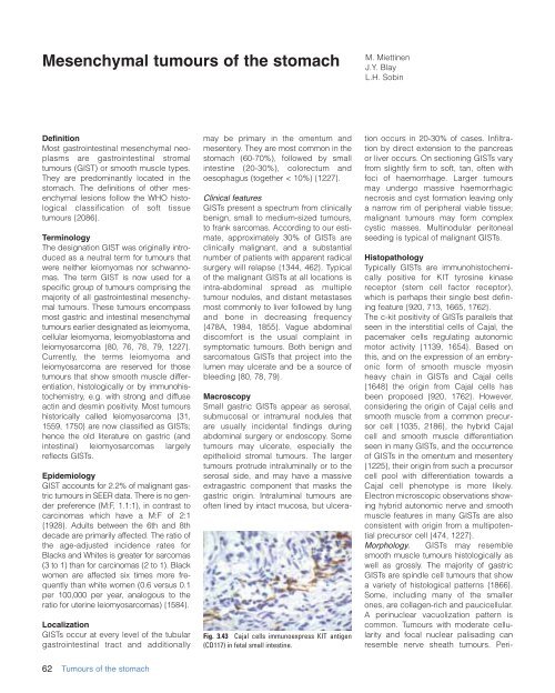
Fig. 3.43 Cajal cells immunoexpress KIT antigen<br />
(CD117) in fetal small intestine.<br />
Macroscopy<br />
Small gastric GISTs appear as serosal,<br />
submucosal or intramural nodules that<br />
are usually incidental findings during<br />
abdominal surgery or endoscopy. Some<br />
tumours may ulcerate, especially <strong>the</strong><br />
epi<strong>the</strong>lioid stromal tumours. The larger<br />
tumours protrude intraluminally or to <strong>the</strong><br />
serosal side, and may have a massive<br />
extragastric component that masks <strong>the</strong><br />
gastric origin. Intraluminal tumours are<br />
<strong>of</strong>ten lined by intact mucosa, but ulceration<br />
occurs in 20-30% <strong>of</strong> cases. Infiltration<br />
by direct extension to <strong>the</strong> pancreas<br />
or liver occurs. On sectioning GISTs vary<br />
from slightly firm to s<strong>of</strong>t, tan, <strong>of</strong>ten with<br />
foci <strong>of</strong> haemorrhage. Larger tumours<br />
may undergo massive haemorrhagic<br />
necrosis and cyst formation leaving only<br />
a narrow rim <strong>of</strong> peripheral viable tissue;<br />
malignant tumours may form complex<br />
cystic masses. Multinodular peritoneal<br />
seeding is typical <strong>of</strong> malignant GISTs.<br />
Histopathology<br />
Typically GISTs are immunohistochemically<br />
positive for KIT tyrosine kinase<br />
receptor (stem cell factor receptor),<br />
which is perhaps <strong>the</strong>ir single best defining<br />
feature {920, 713, 1665, 1762}.<br />
The c-kit positivity <strong>of</strong> GISTs parallels that<br />
seen in <strong>the</strong> interstitial cells <strong>of</strong> Cajal, <strong>the</strong><br />
pacemaker cells regulating autonomic<br />
motor activity {1139, 1654}. Based on<br />
this, and on <strong>the</strong> expression <strong>of</strong> an embryonic<br />
form <strong>of</strong> smooth muscle myosin<br />
heavy chain in GISTs and Cajal cells<br />
{1648} <strong>the</strong> origin from Cajal cells has<br />
been proposed {920, 1762}. However,<br />
considering <strong>the</strong> origin <strong>of</strong> Cajal cells and<br />
smooth muscle from a common precursor<br />
cell {1035, 2186}, <strong>the</strong> hybrid Cajal<br />
cell and smooth muscle differentiation<br />
seen in many GISTs, and <strong>the</strong> occurrence<br />
<strong>of</strong> GISTs in <strong>the</strong> omentum and mesentery<br />
{1225}, <strong>the</strong>ir origin from such a precursor<br />
cell pool with differentiation towards a<br />
Cajal cell phenotype is more likely.<br />
Electron microscopic observations showing<br />
hybrid autonomic nerve and smooth<br />
muscle features in many GISTs are also<br />
consistent with origin from a multipotential<br />
precursor cell {474, 1227}.<br />
Morphology. GISTs may resemble<br />
smooth muscle tumours histologically as<br />
well as grossly. The majority <strong>of</strong> gastric<br />
GISTs are spindle cell tumours that show<br />
a variety <strong>of</strong> histological patterns {1866}.<br />
Some, including many <strong>of</strong> <strong>the</strong> smaller<br />
ones, are collagen-rich and paucicellular.<br />
A perinuclear vacuolization pattern is<br />
common. <strong>Tumours</strong> with moderate cellularity<br />
and focal nuclear palisading can<br />
resemble nerve sheath tumours. Peri-<br />
62 <strong>Tumours</strong> <strong>of</strong> <strong>the</strong> stomach