A comparative structural analysis of direct and indirect shoot ...
A comparative structural analysis of direct and indirect shoot ...
A comparative structural analysis of direct and indirect shoot ...

You also want an ePaper? Increase the reach of your titles
YUMPU automatically turns print PDFs into web optimized ePapers that Google loves.
300 lM AlCl 3 induced enhanced formation <strong>of</strong> lateral roots<br />
(Fig. 2). Thus, the inhibition <strong>of</strong> root elongation was tightly<br />
correlated with, <strong>and</strong> partly compensated by, an increasing<br />
number <strong>of</strong> growing zones with the formation <strong>of</strong> lateral roots.<br />
Histochemical detection <strong>of</strong> aluminium<br />
For histochemical detection <strong>of</strong> aluminium in the roots <strong>of</strong><br />
Arabidopsis plants cultivated on agar plates with different<br />
concentrations <strong>of</strong> aluminium, two different staining methods<br />
were used: haematoxylin staining <strong>and</strong> morin staining (Polle<br />
et al., 1978; Vitorello <strong>and</strong> Haug, 1997). Using both methods,<br />
an aluminium-specific signal was observed mainly in the<br />
apical parts <strong>of</strong> the roots exposed to aluminium for 7 d<br />
(Fig. 3). In the control (Fig. 3), <strong>and</strong> at 10 lM AlCl 3 (data<br />
not shown), aluminium staining by haematoxylin was<br />
not detectable <strong>and</strong> only a weak diffuse fluorescence <strong>of</strong> morin<br />
occurred. At 50 lM AlCl 3 , there was hardly any detection<br />
<strong>of</strong> aluminium in roots by means <strong>of</strong> haematoxylin, while<br />
morin gave bright fluorescence (Fig. 3). At higher concentrations,<br />
haematoxylin started to detect aluminium in the<br />
roots, the pattern <strong>of</strong> staining being similar to the morin fluorescence.<br />
In the roots grown at 100 lM <strong>and</strong> 200 lM<br />
AlCl 3 , both staining methods showed maximum accumulation<br />
<strong>of</strong> aluminium in the root apex. In the plants exposed<br />
to 300 lM AlCl 3, aluminium was abundant not<br />
only in the root apex but it also invaded the root central<br />
cylinder (Fig. 3).<br />
Obviously, the sensitivity <strong>of</strong> morin to aluminium is higher<br />
than that <strong>of</strong> haematoxylin. Thus, morin is considered as<br />
a more suitable tracer dye for aluminium detection in the<br />
cells <strong>of</strong> the Arabidopsis root apex grown on aluminiumsupplemented<br />
agar plates. The critical discrimination limit<br />
in the sensitivity between morin <strong>and</strong> haematoxylin is apparently<br />
at 50 lM AlCl 3 (Fig. 3). Because <strong>of</strong> mild effects on<br />
root growth <strong>and</strong> the capability <strong>of</strong> morin to localize aluminium,<br />
50 lM AlCl 3 was utilized as the indicative testing<br />
concentration for monitoring aluminium effects on<br />
Arabidopsis roots in the following experiments.<br />
Aluminium sensitivity <strong>of</strong> root cells related to aluminium internalization 4205<br />
on this electrophysiological characteristic, all further experiments<br />
were performed separately for DTZ <strong>and</strong> PTZ.<br />
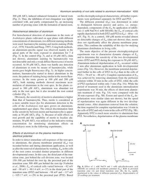
The diffusion potential (E D ) was determined in order<br />
to distinguish between passive <strong>and</strong> active, i.e. energydependent,<br />
components <strong>of</strong> the E m by application <strong>of</strong> inhibitors<br />
(1 mM NaCN+1 mM SHAM); the E m <strong>of</strong> cortical cells<br />
rapidly depolarized in both DTZ <strong>and</strong> PTZ to E D ( 40 to 41<br />
mV, Fig. 4). As a control, 100 lM morin revealed almost<br />
no detectable changes <strong>of</strong> E m (data not shown); thus, morin<br />
does not significantly affect the plasma membrane properties.<br />
This confirms the suitability <strong>of</strong> this dye for studying<br />
aluminium distribution in living cells.<br />
The main objective <strong>of</strong> the present electrophysiological<br />
experiments was to characterize dynamic changes <strong>of</strong> E m<br />
<strong>and</strong> to determine the sensitivity <strong>of</strong> the two developmental<br />
zones (DTZ <strong>and</strong> PTZ) during the exposure to 50 lM AlCl 3 .<br />
Aluminium-induced depolarization <strong>of</strong> E m occurred within<br />
2 min after aluminium application in both developmental<br />
zones (Fig. 5A). However, the membrane potential depolarized<br />
further in the cells <strong>of</strong> DTZ (to E D ) than in the cells <strong>of</strong><br />
PTZ ( 78 mV to 88 mV). Complete repolarization <strong>of</strong> E m<br />
was achieved by removing aluminium from the perfusion<br />
solution within 10 min in the cells <strong>of</strong> DTZ, while the cells<br />
<strong>of</strong> PTZ repolarized within only 3 min (Fig. 5A). While the<br />
period <strong>of</strong> treatment used in the aluminium internalization<br />
experiments was 30 min, the effects <strong>of</strong> short-term aluminium<br />
treatment (5 min; Fig. 5A) were compared with the<br />
30 min exposure (Fig. 5B). Extent <strong>and</strong> speed <strong>of</strong> the E m depolarization<br />
were similar (data not shown), but the speed<br />
<strong>of</strong> repolarization was again different in the two developmental<br />
zones. After aluminium removal from the solution,<br />
the time required for complete repolarization was 14 min in<br />
the cells <strong>of</strong> DTZ <strong>and</strong> only 6 min in the cells <strong>of</strong> PTZ (Fig. 5B).<br />
Apparently DTZ is much more sensitive to aluminium than<br />
PTZ. Nevertheless, depolarization <strong>of</strong> the plasma membrane<br />
Effects <strong>of</strong> aluminium on the plasma membrane<br />
electrical potential<br />
In order to detect immediate cell responses <strong>of</strong> the root apex<br />
to aluminium, the plasma membrane potential (E m ) was<br />
recorded before <strong>and</strong> during aluminium application, as well<br />
as after the removal <strong>of</strong> aluminium by washing. E m in the cells<br />
<strong>of</strong> the root cortex revealed distinct properties in different<br />
developmental zones. In this case, the distal part <strong>of</strong> the transition<br />
zone, just behind the cell division zone, was located<br />
150–300 lm behind the root tip, the proximal part <strong>of</strong> the<br />
transition zone 300–400 lm behind the root tip (Verbelen<br />
et al., 2006). E m <strong>of</strong> cortical cells varied between 82 mV<br />
<strong>and</strong> 98 mV ( 8864 mV, n¼37) in the distal part <strong>of</strong> the<br />
transition zone (DTZ) <strong>and</strong> between 94 mV <strong>and</strong> 117 mV<br />
( 10567 mV, n¼29) in the proximal part (PTZ). Based<br />
Fig. 4. Effect <strong>of</strong> 1 mM NaCN <strong>and</strong> 1 mM salicylhydroxamic acid<br />
(SHAM) on cortical cell membrane potential (E m ). Both in proximal<br />
transition zone (A) <strong>and</strong> distal transition zone (B), the E m rapidly<br />
depolarized to the values <strong>of</strong> diffusion potential (E D ). Representative <strong>of</strong><br />
20 seedlings per treatment.