Chitosan Loaded Mucoadhesive Microspheres of Gliclazide - Journal
Chitosan Loaded Mucoadhesive Microspheres of Gliclazide - Journal
Chitosan Loaded Mucoadhesive Microspheres of Gliclazide - Journal

Create successful ePaper yourself
Turn your PDF publications into a flip-book with our unique Google optimized e-Paper software.
R G U H S<br />
<strong>Journal</strong> <strong>of</strong><br />
Pharmaceutical<br />
Sciences<br />
July - September 2011 / Vol 1 / Issue 2<br />
ISSN: 2249-2208<br />
Abstracted and Indexed in Geneva Foundation for Medical Education and Research (GFMER)<br />
and Pharmaceutical Sciences Open Access Resources (PSOAR)<br />
Rajiv Gandhi University <strong>of</strong> Health Sciences, Karnataka<br />
w w w . r j p s . i n
R G U H S<br />
Dr. Divakar Goli<br />
Principal & Pr<strong>of</strong>essor <strong>of</strong> Biotechnology<br />
Acharya B M Reddy College <strong>of</strong> Pharmacy<br />
Bangalore<br />
Dr. Gopal Krishna Rao<br />
Pr<strong>of</strong>. & Head, Dept. <strong>of</strong> Pharmaceutical Chemistry<br />
Al-Ameen College <strong>of</strong> Pharmacy<br />
Bangalore<br />
Dr. Murugan V.<br />
Principal,Pr<strong>of</strong>essor <strong>of</strong> Pharmaceutical Chemistry<br />
Dayanand Sagar College <strong>of</strong> Pharmacy<br />
Bangalore<br />
Dr. Md. Naseeruddin Inamdar<br />
Pr<strong>of</strong> & Head, Dept. <strong>of</strong> Pharmacology<br />
Al-Ameen College <strong>of</strong> Pharmacy<br />
Bangalore<br />
Dr. Nagaraj<br />
Pr<strong>of</strong>essor<br />
Dept. <strong>of</strong> Pharma Analysis<br />
PES College <strong>of</strong> Pharmacy<br />
Bangalore<br />
<strong>Journal</strong> <strong>of</strong> Pharmaceutical Sciences (RJPS)<br />
EDITORIAL BOARD<br />
Dr. K.S Sriprakash<br />
Vice-Chancellor<br />
RGUHS<br />
Dr. D. Prem Kumar<br />
Registrar<br />
RGUHS<br />
Dr. Niranjan<br />
Registrar (Evaluation)<br />
RGUHS<br />
Editor-in-Chief<br />
Pr<strong>of</strong>. B. G. Shivananda<br />
Principal<br />
Al-Ameen College <strong>of</strong> Pharmacy, Bangalore<br />
Executive Editor<br />
Dr. Roopa S. Pai<br />
Pr<strong>of</strong>essor <strong>of</strong> Pharmaceutics<br />
Al-Ameen College <strong>of</strong> Pharmacy, Bangalore<br />
Associate Editor<br />
Dr. Raju B. Koneri<br />
Dean & Pr<strong>of</strong>essor <strong>of</strong> Pharmacology<br />
Karnataka College <strong>of</strong> Pharmacy, Bangalore<br />
MEMBERS<br />
Dr. Nithin Mahurkar<br />
Pr<strong>of</strong> & Head, Dept. <strong>of</strong> Pharmacology<br />
HKE College <strong>of</strong> Pharmacy<br />
Dr. Raman Dang<br />
Pr<strong>of</strong>essor, Dept. <strong>of</strong> Pharmacognosy<br />
Al-Ameen College <strong>of</strong> Pharmacy<br />
Bangalore<br />
Dr. Rama Raj Urs<br />
Librarian<br />
RGUHS, Bangalore<br />
Pr<strong>of</strong>. Ramesh C.<br />
Board <strong>of</strong> Studies Chairman<br />
Under Graduate<br />
RGUHS<br />
Dr. Sanjay Pai P.N.<br />
Pr<strong>of</strong>essor<br />
Dept. <strong>of</strong> Pharma Chemistry<br />
Goa College <strong>of</strong> Pharmacy<br />
Panaji, Goa<br />
Dr. Pranesh Gudur<br />
Director I/C Prasaranga<br />
RGUHS, Karnataka<br />
Dr. Shoba Rani R. Hiremath<br />
Pr<strong>of</strong> & Head, Dept. <strong>of</strong> Pharmacy Practice<br />
Al-Ameen College <strong>of</strong> Pharmacy<br />
Bangalore<br />
Pr<strong>of</strong>. Dr. Sirse Kranti Kumar<br />
Board <strong>of</strong> Studies Chairman<br />
Post Graduate<br />
RGUHS<br />
Dr. Srinath M.S.<br />
Dean<br />
Faculty <strong>of</strong> Pharmacy<br />
RGUHS<br />
Dr. Swamy P.V.<br />
Pr<strong>of</strong>essor<br />
Dept. <strong>of</strong> Pharmaceutics<br />
HKE College <strong>of</strong> Pharmacy<br />
Dr. Vishwanath B.A<br />
Principal<br />
Bangalore Institute <strong>of</strong> Pharmacy<br />
Education and Research, Bangalore
RJPS<br />
Issn: 2249-2208<br />
R G U H S<br />
<strong>Journal</strong> <strong>of</strong><br />
Pharmaceutical<br />
Sciences<br />
(An Official Publication <strong>of</strong> RGUHS)<br />
July - September 2011 / Vol 1 / Issue 2<br />
Rajiv Gandhi University <strong>of</strong> Health Sciences, Karnataka<br />
4th ‘T’ Block, Jayanagar, Bangalore 560041<br />
Phone: 080-26961934, 26961935, E-mail: rguhsjps@gmail.com<br />
Website: www.rjps.in
RJPS<br />
Editorial Board Welcomes<br />
Dr. K.S. Sriprakash<br />
Vice-Chancellor<br />
RGUHS, Bangalore<br />
Dr. K.S. Sriprakash has taken over charge as 6th Vice-Chancellor <strong>of</strong><br />
RGUHS w.e.f 14-06-2011. He was Director <strong>of</strong> Minto Ophthalmic<br />
Hospital, Regional Institute <strong>of</strong> Ophthalmology, Bangalore and Chief<br />
<strong>of</strong> Department <strong>of</strong> Vitre-retina. He has undergone training in Vitre-<br />
retina in Wills Eye Hospital, USA and Schieie Eye Institute, USA.<br />
Dr. K.S. Sriprakash has 29 years <strong>of</strong> experience and was instrumental<br />
in organising community ophthalmic services.
RGUHS<br />
<strong>Journal</strong> <strong>of</strong> Pharmaceutical<br />
Sciences<br />
Scientific Tools<br />
RJPS<br />
Contents<br />
Preamble<br />
Gowraganahalli Jagadeesh..................................................................................................................................................................................... 96<br />
Creative, Critical Thinking and Logic in Research<br />
Fredricka Reisman ..................................................................................................................................................................................... .. 97 - 102<br />
Review Article<br />
Need for Inclusion <strong>of</strong> Scientific Writing Skill Subjects in Indian Post Graduate Pharmacy Course<br />
Patil J.S, Kotnal R.B, Birajdar R.P, Marapur S.C and Kadam D.V .............................................................................................................. 103 - 106<br />
Research Article<br />
A Novel Spectrophotometric Estimation <strong>of</strong> Pramipexole in Bulk Drug and Formulations<br />
Shobha Manjunath, Satish Middi and Venkatesh Chouhan ........................................................................................................................ 107 - 110<br />
Validated UV-Spectrophotometric Estimation <strong>of</strong> Entecavir in Bulk and Formulations<br />
Malipatil S.M, Bharath S Athanikar and Mogal Dipali. ..................................................................................................................................111 - 116<br />
Antihyperlipidemic Effect <strong>of</strong> Ethanolic Extract <strong>of</strong> Hibiscus rosa sinensis Flowers in Hyperlipidemic Rats<br />
Sikarwar Mukesh S. and Patil M.B ...............................................................................................................................................................117 - 122<br />
A Study on Drug-Drug Interaction <strong>of</strong> Diltiazem with Nateglinide in Diabetic Animals<br />
Suresh D.K, Raza Hasan, Hamza Sheth, Md. Saifuddin Khalid and Mohiuddin M ..................................................................................... 123- 126<br />
Influence <strong>of</strong> Vitamin C with Lansoprazole in Pylorus Ligation Induced Ulcer Model in Rats<br />
Nitin M, Prasad K, Girish M, Ather Javed, Chetan M and Krunal S. ............................................................................................................127 - 130<br />
Assessment <strong>of</strong> Safety and Efficacy <strong>of</strong> Doxycycline and Azithromycin Preparations in Patients with Acne Vulgaris<br />
Mahendra Kumar B.J, Ramakrishna S, Kranti Basavant Patil, Sandeep A, Bhimaray S Krishnagoudar and Katti Ravi Venkappa .......... 131 - 135<br />
Antidiarrhoeal Activity <strong>of</strong> Aqueous Extract <strong>of</strong> Mimosa pudica Leaves<br />
Md. Saifuddin Khalid, Shah Jinesh Kumar, Suresh D.K., Rajnish Kumar Singh, Reddy Narasimha I.V. and Shaikh Azhar Hussain ........ 136 - 140<br />
Assessment <strong>of</strong> Various Combination <strong>of</strong> Drugs Used in Treatment <strong>of</strong> Lower Respiratory Tract Infection<br />
Imran Ahmad Khan, Shobha Rani R.H, Geeta S, Mahvash Iram ............................................................................................................... 141 - 145<br />
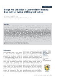
Formulation and Evaluation <strong>of</strong> <strong>Mucoadhesive</strong> Buccal Drug Delivery System <strong>of</strong> Metoprolol Tartrate by Using Central<br />
Composite Design<br />
Prakash Rao B and Gandhi Purvesh ........................................................................................................................................................ 146 - 156<br />
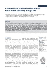
Development and Evaluation <strong>of</strong> <strong>Mucoadhesive</strong> Buccal Films <strong>of</strong> Nebivolol<br />
Bushetti S.S, Mane Prashant P and Kardame S.S ..................................................................................................................................... 157 - 162<br />
<strong>Chitosan</strong> <strong>Loaded</strong> <strong>Mucoadhesive</strong> <strong>Microspheres</strong> <strong>of</strong> <strong>Gliclazide</strong>: In vitro And In vivo Evaluation<br />
Senthil A, Thakkar Hardik R, Ravikumar and Narayanaswamy V.B ...........................................................................................................163 - 171<br />
Effect <strong>of</strong> Different Acids on the Formation <strong>of</strong> E and Z Isomers <strong>of</strong> Dothiepin<br />
Gopal Krishna Rao, Ramesha A.R, Amit Kumar Jain and Sanjay Pai P.N ................................................................................................ 172 - 175<br />
Instructions to Authors
RJPS<br />
RGUHS <strong>Journal</strong> <strong>of</strong> Pharmaceutical Sciences Scientific Tools<br />
Preamble<br />
Scientific research is essentially an intellectual investigation undertaken to gain<br />
new information, close gaps in knowledge, and understand concepts to confirm<br />
an idea. Research, whether basic or applied, should have a reasonable possibility<br />
for success. The thrill <strong>of</strong> scientific discovery and the transfer <strong>of</strong> technology from<br />
the lab bench and its potential application to a patient's bedside is the hallmark<br />
accomplishment <strong>of</strong> one's research career. It is a reward <strong>of</strong> unmatched happiness.<br />
For that to happen, one needs clear vision and imagination.<br />
Research is both a social and cooperative venture. It is important to establish a<br />
positive climate for research by organizing appropriate resources and making<br />
them readily available. To accomplish this, assistance from all corners is needed.<br />
After all, it takes a whole research community to mold a scientist. In addition, it<br />
needs an ever-growing array <strong>of</strong> scientific tools, and the expertise <strong>of</strong> an<br />
experienced mentor to steer the knowledge and enthusiasm <strong>of</strong> a novice<br />
researcher into the proper direction <strong>of</strong> becoming an established research<br />
investigator. In this endeavor, scientific journals, like mentors, play a vital role in<br />
educating researchers at all levels. The objective <strong>of</strong> the newly created section<br />
'Scientific Tools' will deliver concise, but regular, descriptions <strong>of</strong> skills required to<br />
take the journey a step forward in improving one's scientific enterprise.<br />
The first article in the series launches the basic concepts <strong>of</strong> scientific research by<br />
introducing application <strong>of</strong> the creative process needed for correctly choosing a<br />
research topic or idea. Dr. Reisman reviews essentials <strong>of</strong> creativity and critical<br />
thinking in developing many ideas and later how to converge them. We wish to<br />
continue our scientific journey into the areas <strong>of</strong> research processes, research<br />
methods, study designs, data analysis, scientific communication and much more<br />
in an attempt to help everyone who does pr<strong>of</strong>essional business in biomedical<br />
research. Check this section promptly for a description <strong>of</strong> a new tool when each<br />
new issue is received.<br />
Gowraganahalli Jagadeesh<br />
US Food and Drug Administration<br />
Silver Spring, Maryland, USA<br />
gowra.jagadeesh@fda.hhs.gov<br />
RJPS, Jul - Sep, 2011/ Vol 1/ Issue 2
RGUHS <strong>Journal</strong> <strong>of</strong> Pharmaceutical Sciences<br />
Creative, Critical Thinking and Logic in Research<br />
Fredricka Reisman<br />
Goodwin College <strong>of</strong> Pr<strong>of</strong>essional Studies, Drexel/Torrance Center for Creativity and Innovation, Drexel University, Philadelphia, PA 19104,<br />
USA<br />
Creative, Critical Thinking and Logic in Research<br />
In order for me, a western mathematics and creativity<br />
researcher and educator, to respond to the exciting invitation<br />
to write an article dealing with creativity for this prestigious<br />
journal published by the Rajiv Gandhi University <strong>of</strong> Health<br />
Sciences, I needed to investigate the essence and focus <strong>of</strong> the<br />
audience. I needed to become familiar with the context in<br />
which my article was to contribute. My investigation led me to<br />
the meanings behind the Emblem <strong>of</strong> the Rajiv Gandhi<br />
University <strong>of</strong> Health Sciences, which to my delight sets the<br />
stage for creative (Human Energy), critical thinking (the<br />
Human Soul) and logic (Knowledge And Enlightenment) as<br />
cornerstones for pharmaceutical research.<br />
The Emblem<br />
The Emblem <strong>of</strong> The Rajiv Gandhi University <strong>of</strong> Health<br />
Sciences is a symbolic expression <strong>of</strong> the confluence <strong>of</strong> both<br />
Eastern and Western Health Sciences. A central wand with<br />
entwined snakes symbolizes Greek and Roman Gods <strong>of</strong><br />
Health called Hermes. Mercury is adapted as symbol <strong>of</strong><br />
modern Medical Science. The pot above the snake depicts<br />
Amrutha Kalasham <strong>of</strong> Dhanvanthri, the father <strong>of</strong> all health<br />
sciences. The wings above it depicts Human Soul called<br />
Hamsa (Swan) in Indian Philosophy. The rising sun at the top<br />
symbolises knowledge and enlightenment. All <strong>of</strong> them set<br />
inside the state map <strong>of</strong> Karnataka. The two twigs <strong>of</strong> leaves in<br />
RGUHS <strong>Journal</strong> <strong>of</strong> Pharmaceutical Sciences<br />
Received: 14/8/2011, Modified: 27/8/2011, Accepted: 28/8/2011<br />
97<br />
Scientific Tools<br />
Western Philosophy symbolizes Olive branches, which is an<br />
expression <strong>of</strong> Peace, Love And Harmony. In Hindu<br />
Philosophy, it depicts the Vanaspathi (also called as Aushadi)<br />
held in the hands <strong>of</strong> Dhanvanthri, which are the source <strong>of</strong> all<br />
medicines. The lamp at the bottom depicts human energy<br />
(Kundalini). The script “Devahitham Yadayahu” inside the<br />
lamp is taken from Upanishath Shanthi Manthram<br />
(Bhadram Karnebhi Shrunuyanadev…) which says “May we<br />
live the full span <strong>of</strong> our lives allotted by God in perfect health”<br />
which is the motto <strong>of</strong> the Rajiv Gandhi University <strong>of</strong> Health<br />
Sciences.<br />
Link to Creative, Critical Thinking and Logic<br />
This link from emblem to reality is assuring that pharmacy<br />
students, faculty and practitioners, in addition to being<br />
excellent learners and researchers, are also creative problem<br />
solvers, first-rate scientists, and effective clinicians. In looking<br />
at the traits <strong>of</strong> highly creative people listed below, we see that<br />
many <strong>of</strong> these traits are salient to creative science researchers<br />
and practitioners in the Pharma industries. In fact, a recent<br />
1<br />
publication considered the question <strong>of</strong> why an<br />
understanding <strong>of</strong> creativity and critical thinking is important<br />
for biomedical scientists, especially those new to their science<br />
career paths.<br />
“Understanding how the big breakthroughs occur can lead the newly<br />
minted scientist to efficiently engage in creative research that results in a<br />
novel, appropriate and useful discovery, to obtain funding support, and<br />
to navigate the publication channel…”<br />
1<br />
F. K. Reisman, 2010<br />
In addition, a recent survey <strong>of</strong> 1,500 chief executives<br />
2<br />
conducted by IBM's Institute for Business Value identified<br />
"creativity" as the most important leadership competency for<br />
corporate success <strong>of</strong> the future. Note that the desired<br />
competency <strong>of</strong> leaders is “creativity----not operational<br />
effectiveness, managerial discipline, influence, or even<br />
dedication.” Until recently, creativity was viewed as an<br />
essential element <strong>of</strong> research or product development, not the<br />
crucial characteristic <strong>of</strong> leadership.<br />
Traits <strong>of</strong> Highly Creative People<br />
Following are traits representative <strong>of</strong> highly creative people<br />
which embody members <strong>of</strong> the pharmaceutical industries<br />
and that form an assessment checklist as shown in Table 1:<br />
Next, in Table 2 are evaluation criteria for assessing creative<br />
RJPS, Jul - Sep, 2011/ Vol 1/ Issue 2
Table 1: Creative Traits Assessment<br />
Creative Trait<br />
1. A high level <strong>of</strong> curiosity<br />
2. Willingness to learn from experience<br />
3. Preparedness to take risks<br />
4. Persistence in situations <strong>of</strong> failure<br />
5. High levels <strong>of</strong> energy<br />
6. Tolerate contradictions, ambiguities, and uncertainties in work<br />
7. Resist premature closure<br />
8. May see problems or challenges as more complex because <strong>of</strong> seeing more alternatives<br />
9. Embrace change<br />
1 2 3 4<br />
Table 2. Evaluation Form Used by Preceptors or Supervisors<br />
Evaluation <strong>of</strong> Problem-Solving Abilities Instructions: Please provide the following information on<br />
your students or employee. Complete a separate evaluation form for each individual.<br />
Preceptor/Supervisor: ___________________<br />
Student/Employee: __________________<br />
Date:___________________<br />
1. Please rate individual's overall problem (clinical or nonclinical)-solving abilities by circling the letter that<br />
most closely describes the student/employee.<br />
a. High, able to solve difficult problems, equivalent to an experienced researcher or pharmacist-clinician.<br />
b. Very good, able to solve moderate problems, but with some inadequacies with difficult problems.<br />
c. Good, able to solve simple and moderate problems, but unable to solve difficult problems.<br />
d. Fair, able to solve simple problems but unable to solve problems <strong>of</strong> greater difficulty.<br />
e. Poor, unable to solve simple problems.<br />
Fredricka Reisman - Creative, Critical Thinking and Logic in Research<br />
Please evaluate yourself or other individuals on each <strong>of</strong> the following traits <strong>of</strong> highly creative people, using the scale below. Check ( ) the<br />
number corresponding to your evaluation. 4 = Outstandingly creative, 3 = Competently creative majority <strong>of</strong> time, 2 = Likehood <strong>of</strong> improvement<br />
with training, 1 = Lacks evidence <strong>of</strong> creative thinking<br />
Nonclinical/Clinical Problem-solving Competence 1 2 3 4<br />
a. Recognizing the existence <strong>of</strong> a problem.<br />
b. Defining the nature/requirements <strong>of</strong> a problem.<br />
c. Generating more than one set <strong>of</strong> steps that may solve a problem.<br />
d. Knowledge acquisition to solve a problem.<br />
e. Organizing information about a problem.<br />
f. Critical and logical thinking process related to a problem.<br />
g. Allocating mental and physical resources to solving a problem.<br />
h. Monitoring the outcome related to the solution <strong>of</strong> a problem.<br />
i. Personal attributes required for problem-solving (e.g., values, attitudes, emotions, confidence).<br />
Please evaluate student/employee on each <strong>of</strong> the following aspects <strong>of</strong> nonclinical or clinical problem solving, using the scale below. Check<br />
( ) the number corresponding to your evaluation. 4 = Outstanding, 3 = Competent, 2 = Improvement needed, 1 = Incompetent<br />
98
problem solving in pharmacy students or employees by their<br />
3<br />
preceptors or supervisors :<br />
A mobile-based self-assessment is the Reisman Diagnostic<br />
4<br />
Creativity Assessment (RDCA) . The RDCA is a free Apple<br />
application that may be downloaded to an iPhone, iPad or<br />
iTouch via iTunes. The RDCA is built upon 11 creativity<br />
factors, shown in Table 3, that are most prevalent in the<br />
research literature as representative <strong>of</strong> creative thinking. The<br />
RDCA is built upon the Torrance Tests <strong>of</strong> Creative Thinking<br />
5<br />
(TTCT) , which in turn stems from Guilford's creativity<br />
6<br />
research . The TTCT remains the most widely used test <strong>of</strong><br />
creativity and the most referenced <strong>of</strong> all creativity tests.<br />
However, it must be scored by trained evaluators, takes two<br />
hours for administration, focuses on prediction <strong>of</strong> creative<br />
performance, and is costly.<br />
The RDCA may be self - scored, takes about 10 minutes to<br />
complete, and at this point in time is free. This is a self-report<br />
Likert-type assessment designed to be used diagnostically to<br />
identify one's creative strengths, rather than to predict<br />
creativity. The RDCA assesses an individual's self-perception<br />
on 11 major creativity factors. The results may be used to<br />
determine which factors already are strong, which factors one<br />
personally wishes to strengthen, which they are satisfied with,<br />
and which are most important for strengthening their<br />
creativity through selected exercises.<br />
You can decide if you wish to strengthen an area by practice.<br />
For example, to increase fluency, practice generating many<br />
scenarios, such as brainstorming possible drug trial outcomes,<br />
identifying possible contraindications, etc., within a short<br />
timeframe such as three minutes. To increase flexibility,<br />
generate many categories <strong>of</strong> possible trial techniques, such as<br />
realigning the trial expectations, reallocating resources, or<br />
redefining success. To increase originality, practice coming up<br />
with many possible trial scenarios, based on the trial research<br />
to date. To increase elaboration, add detail to possible<br />
resolutions.<br />
Fredricka Reisman - Creative, Critical Thinking and Logic in Research<br />
The RDCA is electronically scored and you may email<br />
yourself your results. The 11 creativity factors assessed and<br />
their definitions are shown in Table 3.<br />
Examples <strong>of</strong> Integrating Creativity into Pharma<br />
industry<br />
The latest trends within the pharmaceutical industries engulf<br />
creativity from many perspectives:<br />
Ÿ Marketing, drug-delivery systems and package design that<br />
please the customer;<br />
Ÿ Making new and original connections among pharma<br />
products and diseases;<br />
Ÿ Interacting with patients;<br />
Ÿ Dealing with challenges resulting from patents expiring<br />
and generic drugs flooding the market;<br />
Ÿ Pharmacoeconomics <strong>of</strong> a new drug; and<br />
Ÿ Investigation <strong>of</strong> stem cell biology whereby small molecules<br />
that target cancer stem cells may lead to a potential cancer<br />
therapy innovation including pan-active compounds such<br />
as some kinase inhibitors.<br />
In India, AstraZeneca packages its cholesterol lowering drug<br />
®<br />
Crestor in transparent packs so that patients can visually<br />
®<br />
examine the pills. New Arimidex packaging resembles a<br />
cosmetics case because the old version reminded women that<br />
they were cancer patients, not cancer survivors. Proteus<br />
Biomedical's intelligent pills contain soluble microchips linked<br />
to detectors that remind patients if they have not swallowed<br />
their medication.<br />
Six Elements for Establishing Corporate Creativity<br />
7<br />
Robinson & Stern presented six conditions for a creative<br />
corporate environment that may be applied to the Pharma<br />
Table 3: RDCA Factors and Definitions<br />
Creativity Factor Definition<br />
Originality Unique and novel<br />
Fluency Generates many ideas<br />
Flexibility Generates many categories <strong>of</strong> ideas<br />
Elaboration Adds detail<br />
Tolerance <strong>of</strong> Ambiguity Comfortable with the unknown<br />
Resistance to premature closure Keeps an open mind<br />
Convergent Thinking Comes to closure, evaluative, critical, logical thinking<br />
Divergent thinking Generates many solutions (related to fluency)<br />
Risk Taking Adventuresome<br />
Intrinsic Motivation Inner drive<br />
Extrinsic Motivation Needs reward or reinforcement<br />
99<br />
RJPS, Jul - Sep, 2011/ Vol 1/ Issue 2
industries. The first element is alignment that involves ensuring<br />
that the interests and actions <strong>of</strong> all employees are directed<br />
toward a company's key goals. Companies can function with<br />
relatively poor alignment, but they cannot be consistently<br />
creative unless they are strongly aligned. The effects <strong>of</strong><br />
alignment on corporate creativity are apparent when a<br />
company is either well aligned or misaligned.<br />
The second element <strong>of</strong> corporate creativity is self-initiated<br />
activity that allows employees to pick a problem they are<br />
interested in and feel able to solve it. The previous examples <strong>of</strong><br />
integrating creativity into Pharma worksites are indicative <strong>of</strong><br />
someone realizing that knowledge <strong>of</strong> the psychological and<br />
personality needs <strong>of</strong> customers are as important as the<br />
medicinal chemistry expertise, and resulting in focus on<br />
design in packaging and marketing.<br />
The third element <strong>of</strong> corporate creativity is un<strong>of</strong>ficial activity<br />
that occurs in the absence <strong>of</strong> direct <strong>of</strong>ficial support, and with<br />
the intent <strong>of</strong> doing something new and useful. When an idea is<br />
new to a company, it is <strong>of</strong>ten resisted and opposed. This is<br />
analogous to Sternberg and Lubart's Investment Theory <strong>of</strong><br />
8<br />
Creativity :<br />
Investment Theory asserts that creative thinkers are like good investors:<br />
They buy low and sell high. Whereas investors do so in the world <strong>of</strong><br />
finance, creative people do so in the world <strong>of</strong> ideas. Creative people generate<br />
ideas that are like undervalued stocks (stocks with a low price-to-earnings<br />
ratio), and the public generally rejects both the stocks and the ideas. When<br />
creative ideas are proposed, they <strong>of</strong>ten are viewed as bizarre, useless, and<br />
even foolish, and summarily are rejected. The person proposing them <strong>of</strong>ten<br />
is regarded with suspicion and perhaps even with disdain and derision.<br />
Un<strong>of</strong>ficial activity gives ideas a safe haven where they have the<br />
chance to develop until they are strong enough to overcome<br />
that resistance. The introduction <strong>of</strong> packaging design in<br />
®<br />
placing Crestor in transparent packs was a creative approach<br />
to <strong>of</strong>fset the problem <strong>of</strong> drug counterfeiting.<br />
The fourth element is serendipity. A serendipitous discovery is<br />
one made by fortunate accident in the presence <strong>of</strong> wisdom or<br />
insight. Creativity <strong>of</strong>ten involves recombining or making<br />
connections between things that may seem unconnected. The<br />
®<br />
Arimidex packaging came about from connecting a woman's<br />
emotional state as a cancer survivor and her aesthetic nature<br />
and sense <strong>of</strong> fashion. Serendipity has played a significant role<br />
throughout the history <strong>of</strong> drug discovery, especially in<br />
9<br />
cardiovascular medicine . The anticoagulant properties <strong>of</strong><br />
dicoumarol were discovered after farmers observed that their<br />
cattle were dying <strong>of</strong> internal hemorrhaging after feeding on<br />
10<br />
rotting sweet clover and digitoxin was identified when the<br />
condition <strong>of</strong> a patient suffering from congestive heart failure<br />
dramatically improved after being given an herbal remedy<br />
Fredricka Reisman - Creative, Critical Thinking and Logic in Research<br />
100<br />
11<br />
containing extracts <strong>of</strong> the foxglove plant . In fact, many <strong>of</strong><br />
the most effective pharmacological agents in use today arose<br />
through serendipity.<br />
The fifth element <strong>of</strong> corporate creativity is diverse stimuli. A<br />
stimulus may provide fresh insight into something a person<br />
has already set out to do, or it may change their course <strong>of</strong><br />
action. It is important for an organization to provide<br />
opportunities for its employees to tell others about the stimuli<br />
they have received and the possibilities these stimuli suggest to<br />
them. It is here that the real leverage lies as in the cases where<br />
design and creativity became important industry changers in<br />
how they packaged and marketed their Pharma products.<br />
The sixth and final element <strong>of</strong> corporate creativity is withincompany<br />
communication; especially, unanticipated<br />
communication. Every organization carries out planned<br />
activities and establishes lines <strong>of</strong> communication to support<br />
them. But these <strong>of</strong>ficial channels are <strong>of</strong> limited usefulness for<br />
corporate creativity. Corporations need to promote<br />
unanticipated exchanges <strong>of</strong> information. A company's<br />
creative potential needs systems in place to promote<br />
unanticipated exchanges <strong>of</strong> information for these illuminate<br />
creative and serendipitous connections.<br />
Critical Thinking and Logic as Essentials <strong>of</strong><br />
Creative Thinking<br />
Usually, creative thinking is associated with brainstorming<br />
(generating many ideas), novelty, and uniqueness <strong>of</strong> ideas.<br />
Critical thinking is analytical, judgmental and involves<br />
evaluating choices before making a decision. When you are<br />
thinking critically, you are using logic, reason and convergent-<br />
1<br />
type thinking. As I point out in another publication :<br />
“…creativity involves creative thinking as a process <strong>of</strong><br />
sequential interaction <strong>of</strong> two types <strong>of</strong> thinking – divergence<br />
and convergence as depicted in Figure 1. Divergent thinking is<br />
the ability to elaborate and think <strong>of</strong> diverse and original ideas<br />
with fluency and speed (e.g., brainstorming). Convergent<br />
thinking involves narrowing ideas by evaluating the previously<br />
generated ideas that emerged in the divergent portion <strong>of</strong> the<br />
sequence (e.g., settling upon an idea from a selection <strong>of</strong><br />
1<br />
ideas).” F. K. Reissman, 2010<br />
Creative Ideas Versus Innovative Ideas<br />
First, let's distinguish between creativity and innovation.<br />
Creativity involves generating unique, original and novel<br />
ideas. Innovation is the implementation <strong>of</strong> these ideas. Merely<br />
generating ideas without bringing them to fruition in the form<br />
<strong>of</strong> a product or service is uneconomical and a waste <strong>of</strong> talent.<br />
These two parameters play important roles in choosing a<br />
research project, identifying a thesis topic, or improving<br />
RJPS, Jul - Sep, 2011/ Vol 1/ Issue 2
Figure 1. Creative thinking process<br />
Divergent<br />
customer service in a clinical or retail Pharma setting.<br />
One <strong>of</strong> the most difficult things in research as in any other<br />
endeavour is identifying a research question, a real problem<br />
(as opposed to a superficial or incorrect problem), or the cause<br />
<strong>of</strong> an organizational dilemma. Key is having in-depth<br />
knowledge related to the problem - <strong>of</strong>ten the salient issue<br />
arises from a knowledge-base developed in writing a review <strong>of</strong><br />
the relevant literature.<br />
Thus, how many have said they "invented" the latest new<br />
gadget or fad, only they never found the time to actually<br />
develop the product. They had the idea, but never took the<br />
idea to fruition. That's the difference between creativity and<br />
innovation.<br />
Elements <strong>of</strong> Creativity<br />
12<br />
Strong and Davis list four creativity elements; namely,<br />
valuable (perceived as having worth and genuinely contributing<br />
to society), intentional (result <strong>of</strong> a deliberate effort), novel (new or<br />
has at least some element <strong>of</strong> originality), excellent (significant<br />
effort expended to make it the best it can be). Thus, the<br />
creative product or service must be new and judged to be<br />
valuable according to designated criteria. Further, creative<br />
products are the result <strong>of</strong> purposive behaviour and to become<br />
13<br />
excellent, creative effort takes time . It is noted that creativity<br />
usually is not purely original, but rather that it stems from<br />
some prior knowledge and something is considered creative<br />
through implementation (innovation) and enhancement,<br />
although not necessarily a completely new idea. For example,<br />
the Romans improved upon Greek culture, and the Greeks in<br />
12<br />
turn built upon Mesopotamian and Egyptian cultures .<br />
Elements <strong>of</strong> Innovation<br />
A broadly accepted definition for innovation is: To turn a creative<br />
idea into products and services <strong>of</strong> value and pr<strong>of</strong>it. The basic goal <strong>of</strong><br />
all innovation is positive change, to make someone or<br />
something better. There are two basic types <strong>of</strong> innovation.<br />
Incremental Innovation: Also called continuous innovation, this<br />
Fredricka Reisman - Creative, Critical Thinking and Logic in Research<br />
Convergent<br />
Divergent Convergent Divergent Convergent<br />
type improves upon existing products/services. From a result<br />
standpoint, incremental innovations can range from very<br />
small to huge increases in productivity, revenues and pr<strong>of</strong>its.<br />
Breakthrough (Radical, Disruptive) Innovation: This type <strong>of</strong><br />
innovation develops new products/services that do not exist.<br />
Many times this type <strong>of</strong> innovation emerges from scientific<br />
discoveries or R&D organizations. But, while a breakthrough<br />
innovation might mean getting a patent, it does not guarantee<br />
huge pr<strong>of</strong>its.<br />
Out <strong>of</strong> many hundreds <strong>of</strong> creative ideas, only a few may ever<br />
be implemented. For those precious few, we know them as<br />
innovation - or simply, applied creativity.<br />
So creativity is the idea, and innovation is the idea applied or<br />
implemented.<br />
Future for Creative, Critical Thinking and Logic in<br />
India's Pharmaceutical Industries<br />
In September 2004, a global innovation survey by the<br />
14<br />
Economist Intelligence Unit identified India “as an R&D<br />
hotspot, defined as a place where (1) companies are able to tap<br />
into existing scientific and technical expertise networks, (2)<br />
there are good links to academic research facilities, (3) the<br />
environment supports innovation and (4) it is easy to<br />
commercialize.” The Economist further states:<br />
Costs <strong>of</strong> pharmaceutical innovation in India are estimated as low as oneseventh<br />
<strong>of</strong> their levels in Europe, and the country's clinical research<br />
industry is currently worth $100 million growing around 40 to 50<br />
percent annually, although some forecasts say it could be worth as much as<br />
$1 billion to Indian firms in 2008.<br />
The research enterprise in India is exemplified by numerous<br />
15<br />
R & D investments including the following :<br />
Ÿ AstraZeneca is conducting research into tuberculosis (TB) at the<br />
AstraZeneca Research Foundation India in Bengaluru. India's estimated<br />
8.5 million TB patients mean clinical trials can be conducted easily and<br />
economically.<br />
101 RJPS, Jul - Sep, 2011/ Vol 1/ Issue 2
Ÿ GSK and Ranbaxy are partnering where GSK will provide drug<br />
research leads and Ranbaxy will conduct preclinical studies; GSK will<br />
take the drug through human trials.<br />
Ÿ Pfizer is exploring setting up an Academy for Clinical Research in<br />
Mumbai since costs <strong>of</strong> clinical trials in India are around one-tenth the<br />
U.S.<br />
In addition to drug studies, investigated is the correlation<br />
16<br />
between brain chemistry and creative cognition , the<br />
17<br />
relationship between creativity and academic achievement ,<br />
18-21<br />
and the relationship between creativity and intelligence .<br />
New technologies are opening pathways for<br />
biochemical and neurological research as these<br />
impact creativity research, and the role <strong>of</strong> creativity<br />
22<br />
in design and marketing research is at the<br />
forefront <strong>of</strong> the Pharma industries. As Einstein<br />
cautioned: “you cannot solve problems by using the kind <strong>of</strong><br />
thinking that produced the problem in the first place.”<br />
REFERENCES<br />
1. Reisman FK. Creative and critical thinking in biomedical research. In:<br />
Jagadeesh G, Murthy S, Gupta YK, Prakash A, editors. Biomedical<br />
research: from ideation to publication. New Delhi: Wolters Kluwer<br />
Health, Lippincott Williams & Wilkins; 2010. 3-17.<br />
2. IBM 2010 Global CEO Study. Creativity selected as most crucial factor<br />
for future success [online]. [Cited 2011 August 10]. Available from:<br />
URL: http://www-03.ibm.com/press/us/en/pressrelease/31670.wss<br />
3. Adamcik B, Hurley S, Erramouspe J. Assessment <strong>of</strong> pharmacy<br />
students' critical thinking and problem-solving abilities. Am J Pharm<br />
Edu 1996;60:256-64.<br />
4. Reisman FK. Reisman diagnostic creativity assessment (RDCA).<br />
Apple Application via iTunes. 2011.<br />
5. Torrance EP. Torrance tests <strong>of</strong> creative thinking. Bensenville, Illinois,<br />
USA: Scholastic Testing Service; 1974.<br />
6. Guilford JP. The nature <strong>of</strong> human intelligence. New York, NY:<br />
McGraw-Hill;1967.<br />
7. Robinson A, Stern S. Corporate creativity. San Francisco, CA: Berrett-<br />
Koehler;1997.<br />
8. Sternberg RJ, Lubart TI. Defying the crowd: cultivating creativity in a<br />
culture <strong>of</strong> conformity. New York, NY: Free Press; 1995.<br />
9. Schlueter PJ, Peterson RT. Basic science for clinicians systematizing<br />
serendipity for cardiovascular drug discovery. Circulation<br />
2009;120:255-63.<br />
Fredricka Reisman - Creative, Critical Thinking and Logic in Research<br />
10. Mueller RL, Scheidt S. History <strong>of</strong> drugs for thrombotic disease:<br />
discovery, development, and directions for the future. Circulation<br />
1994;89:432-49.<br />
11. Norman JN. William Withering and the purple foxglove: a<br />
bicentennial tribute. J Clin Pharmacol 1985;25:479-83.<br />
12. Strong B, Davis M. The history <strong>of</strong> creativity in the arts, science and<br />
nd<br />
technology: 1500-present. 2 edition. Dubuque, Iowa: Kendall Hunt<br />
Publishing Company; 2011.<br />
13. Wallace DB, Gruber HE. Creative people at work. UK:Oxford<br />
University Press, 1989.28-9.<br />
14. Reddy P. Global Innovation in Emerging Economies. New York:<br />
Routledge; 2011.108.<br />
15. Gopi PG, Subramani R, Santha, T. et al. Estimation <strong>of</strong> burden <strong>of</strong><br />
tuberculosis in India for the year 2000. Ind J Med Res 2005;122:243-<br />
8.<br />
16. Jung RE, Gasparovic C, Chavez, RS. et al. Biochemical support for<br />
the “threshold” theory <strong>of</strong> creativity: a magnetic resonance<br />
spectroscopy study. J Neurosci 2009;29:5319 –25.<br />
17. Naderi H, Abdullah R, Aizan HT, Sharir J, Kumar V. Relationship<br />
between creativity and academic achievement: a study <strong>of</strong> gender<br />
differences. J Am Sci 2010;6:181-90<br />
18. Weisberg RW. Creativity: beyond the myth <strong>of</strong> genius. New York:WH<br />
Freeman & co; 1993.<br />
19. Sternberg RJ. Wisdom, intelligence, and creativity synthesized.<br />
Cambridge: Cambridge university press; 2003.<br />
20. Kassem A. The creativity chemical [online]. 2011 [Cited 2011 August<br />
11]. Available from: URL: http://theilluminatedbrain.com/drugschemicals/the-creativity-chemical.<br />
21. Kharkhurin AV. The Impact <strong>of</strong> Culture on the Relationship between<br />
Bilingualism and Creative Potential [online]. [Cited 2011 August 11].<br />
Available from: URL: http://academic.brooklyn.cuny.edu/psych/<br />
tovyharhur/research/publications/jccp1.pdf<br />
22. Jack A. A pharmaceutical experiment in design [online]. 2010 [Cited<br />
2011 August 11]. Available from: UR: http://www.ft.com/cms/s/0/<br />
4ed58ce0-b091-11df-8c04-00144 feabdc0. html#ixzz1U5wNrIvd.<br />
Address for Correspondence<br />
Fredricka K. Reisman, Ph.D., Pr<strong>of</strong>essor, Goodwin College <strong>of</strong> Pr<strong>of</strong>essional<br />
Studies, Director, Drexel/Torrance Center for Creativity and Innovation, Drexel<br />
University, 3001 Market Street Suite 110, Philadelphia, PA 19104, USA<br />
President, American Creativity Association<br />
E-mail: reismafk@drexel.edu<br />
102 RJPS, Jul - Sep, 2011/ Vol 1/ Issue 2
A B S T R A C T<br />
RGUHS <strong>Journal</strong> <strong>of</strong> Pharmaceutical Sciences<br />
Need for Inclusion <strong>of</strong> Scientific Writing Skill Subjects in Indian Post Graduate<br />
Pharmacy Course<br />
1 2 1 1 1<br />
Patil J.S* , Kotnal R.B , Birajdar R.P , Marapur S.C and Kadam D.V<br />
1 2<br />
Dept. <strong>of</strong> Pharmaceutics, Dept. <strong>of</strong> Pharmaceutical Chemistry, B.L.D.E.A's College <strong>of</strong> Pharmacy, BLDE University Campus,<br />
Bijapur- 586103, Karnataka, India.<br />
This article emphasize more on why we need stuffing <strong>of</strong> scientific writing skills in curriculum rather than how to write scientific articles.<br />
Writing scientific articles is a great obstacle for many people, or perhaps for most people. Scientific writing provides vital information with<br />
the creation and dissemination <strong>of</strong> research knowledge. In recent years, most <strong>of</strong> the Indian pharmacy research students are unable to<br />
publish their scientific data in peer reviewed journals. Reasons for this failure are numerous, few among these are not knowing how to<br />
begin, poor language and drafting skills. Lacking in basics <strong>of</strong> scientific writing skills and no motivation by the research<br />
guides/supervisors is the matter <strong>of</strong> concern today. In India, all the universities <strong>of</strong>fering post graduate (PG) course in pharmacy are<br />
concentrating more on teaching the subject contents <strong>of</strong> respective specializations. The PG course is a blend <strong>of</strong> study <strong>of</strong> specialized<br />
subjects in part- I and research programme in part-II. The students need to write thesis <strong>of</strong> their dissertation work at the end. For writing the<br />
thesis students require scientific writing knowledge. Hence, it necessitates studying the subjects on scientific writing skills at part-I level.<br />
In this present discussion an attempt was made to explore the reasons which emphasize the inclusion <strong>of</strong> scientific writing subject/s in PG<br />
course and also tried to suggest the possible course structure which may become more appropriate for our present context.<br />
Keywords: Scientific writing, publication, course structure, post graduate, pharmacy.<br />
INTRODUCTION<br />
Good scientific writing skills open up many opportunities to<br />
the researcher: publications, conference or seminar<br />
attendance. They also lead to better patents, better research<br />
partnerships and better funded research. Clarity and<br />
efficiency in scientific writing bears witness to the quality <strong>of</strong> a<br />
researcher; it influences career promotion. As we are seeing<br />
many avenues for scientific writing, medical writing in<br />
healthcare, medical and R & D sector viz; pharmaceutical<br />
industry and Clinical Research centers. It is essential to have<br />
subject in the post graduate level <strong>of</strong> pharmacy course. For a<br />
researcher, scientific writing is a rewarding knowledge both<br />
pr<strong>of</strong>essionally and socially. The idea and skill <strong>of</strong> writing help<br />
to publish scientific data in the peer reviewed journals.<br />
Publication <strong>of</strong> research articles is the measure <strong>of</strong> his/her<br />
productivity which may lead to upgrade the pr<strong>of</strong>essional<br />
1,2<br />
status especially for those from academic field . Publishing<br />
the research results in scientific journals reaches the audience<br />
in larger extent and it contributes a significant influence on<br />
career development. The scientific writing is a well written<br />
report describing the results <strong>of</strong> overall original research work.<br />
Publication is the crucial end point <strong>of</strong> a research work to share<br />
RGUHS <strong>Journal</strong> <strong>of</strong> Pharmaceutical Sciences<br />
Received: 3/1/2011, Modified: 3/8/2011, Accepted: 1/9/2011<br />
103<br />
Review Article<br />
important information with the scientific community which<br />
results in personal contentment and pr<strong>of</strong>essional<br />
3<br />
advancement . Writing is a skill born from practice. Before<br />
becoming a good writer it is essential to become an avid and<br />
careful reader. Most reputed institutes consider quality<br />
publications as a measure <strong>of</strong> research productivity and basic<br />
indicators <strong>of</strong> accountability. In recent days, number <strong>of</strong> good<br />
publications by Indian pharmaceutical scientists in reputed<br />
journals is drastically declining. The reasons for such failure<br />
are plenty. But this is really a serious concern and our<br />
educators need to think urgently to rectify this problem.<br />
Although, few research/academic institutions routinely<br />
conducting short term workshops and seminars on scientific<br />
writing skills in their institutions but the number <strong>of</strong> virtual<br />
beneficiaries are less. Most <strong>of</strong> the pharmacy institutions<br />
<strong>of</strong>fering PG programme in India are teaching basic subjects<br />
<strong>of</strong> the respective specialization. Although a great deal <strong>of</strong><br />
importance is given to teach such specialization subjects, no<br />
attention is paid in teaching scientific writing skills. It may be<br />
said without any exaggeration that scientific writing skill is one<br />
among the most crucial problems that plague the research<br />
oriented educational scene in India and there is almost no<br />
place for learning, coaching and motivation for scientific<br />
writing skills which is essential for PG students. In most <strong>of</strong> the<br />
institutions almost 90% students leave the college after<br />
completion <strong>of</strong> the course without even communicating their<br />
RJPS, Jul - Sep, 2011/ Vol 1/ Issue 2
Patel J.S et al./ Need For Inclusion <strong>of</strong> Scientific Writing Skill Subjects in Indian Post Graduate Pharmacy Course<br />
scientific results for possible publication. Further, it is a matter<br />
<strong>of</strong> great regret that, scientific writing is not considered as one<br />
<strong>of</strong> the essential study tool for PG pharmacy students. That is<br />
why even though a large number <strong>of</strong> PG students are coming<br />
out every year, the number <strong>of</strong> publications in peer reviewed<br />
journals are very less. This aspect not only reducing the<br />
pr<strong>of</strong>essional reputation and recognition but also reduces the<br />
scope <strong>of</strong> pharmacy post graduates heading high academic<br />
positions, editorial and scientific review opportunities.<br />
Most modern universities in the world have begun to modify<br />
the course structure or entire study programme in various<br />
disciplines according to the present need and importance.<br />
Instructors tailor such course to many individuals, including<br />
4 5<br />
medical students , osteopathic residents and scientific<br />
6<br />
researchers . Even in India, students studying in<br />
universities/institutions exposing to learning scientific writing<br />
skills through seminars and workshops in their campus have<br />
the benefit <strong>of</strong> a better eminence. However, most pharmacy<br />
institutions still continue with primitive and callous fashion <strong>of</strong><br />
teaching and not thinking on importance and significances <strong>of</strong><br />
scientific writing skills. This seems that there is need for<br />
inclusion <strong>of</strong> subject/s on scientific writing skills as a part <strong>of</strong><br />
PG curriculum. Thus, the present study was done with the<br />
aim <strong>of</strong> discussing the need for inclusion <strong>of</strong> such subject/s with<br />
a possible proposal <strong>of</strong> course design.<br />
“But in science the credit goes to the man (or woman) who convinces the<br />
world, not to the man (or woman) to whom the idea first occurs”. Sir<br />
Francis Darwin.<br />
Reasons for inclusion <strong>of</strong> scientific writing subject/s<br />
Motives for the publication vary widely. Some students having<br />
a special driving force and well guided by their research<br />
supervisors are finally publish their scientific results in reputed<br />
journals. But this is all depends on special talent and skills.<br />
Scientific writing is easier when it is an integral part <strong>of</strong> the<br />
study and it is harder when it require a student to think and<br />
prepare a scientific paper. To motivate all the students towards<br />
publication habits it is essential to make scientific writing as a<br />
curriculum part. In the present Indian context, motives to<br />
scientific writing and publishing habits are poor. In the present<br />
discussion we focused mainly on the reasons which made to<br />
discourage the publishing habits in our PG students. These<br />
include,<br />
In recent days, pr<strong>of</strong>essional and technical teaching<br />
community suffering badly with grammatical English<br />
language. This is because most <strong>of</strong> us not considering that the<br />
language is necessarily grammatical in pr<strong>of</strong>essional teaching<br />
field. This situation lacking behind in flourished writing<br />
activities.<br />
104<br />
A poor motives and encouragement to the student<br />
community pertaining to scientific writing and publishing<br />
habits. This may be because <strong>of</strong> the fact that the teachers/<br />
research guides themselves hesitate to write the papers due to<br />
their poor language and lack <strong>of</strong> writing skills.<br />
Though some people interest to publish the papers <strong>of</strong> their<br />
research results, a primary obstacle is how to begin, even<br />
though the approaches to and procedure for writing a<br />
7<br />
scientific paper is well defined .<br />
A poor or no clear vision for the institutions which are<br />
currently working on commercial base, such institutions has<br />
absolutely failed to attract a talented research supervisor who<br />
has basics <strong>of</strong> teaching and interest in research. This ultimately<br />
failed to achieve minimum scientific and educational<br />
standards in our research based educational system. The<br />
Indian universities seem to have not made publication as<br />
mandatory requirement for the PG students in the<br />
curriculum, although it is already exist in some universities for<br />
Ph D programme.<br />
Benefits <strong>of</strong> scientific writing knowledge and<br />
publications<br />
Scientific writing skill and research publications give publicity<br />
to the scientist which may help him in many <strong>of</strong> the following<br />
ways. The recognition and publicity that gained by<br />
publications may result in getting consulting work and<br />
assignments such as resource person. Publishing the research<br />
results in scientific journals contributes a significant influence<br />
on career development. Publication may lead to pr<strong>of</strong>essional<br />
recognition and job promotion. Publication is the crucial end<br />
point <strong>of</strong> a research work which results in personal satisfaction<br />
and pr<strong>of</strong>essional advancement.<br />
In the recruitment <strong>of</strong> faculty most reputed institutes consider<br />
quality publications as a measure <strong>of</strong> research productivity and<br />
basic indicators <strong>of</strong> accountability. Good scientific writing<br />
skills can bring many personal rewards such as getting<br />
research grants.<br />
Suggested course design<br />
Acquiring good writing skill is a difficult task for the student<br />
community especially those who have not studied the 'English'<br />
as a first language. An issue always faced in teaching any<br />
specializations <strong>of</strong> pr<strong>of</strong>essional communication is the bridge<br />
between technical knowledge and rhetorical skills. Teaching<br />
subjects like scientific writing skills is always suggested to<br />
concentrate on coordinating the theoretical knowledge and<br />
rhetorical skills, there by students are made more skillful<br />
candidates both in theory as well as in practical. All research<br />
process always begins with a standard protocol and concludes<br />
RJPS, Jul - Sep, 2011/ Vol 1/ Issue 2
with writing the scientific data in a systematic manner. A<br />
scientific writing describing an instance <strong>of</strong> the scientific<br />
process reflects the way that experiments are devised and<br />
8, 9<br />
carried out . In this context, Indian universities are required<br />
to design the practical oriented course structure on scientific<br />
writing and research methodology subjects for post graduate<br />
pharmacy students. For this, we have to begin with<br />
constituting an expert committee comprising renowned<br />
educationists and subject experts who can design and tailor<br />
such subject suitable for our PG students. The Educationists<br />
constructing syllabi for such subjects have to describe the<br />
benefits and contributions <strong>of</strong> the subjects for their career<br />
advancement in a clear way. The course design proposed by us<br />
comprise two distinct parts covering the study on theoretical<br />
aspects as well as practical assignments and workshops<br />
pertaining to impart the knowledge <strong>of</strong> scientific writing skills.<br />
Section- I: Theory Part<br />
Patel J.S et al./ Need For Inclusion <strong>of</strong> Scientific Writing Skill Subjects in Indian Post Graduate Pharmacy Course<br />
Theory part comprises the comprehensive contents on<br />
fundamentals and applied aspects <strong>of</strong> scientific writing skills.<br />
The course structure has different sections;<br />
Start with study <strong>of</strong> literature review procedures; because<br />
research programmes always begin with thorough literature<br />
review which provides a strong base for the proposed study.<br />
The next important section is study on writing skills <strong>of</strong><br />
research protocol, because research protocol is a brief plan <strong>of</strong><br />
proposed work with established methods needs to submit for<br />
approval <strong>of</strong> the study. Hence, it is essential to study the design<br />
and writing skills <strong>of</strong> research protocol along with study on<br />
planning and execution <strong>of</strong> the research programme.<br />
The theory part also contains a detailed study on abstract<br />
writing, knowledge on anatomy <strong>of</strong> a good abstract, use <strong>of</strong><br />
rhetorical language in framing the abstract as well as whole<br />
scientific paper, criteria to identify the target journal, type <strong>of</strong><br />
manuscript and authorship.<br />
Further, the course also cover chapters on fundamental rules<br />
and techniques involved in framing the result and discussion<br />
parts <strong>of</strong> a scientific writing, statistical analysis <strong>of</strong> the data<br />
obtained, and use <strong>of</strong> various statistical s<strong>of</strong>twares.<br />
This part also includes the systematic study <strong>of</strong> bibliographic<br />
writing because it is vital part <strong>of</strong> a scientific writing. Different<br />
journal follow unique reference style. The study covers<br />
introduction <strong>of</strong> various reference styles. Hence, a thorough<br />
study on bibliographic writing helps the students in preparing<br />
the thesis or scientific paper.<br />
Finally the course also cover study on preparation <strong>of</strong> posters<br />
and power point slides for oral presentation. Study <strong>of</strong> basic<br />
procedures involved in the preparation <strong>of</strong> poster and oral<br />
105<br />
presentations at various scientific conferences, seminars and<br />
conventions.<br />
To make all these study components more familiar to students<br />
the commonest ways <strong>of</strong> teaching and to explore their views<br />
are in-depth counseling and group discussions. In depth<br />
counseling as one-to-one basis and group discussions are<br />
suggested to cover in the syllabus with respect to the study<br />
content.<br />
In present day academic scenario it is worth important to<br />
follow the ethical principles in scientific publication rather<br />
than publishing good number <strong>of</strong> papers. Hence, it is very<br />
much essential to have a chapter on ethical principles to be<br />
stringently followed by every research students while involving<br />
in the scientific writing work.<br />
To ensure successful teaching <strong>of</strong> this course, a periodic<br />
internal assessment and university examinations are to be<br />
conducted through a systematic evaluation procedures.<br />
Section-II: Practical Part<br />
In the practical part <strong>of</strong> the subject design the vigorous<br />
practical assignments and workshops are suggested to<br />
conduct. Students receive a thorough introduction to the<br />
various theoretical aspects on scientific writing that provides<br />
the necessary context for practical writing assignments that<br />
complements the lectures and discussions.<br />
A definite number <strong>of</strong> students in a group are to be allotted the<br />
specific scientific writing assignments. In these assignments<br />
students are supplied the results <strong>of</strong> different published papers<br />
asking them to interpret the data and write the results and<br />
discussion.<br />
The journal articles provide source material from which the<br />
10<br />
students can craft the research report using a template<br />
provided in the theory class. Each assignments challenges the<br />
students to selectively organize the information found in<br />
published results.<br />
The practical training provides student to practice the<br />
abstract, methodology, result-discussion, and reference<br />
writing. A series <strong>of</strong> stringent practical workshops are to be<br />
recommended on various aspects <strong>of</strong> scientific writing<br />
including the preparation <strong>of</strong> a scientific paper for publication<br />
as well as thesis writing exercises.<br />
The students are made much aware about the existing<br />
reputed national and international journals and their<br />
reputation, importance <strong>of</strong> impact factor and its calculation<br />
and study on better understanding <strong>of</strong> instructions to authors<br />
followed by peer reviewed journals.<br />
The institutes are also suggested to invite the reputed experts<br />
RJPS, Jul - Sep, 2011/ Vol 1/ Issue 2
working at editorial levels <strong>of</strong> various journals as resource<br />
persons to provide valuable knowledge pertaining to scientific<br />
writing skills.<br />
During the study course, the students are to be motivated to<br />
publish at least a review article or short communication in a<br />
reputed peer reviewed journal.<br />
At the end <strong>of</strong> the programme when the students start their<br />
research work in part II <strong>of</strong> the course the knowledge <strong>of</strong><br />
scientific writing skills studied in part I make them more<br />
confident to proceed. Finally the students are expected to be<br />
able to write a scientific paper in a lucid and elegant manner<br />
after completion <strong>of</strong> their research programme.<br />
CONCLUSION<br />
Patel J.S et al./ Need For Inclusion <strong>of</strong> Scientific Writing Skill Subjects in Indian Post Graduate Pharmacy Course<br />
Writing is not an easy task for most students. The ability to<br />
write in 'second' language is mainly depends on ability <strong>of</strong> a<br />
student to understand and use grammar. Students who<br />
studied English as a second language <strong>of</strong>ten commit many<br />
errors while writing. This may be due to incomplete<br />
knowledge <strong>of</strong> the English language and its complexities. This<br />
is exactly true in case <strong>of</strong> Indian students who pursue post<br />
graduate study in any <strong>of</strong> the pr<strong>of</strong>essional courses. Hence,<br />
there is an urgent need to introduce the subjects on scientific<br />
writing skills at the post graduate level in pharmacy education<br />
which in turn not only help them to write scientific papers in a<br />
elegant manner but also helps to establish the good and<br />
acceptable writing and communication skills along with<br />
enhancing credibility, competence and pr<strong>of</strong>essionalism<br />
among the budding research scientists.<br />
106<br />
REFERENCES<br />
1. Hamilton C W. How to write and publish scientific papers:<br />
Scribing information for pharmacists. Am J Hosp Pharm 1992;<br />
49:2477-84.<br />
2. Fye W B. Medical authorship: traditions, trends, and tribulations. Ann<br />
Intern Med 1990; 113:317-25.<br />
3. Richard D B. Anatomy <strong>of</strong> research paper. Respiratory Care 2004;<br />
49(10):1222-8.<br />
4. Tollan A, Magnus J H. Writing a scientific paper as a part <strong>of</strong> medical<br />
curriculum. Med Educ 1993; 27(5):461-4.<br />
5. Coleridge S T. Teaching residents to write a research paper. J Am<br />
Osteopath Assoc 1993; 93(9):936-40.<br />
6. Stephens P A, Campbell J M. Scientific writing and editing: a new role<br />
for the library. Bull Med Libr Assoc 1995; 83(4):478-82.<br />
7. Hulth E J. How to write and publish papers in the medical sciences.<br />
2nd ed. Baltimore: Williams and Wilkins; 1990.<br />
8. Charles w. Van W III. Writing a scientific paper. Nutr Clinic Pract 2007;<br />
22:636-40.<br />
9. Van way C W III. On scientific writing. J Parenter Enteral Nutr 2007;<br />
31:449-50.<br />
10. Bone R J. Appendix A. Template for a clinical trial report. In: medical<br />
writing in drug development: A practical guide for pharmaceutical<br />
research. New York: Haworth Press; 1998.<br />
Address for Correspondence<br />
J S Patil, Dept. <strong>of</strong> Pharmaceutics, BLDEA's College <strong>of</strong> Pharmacy, BLDE<br />
University Campus, Bijapur-586 103, Karnataka, India.<br />
E-mail: pharmajspatil@gmail<br />
RJPS, Jul - Sep, 2011/ Vol 1/ Issue 2
A B S T R A C T<br />
RGUHS <strong>Journal</strong> <strong>of</strong> Pharmaceutical Sciences<br />
A Novel Spectrophotometric Estimation <strong>of</strong> Pramipexole in Bulk Drug and<br />
Formulations<br />
Shobha Manjunath*, Satish Middi and Venkatesh Chouhan<br />
Department <strong>of</strong> Pharmaceutical Analysis, H.K.E. Society's College <strong>of</strong> Pharmacy, Gulbarga- 585105, Karnataka State (India)<br />
Three simple, sensitive and selective spectrophotometric methods have been developed and validated for the estimation <strong>of</strong><br />
Pramipexole in bulk drug and pharmaceutical formulations. In Method A Pramipexole exhibits absorption maximum at 261.8 nm in<br />
ethanol, in Method B it shows a sharp peak at 249.4 nm in first order derivative spectrum with n=1 and Method C is based on calculation<br />
<strong>of</strong> area under curve(AUC) for analysis <strong>of</strong> Pramipexole in the wavelength range <strong>of</strong> 255-265 nm. The drug follows the Beer-Lambert's law<br />
-1<br />
in the concentration range <strong>of</strong> 9-45 µg mL for the three methods. Results <strong>of</strong> the analysis were validated statistically and by recovery<br />
studies it was found to be satisfactory.<br />
Keywords: Pramipexole, UV Spectrophotometry, Derivative Spectroscopy, Area Under Curve.<br />
INTRODUCTION<br />
Pramipexole is a new drug in therapy <strong>of</strong> Parkinson's disease.<br />
Chemically it is (s)-2-amino-4,5,6,7-tetrahydro-6-<br />
(propylamino) benzothiazole, a non-ergotine dopamine<br />
agonist, initially introduced for the treatment <strong>of</strong> early and<br />
advanced parkinson's disease and recently approved in US<br />
and Europe for the treatment <strong>of</strong> idiopathic restless legs<br />
1<br />
syndrome in adults . Parkinson's disease is chronic<br />
neurodegenerative disease characterized by bradykinesia,<br />
predominantly affecting the elderly. It occurs when certain<br />
nerve cells (neurons) in a part <strong>of</strong> brain called substantia nigra<br />
die or become impaired. Normally, these neurons produce a<br />
vital chemical known as dopamine which allows smooth,<br />
2<br />
coordinated function <strong>of</strong> the body's muscles and movement .<br />
It is not <strong>of</strong>ficial in any <strong>of</strong> the Pharmacopeias. It is listed in the<br />
3 4<br />
Merck Index and Martindale: The complete drug reference .<br />
5<br />
Literature survey reveals that a few methods based on HPLC ,<br />
6<br />
LC-MS are reported for the determination <strong>of</strong> Pramipexole in<br />
biological fluids. Analytical procedures were reported for the<br />
determination <strong>of</strong> dissociation constant values <strong>of</strong> Pramipexole<br />
7 8<br />
by HPLC method, an RP-HPLC and simple VISIBLE<br />
9<br />
spectrophotometric method have also been reported for<br />
estimation <strong>of</strong> Pramipexole in bulk drug and formulations.<br />
Since no UV method has been reported, the objective <strong>of</strong> the<br />
work is to develop new UV spectrophotometric method for its<br />
estimation in bulk drug and pharmaceutical formulations<br />
with good accuracy, simplicity, precision and economy.<br />
RGUHS <strong>Journal</strong> <strong>of</strong> Pharmaceutical Sciences<br />
Received: 29/1/2011, Modified: 27/2/2011, Accepted: 1/3/2011<br />
107<br />
Original Research Article<br />
MATERIALS AND METHODS<br />
Pure sample <strong>of</strong> Pramipexole was supplied as gift sample by<br />
Sun Pharmaceutical Ltd, Jammu and Kashmir, India and was<br />
used as received. Ethanol was used as solvent (precaution was<br />
taken by keeping the lid on volumetric flasks and caps on<br />
cuvetts to prevent evaporation <strong>of</strong> ethanol). Shimadzu-1700<br />
UV-visible spectrophotometer was used with 1cm matched<br />
quartz cells. Tablets <strong>of</strong> 1mg strength were procured from local<br />
pharmacy <strong>of</strong> two brands that is Parpex and Pramipex.<br />
Accurately about 10 mg <strong>of</strong> the pure drug was weighed and<br />
dissolved in sufficient quantity <strong>of</strong> distilled ethanol and the<br />
volume was made upto 100 ml with distilled ethanol to give<br />
-1<br />
standard stock solution (100 µg mL ).<br />
Method A: UV Spectrophotometry<br />
Aliquots <strong>of</strong> standard stock solution were pipetted out and<br />
suitably diluted with distilled ethanol to get the final<br />
-1<br />
concentration <strong>of</strong> 9, 18, 27, 36 and 45 µg mL <strong>of</strong> standard<br />
solutions. The absorbance <strong>of</strong> the solutions was measured at<br />
261.8 nm against solvent blank (Fig. 1). A calibration curve<br />
was plotted taking the absorbance against the concentration<br />
<strong>of</strong> the standard solutions (Graph 1). The amount <strong>of</strong> drug was<br />
computed from calibration curve.<br />
Method B: First order derivative spectra<br />
Aliquots <strong>of</strong> standard stock solution were pipetted out and<br />
suitably diluted with distilled ethanol to get the final<br />
-1<br />
concentration <strong>of</strong> 9, 18, 27, 36 and 45 µg mL <strong>of</strong> standard<br />
solutions. The solutions were scanned in the spectrum mode<br />
from 400-200 nm wavelength range and the first order<br />
derivative spectra were obtained at 249.4 nm (Fig. 2). The<br />
absorbance difference at n=1 was calculated by the inbuilt<br />
RJPS, Jul - Sep, 2011/ Vol 1/ Issue 2
s<strong>of</strong>tware <strong>of</strong> the instrument which is directly proportional to<br />
the concentration <strong>of</strong> the standard solutions (Graph.2). The<br />
method was applied for the known concentration and was<br />
found to be satisfactory for the analysis <strong>of</strong> tablet formulations.<br />
Method C: Area Under Curve (AUC)<br />
Shobha Manjunath et al./ A Novel Spectrophotometric Estimation <strong>of</strong> Pramipexole in Bulk Drug and Formulations<br />
This method is applicable when there is no sharp peak or<br />
broad spectra are obtained. It involves the calculation <strong>of</strong><br />
integrated value <strong>of</strong> absorbance with respect to the wave<br />
length between two selected wave lengths λ1 and λ 2.<br />
Area<br />
calculation processing item calculates the area bound by the<br />
curve and the horizontal axis. The horizontal axis is selected<br />
by entering the wavelength range over which the area has to<br />
be calculated. This wave length range is selected on the basis<br />
<strong>of</strong> repeated observations so as to get the linearity between<br />
AUC and concentrations. Suitable dilutions <strong>of</strong> standard stock<br />
-1<br />
solution (100 µg mL ) <strong>of</strong> the drug were prepared and scanned<br />
in the spectrum mode from the wavelength range 400-200 nm<br />
(Fig. 3). From the spectrum, AUC in the range <strong>of</strong> 255-265 nm<br />
was selected for analysis. The calibration curve was plotted as<br />
-1<br />
concentration (9-45 µg mL ) against AUC (Graph 3). The<br />
method was checked by analyzing the samples with known<br />
concentration for the AUC. As per the results obtained were<br />
satisfactory, the method was applied for pharmaceutical<br />
formulations.<br />
For the estimation <strong>of</strong> Pramipexole in tablet formulations by<br />
the three methods, fifty tablets <strong>of</strong> each brand were weighed<br />
and triturated to fine powder. Tablet powder equivalent to<br />
5mg <strong>of</strong> Pramipexole was weighed and dissolved and further<br />
diluted with sufficient quantity <strong>of</strong> distilled ethanol to get stock<br />
-1<br />
solution <strong>of</strong> concentration 100 µg mL . For method A, analysis<br />
<strong>of</strong> Pramipexole in both tablet formulations Parpex (T 1)<br />
and<br />
Pramipex (T 2 ) was done using various sample solutions at<br />
261.8 nm against reagent blank in quantitation mode for six<br />
times. For method B, analysis <strong>of</strong> T 1 and T2 was done for first<br />
order derivative spectra using various sample solutions at<br />
249.4 nm against reagent blank for six times. For method C<br />
the sample solutions were scanned in the spectrum mode and<br />
AUC calculations were done in the wavelength range <strong>of</strong> 255-<br />
265 nm for both the tablet formulation T 1 and T 2 for six times<br />
and concentrations calculated using the calibration curve.<br />
Methods A, B and C were validated for linearity, accuracy.<br />
RESULTS AND DISCUSSION:<br />
The Optical Characteristics such as Absorption maxima,<br />
Beer's law limits, Molar absorptivity are presented in (Table1).<br />
The regression analysis using the method <strong>of</strong> least squares was<br />
made for the slope (b), intercept (a) and correlation coefficient<br />
(r) obtained from different concentrations and the results are<br />
summarized in (Table 1). The percent relative standard<br />
deviation and percent range <strong>of</strong> error (0.05 and 0.01 level <strong>of</strong><br />
confidence limits) calculated from the eight measurements, ¾<br />
<strong>of</strong> the upper Beer's law limits <strong>of</strong> Pramipexole are given in<br />
(Table 1).<br />
Relative standard deviation for analysis <strong>of</strong> six replicate<br />
samples <strong>of</strong> two brands T 1 and T 2 in method A, B and C are<br />
given in (Table 2). The percentage recovery was found to be<br />
within range. The results obtained by proposed method are in<br />
good agreement with the label claims (Table 2).<br />
Table 1: Optical Characteristics and Other Parameters<br />
Parameters Method A Method B Method C<br />
λl max (nm) 261.8 249.4 255-265<br />
-1<br />
Beer ’ s Law Limit (µg mL ) (C) 9 – 45 9 - 45 9 – 45<br />
-1 -1<br />
Molar absorptivity (L mole cm )<br />
3<br />
5.050 x 10<br />
3<br />
4.349 x 10<br />
4<br />
4.7603 x 10<br />
Regression Equation (Y*) Slope (b) 0.0165 0.014 0.001<br />
Intercept (a) 0.0006 0.013 -3.398<br />
Correlation Coefficient ® 0.9998 1.003 1.822 %<br />
RSD<br />
Range <strong>of</strong> errors**<br />
0.1983 0.0527 0.0278<br />
Confidence limits with 0.05 level ±0.0007 ±0.0001 ±0.298<br />
Confidence limits with 0.01 level ±0.0011 ±0.0002 ±0.441<br />
-1<br />
Limit <strong>of</strong> Detection (LOD)( µg mL ) 0.1783 0.0482 0.013<br />
-1<br />
Limit <strong>of</strong> Quantification (LOQ) ( µg mL ) 0.5404 0.1463 0.041<br />
-1<br />
Y*= bC+a where C is the concentration <strong>of</strong> Pramipexole in µg mL and Y is the absorbance at the respective λ max.<br />
** For eight measurements.<br />
108<br />
RJPS, Jul - Sep, 2011/ Vol 1/ Issue 2
Shobha Manjunath et al./ A Novel Spectrophotometric Estimation <strong>of</strong> Pramipexole in Bulk Drug and Formulations<br />
Table 2: Evaluation <strong>of</strong> Pramipexole in Pharmaceutical Preparations<br />
Method Brand Labelled amount (mg) Amount obtained (mg)* %Recovery<br />
A T1 1 0.986 99.73<br />
T2 1 0.983 99.62<br />
B T1 1 0.989 99.83<br />
T2 1 0.987 99.82<br />
C T1 1 0.991 99.87<br />
T2 1 0.986 99.73<br />
*Average <strong>of</strong> Six determinations<br />
Fig.1: Selection <strong>of</strong> Wavelength for Pramipexole. Method A<br />
Graph.1: Calibration curve <strong>of</strong> Pramipexole Method A<br />
Fig.2: First order derivative spectrum <strong>of</strong> Pramipexole with<br />
n=1. Method B<br />
109<br />
Graph.2: Calibration Curve <strong>of</strong> Pramipexole Method B<br />
Fig.3: Wavelength range selected for AUC. Method C<br />
Graph.3: Calibration Curve <strong>of</strong> Pramipexole Method C<br />
RJPS, Jul - Sep, 2011/ Vol 1/ Issue 2
CONCLUSION<br />
Methods A, B and C for the estimation <strong>of</strong> Pramipexole in<br />
tablet dosage form were found to be simple, accurate. Beer-<br />
Lambert's law was obeyed in the concentration range <strong>of</strong> 9-<br />
45µg/ml for the three methods. The values <strong>of</strong> standard<br />
deviation were satisfactory. As the drug Pramipexole showed a<br />
broad spectrum, the derivative spectroscopy method applied<br />
has advantage that it locates the hidden peaks in the normal<br />
spectrum when the spectrum is not sharp and it also<br />
eliminates the interference caused by the excipients and the<br />
degradation products present, if any, in the formulation. The<br />
AUC method is also advantageous as it is applicable to the<br />
drugs which show the broad spectra without a sharp peak.<br />
Hence these methods can be useful in the routine analysis <strong>of</strong><br />
Pramipexole in bulk drug and formulations.<br />
ACKNOWLEDGEMENT<br />
We are thankful to Sun Pharmaceuticals Ltd, Jammu and<br />
Kashmir for providing us the gift sample <strong>of</strong> the pure drug and<br />
to the Principal, H.K.E.Society's college <strong>of</strong> pharmacy,<br />
Gulbarga for providing research facilities.<br />
REFERENCES<br />
Shobha Manjunath et al./ A Novel Spectrophotometric Estimation <strong>of</strong> Pramipexole in Bulk Drug and Formulations<br />
1. McCormack PL, Siddiqui MAA, CNS drugs.2007;21(5):p.429-37.<br />
110<br />
2. Mierau J, Schneider FJ, Ensinger HA, Chio CL, Lajiness ME, Huff RM.<br />
Eur j pharmacol.1995;290(1):29-36.<br />
3. O' Neil M.J, editor. The Merck Index: An Encyclopedia <strong>of</strong> Chemicals,<br />
th<br />
Drug and Biologicals. 14 ed. Merck & Co. Inc 2006; 7707.<br />
th<br />
4. Sweetman SC, editor. Martindale: The Complete Drug Reference, 35<br />
ed. London: Pharmaceutical press.2007:731.<br />
5. Lau YY, Hanson GD, Ichhpurani N. J.chromatography. B Biomed Appl<br />
1996;638(2): 217-23.<br />
6. Lau YY, Hanson GD, Ichhpurani N, Selenka JM, Talaat R. J<br />
Chromatogr. B.Biomed.Appl. 1996; 638(2): 209-16.<br />
7. Jancic B, Medenica M, Ivanovic D, Malenovic A. Chromatographia.<br />
2007;65(9-10): 633-5.<br />
8. VLN.SeshagiriRao J, Anantha Kumar D, Shaik mastanamma,<br />
Srilakshmi K. Int.J.Chem.sci. 2009;7(4):2789-94.<br />
9. Gurupadayya BM, Vishwajith V, Srujana N. World j chem. 2009;<br />
4(2):157-60.<br />
Address for Correspondence<br />
Shobha Manjunath, Department <strong>of</strong> Pharmaceutical Analysis, H.K.E.<br />
Society's College <strong>of</strong> Pharmacy, Gulbarga- 585105, Karnataka, India<br />
E-mail: Shobamanjunath8@gmail.com<br />
RJPS, Jul - Sep, 2011/ Vol 1/ Issue 2
A B S T R A C T<br />
RGUHS <strong>Journal</strong> <strong>of</strong> Pharmaceutical Sciences<br />
Validated UV-Spectrophotometric Estimation <strong>of</strong> Entecavir in Bulk and<br />
Formulations<br />
Malipatil S.M*, Bharath S Athanikar and Mogal Dipali<br />
Original Research Article<br />
Department <strong>of</strong> Pharmaceutical Analysis, H.K.E.S's Matoshree Taradevi Rampure Institute <strong>of</strong> Pharmaceutical Sciences, Gulbarga-585105,<br />
Karnataka, India<br />
Three new, simple and cost effective UV-spectrophotometric methods were developed for the estimation <strong>of</strong> Entecavir in bulk and<br />
pharmaceutical formulations. Entecavir was estimated at 253.6 nm in UV-spectroscopic method (Method A), 242.6 nm in first order<br />
derivative spectroscopy (Method B) and scanned at 258.0 - 248.0 nm in Area Under Curve method (Method C). Linearity range was<br />
2 2 2<br />
found to be 2-18 μg/ml (Correlation coefficient r = 0.9999 in method A, r = 0.9999 in method B and r = 0.9998 in method C) in all the three<br />
4 -1 -1 4 -1 -1 4 -1<br />
methods. The molar absorptivity was found to be 1.5x10 L mol cm in method A, 1.108X10 L mol cm in method B and 3.17x10 L mol<br />
-1<br />
cm in in method C. These methods were tested and validated for various parameters according to ICH guidelines. The proposed<br />
methods were successfully applied for the determination <strong>of</strong> Entecavir in pharmaceutical formulation (tablets). The results demonstrated<br />
that the procedure is accurate, precise and reproducible (% relative standard deviation
tablet dosage form. For the selection <strong>of</strong> solvent the criteria<br />
employed were sensitivity <strong>of</strong> the method, ease <strong>of</strong> sample<br />
preparation, solubility <strong>of</strong> the drug, cost <strong>of</strong> the solvent and<br />
applicability <strong>of</strong> the method to various purposes. Absorbance<br />
<strong>of</strong> the Entecavir in selected solvent at respective wavelength<br />
was determined and molar absorptivity and Sandell's<br />
sensitivity was calculated according to the standard formulae<br />
(Table 1).<br />
Calibration standards<br />
Accurately about 100 mg <strong>of</strong> the pure drug was weighed and<br />
dissolved in double distilled water and the volume is made up<br />
to the volume 100 ml to give standard stock solution<br />
(1000μg/mL) and from this solution 10ml <strong>of</strong> sample was<br />
transferred in to separate 100ml volumetric flask and the<br />
volume was made up to the mark 100ml with double distilled<br />
water to get concentration 100 µg/ml as a second stock.<br />
Aliquots <strong>of</strong> standard stock solution were pipetted out and<br />
suitably diluted with double distilled water to get the final<br />
concentration <strong>of</strong> 2, 4, 6, 8, 10, 12, 14, 16 and 18 μg/ml <strong>of</strong><br />
standard stock solution.<br />
UV-Spectrophotometric method (Method A)<br />
Malipatil S.M et al./ Validated UV-Spectrophotometric Estimation <strong>of</strong> Entecavir in Bulk and Formulations<br />
For the selection <strong>of</strong> analytical wavelength, 10 μg/ml working<br />
standard solution was prepared by appropriate dilution <strong>of</strong><br />
standard stock solution in double distilled water and double<br />
distilled water was used as blank solution. This solution was<br />
scanned in the spectrum mode from 400 nm to 200 nm. From<br />
the spectra <strong>of</strong> drug (Fig. 2), λ max <strong>of</strong> Entecavir, 253.6 nm was<br />
selected for the analysis. A calibration curve was plotted by<br />
taking the absorbance against the concentration <strong>of</strong> standard<br />
stock solutions (Graph 1). By using the calibration curve, the<br />
concentration <strong>of</strong> the sample solution can be determined.<br />
First order derivative spectroscopy (Method B)<br />
In this method, 10 μg/ml working standard solution was<br />
prepared by appropriate dilution <strong>of</strong> standard stock solution in<br />
double distilled water and double distilled water was used as<br />
blank solution. This solution was scanned in the spectrum<br />
mode from 400 nm to 200 nm wavelength ranges and the first<br />
order derivative spectra were obtained at n=1, a sharp peak at<br />
242.6 nm (Fig. 3). The absorbance difference at n=1 (dA/dλ)<br />
was calculated, which is directly proportional to the<br />
concentration <strong>of</strong> the standard stock solution. A calibration<br />
curve was plotted by taking the absorbance difference (dA/dλ)<br />
against the concentration <strong>of</strong> standard stock solutions (Graph<br />
2). By using the calibration curve, the concentration <strong>of</strong> the<br />
sample solution can be determined.<br />
Table 1: Optical characteristics, statistical data <strong>of</strong> the correlation coefficient and validation parameters for<br />
Entecavir by UV spectroscopy, first order derivative & AUC method<br />
Parameters Method A Method B Method C<br />
λ max 253.6 nm 242.6 nm 258.0-248.0<br />
-1 -1<br />
Molar absorptivity (L mol cm )<br />
Regression equation (Y = a+bc)<br />
4<br />
1.5x10<br />
4<br />
1.108x10<br />
4<br />
3.17x10<br />
Slope(b) 0.0548 0.0040 0.2450<br />
Intercept(a) -0.0007 -0.0002 0.0615<br />
Standard deviation 0.001 0.0004 0.0001<br />
RSD% 0.1824 1.1503 0.00402<br />
2<br />
Correlation coefficient (r ) 0.9999 0.9999 0.9998<br />
Sandell ’ s Sensitivity<br />
-2<br />
(µg cm / 0.001 abs unit)<br />
% Range <strong>of</strong> errors<br />
0.003 0.0011 0.0012<br />
Confidence limit with 0.05 level ±0.000788 ±0.000364 ±0.0000788<br />
Confidence limit with 0.01 level<br />
Validation parameters<br />
±0.00166 ±0.000539 ±0.000116<br />
Selectivity<br />
and Specificity (t-test)<br />
0.305 to 0.290 0.0157 to 0.0255 1.731 to 1.146<br />
Linearity (µg / ml) 2-18 µg/ml 2-18 µg/ml 2-18 µg/ml<br />
Limit <strong>of</strong> detection [LOD] (µg / ml) 0.0601 0.1823 0.001346<br />
Limit <strong>of</strong> quantitation [LOQ] (µg / ml) 0.1234 0.3740 0.004081<br />
Robustness (mean % recovery ± S.D.) 100.98 ± 0.001 103.41 ± 0.004 101.86 ± 0.0001<br />
Y=bC+a where C is the concentration <strong>of</strong> Entecavir in µg/ml and Y is absorbance unit.<br />
(Each value is a result <strong>of</strong> nine separate determinations)<br />
112<br />
RJPS, Jul - Sep, 2011/ Vol 1/ Issue 2
Area Under Curve method (Method C)<br />
Malipatil S.M et al./ Validated UV-Spectrophotometric Estimation <strong>of</strong> Entecavir in Bulk and Formulations<br />
This method is applicable when there is no sharp peak or<br />
when broad spectra are obtained. It involves the calculation <strong>of</strong><br />
integrated value <strong>of</strong> absorbance with respect to the wavelength<br />
between two selected wavelengths λ 1 and λ 2.<br />
Area calculation<br />
processing item calculates the area bound by the curve and the<br />
horizontal axis. The horizontal axis is selected by entering<br />
wavelength range over which the area has to be calculated.<br />
This wavelength range is selected on the basis <strong>of</strong> repeated<br />
observations so as to get the linearity between area under<br />
10<br />
curve and concentration. For the selection <strong>of</strong> analytical<br />
wavelength, 10 μg/ml working standard solution was<br />
prepared by appropriate dilution <strong>of</strong> standard stock solution in<br />
double distilled water and double distilled water was used as<br />
blank solution. This solution was scanned in the spectrum<br />
mode from 400 nm to 200 nm (Fig. 4). From the spectra <strong>of</strong><br />
drug, area under the curve in the range <strong>of</strong> 258.0-248.0 nm<br />
was selected for the analysis. The calibration curve was<br />
plotted as AUC against concentration <strong>of</strong> standard stock<br />
solution (Graph 3). By using the calibration curve, the<br />
concentration <strong>of</strong> the sample solution can be determined.<br />
ESTIMATION OF ENTECAVIR FROM TABLET<br />
DOSAGE FORMS<br />
For the estimation <strong>of</strong> Entecavir in tablet formulation by three<br />
methods, 25 tablets <strong>of</strong> each brand were weighed and<br />
triturated to fine powder. Tablet powder equivalent to 25mg<br />
<strong>of</strong> Entecavir was weighed and dissolved and further dilution<br />
was carried out with quantity sufficient <strong>of</strong> double distilled<br />
water. This was then filtered through the Whatmann filter<br />
paper no. 41 to get the stock solution <strong>of</strong> concentration 100<br />
Table 2: Accuracy and precision data for the developed methods<br />
Level<br />
Method A<br />
Range (µg/ml)<br />
a<br />
S.D<br />
b<br />
% RSD<br />
LQC 2.0-4.0 0.0004 0.3071<br />
MQC 8.0-10.0 0.0011 0.2446<br />
HQC<br />
Method B<br />
16.0-18.0 0.0008 0.0969<br />
LQC 2.0-4.0 0.00007 0.2551<br />
MQC 8.0-10.0 0.00004 0.3718<br />
HQC<br />
Method C<br />
16.0-18.0 0.00003 0.7774<br />
LQC 2.0-4.0 0.000053 0.00659<br />
MQC 8.0-10 0.000149 0.006609<br />
HQC 16.0-18.0 0.000035 0.000839<br />
a b<br />
standard deviation, % relative standard deviation LQC Lower<br />
Quantifiable Concentration, MQC Middle Quantifiable<br />
Concentration, HQC Higher Quantifiable Concentration (Each<br />
value is a result <strong>of</strong> six separate determinations)<br />
μg/ml. Further this stock solution was suitably diluted to get<br />
10 μg / ml and these samples were analysed using proposed<br />
methods (Table 4).<br />
ANALYTICAL VALIDATIONS<br />
Specificity and Selectivity<br />
Entecavir solutions (10 μg/ml) were prepared in double<br />
distilled water along with and without common excipients<br />
[lactose monohydrate, microcrystalline cellulose,<br />
crospovidone, povidone, and magnesium stearate, titanium<br />
Table 3: Results <strong>of</strong> intermediate precision study<br />
Concentration Inter-day repeatability Intra-day repeatability Inter-instrument*<br />
(in µg/ml) % RSD (N=8) % RSD (N=8) repeatability % RSD (N=8)<br />
Day 1 Day 2 Day 3<br />
Method A<br />
2 0.7932 0.6903 0.4764 0.50 0.73<br />
10 0.1363 0.2140 0.098 2 0.11 0.29<br />
18<br />
Method B<br />
0.0752 0.1181 0.0767 0.10 0.15<br />
2 0.1234 1.3137 1.7788 0.1238 0.62<br />
10 0.2885 3.4077 2.2308 0.2880 0.55<br />
18<br />
Method C<br />
0.9855 1.0137 1.2948 0.9855 0.49<br />
2 0.0279 0.0203 0.0130 0.013 0.021<br />
10 0.0602 0.0466 0.0029 0.002 0.008<br />
18 0.0059 0.0025 0.0026 0.001 0.002<br />
* Instrument 1: Shimadzu UV-Visible spectrophotometer 1700, Instrument 2: Systronic-118 UV-Visible spectrophotometer<br />
113<br />
RJPS, Jul - Sep, 2011/ Vol 1/ Issue 2
dioxide, hypromellose, polyethylene glycol 400, polysorbate<br />
80, and iron oxide red (used only in 1mg tablets)]. All the<br />
solutions were scanned in the range <strong>of</strong> 400-200 nm and<br />
checked for change in absorbance at respective wavelengths.<br />
In a separate study, drug concentration <strong>of</strong> 10 μg / ml was<br />
prepared independently from pure drug stock and<br />
commercial sample stock in selected media and analysed (N =<br />
5). Paired t-test at 95% level was performed to compare the<br />
means <strong>of</strong> absorbance (Table 1).<br />
Accuracy<br />
As a part <strong>of</strong> determining accuracy <strong>of</strong> the proposed methods,<br />
different levels <strong>of</strong> drug concentrations (LQC, MQC and<br />
HQC) were prepared from independent stock solution and<br />
analysed (N=6). Accuracy was assessed as the standard<br />
deviation and percentage relative standard deviation studies<br />
were found to be satisfactory (Table 2). To give additional<br />
support to accuracy <strong>of</strong> the developed assay method, standard<br />
addition method was done. The percent recovery <strong>of</strong> the<br />
added pure drug was calculated as,<br />
% Recovery = [(C - C ) / C ] X 100<br />
v u a<br />
Malipatil S.M et al./ Validated UV-Spectrophotometric Estimation <strong>of</strong> Entecavir in Bulk and Formulations<br />
Where C is the total drug concentration measured after<br />
v<br />
standard addition<br />
C is the drug concentration in the formulation<br />
u<br />
C is the drug concentration added to the formulation (Table<br />
a<br />
4).<br />
Precision<br />
Repeatability was determined by using different levels <strong>of</strong> drug<br />
concentrations (same concentration levels taken in accuracy<br />
study), prepared from independent stock solution and<br />
analysed (N=6) (Table 2). Inter-day and intra-day variation<br />
and instrument variation were taken to determine<br />
intermediate precision <strong>of</strong> the proposed methods (N=6). The<br />
% relative standard deviation <strong>of</strong> the predicted concentrations<br />
from the regression equation was taken as precision (Table 3).<br />
Linearity<br />
To establish linearity <strong>of</strong> the proposed method, nine separate<br />
series <strong>of</strong> solutions <strong>of</strong> the drug (2-18μg / ml) were prepared<br />
from the stock solution and analysed.<br />
Limit Of Detection (LOD) and Limit Of<br />
Quantitation (LOQ)<br />
The LOD and LOQ <strong>of</strong> the Entecavir by proposed methods<br />
were determined by using calibration standards. LOD and<br />
LOQ were calculated as 3.3σ/S and 10σ/S, respectively,<br />
where S is the slope <strong>of</strong> the calibration curve and σ is standard<br />
deviation <strong>of</strong> the y-intercept <strong>of</strong> regression equation (Table 1).<br />
Table 4: Results <strong>of</strong> standard addition method<br />
Method Concentration Concentration % level <strong>of</strong> Total % Analytical<br />
<strong>of</strong> drug in <strong>of</strong> pure drug pure drug concentration <strong>of</strong> recovery ± SD<br />
formulation added (µg/ml) added drug found<br />
(µg/ml) (µg/ml)<br />
Method A 10 8 80 17.98 99.75 ± 0.01<br />
10 10 100 20.12 101.20 ± 0.15<br />
10 12 120 22.24 102.00 ± 0.01<br />
Method B 10 8 80 18.02 100.25 ± 0.01<br />
10 10 100 19.90 99.90 ± 0.52<br />
10 12 120 21.88 99.00 ± 0.08<br />
Method C 10 8 80 18.10 101.25 ± 0.12<br />
10 10 100 20.21 102.10 ± 0.10<br />
10 12 120 22.27 102.25 ± 0.03<br />
(Each value is a result <strong>of</strong> three separate determinations)<br />
Table 5: Application <strong>of</strong> the proposed spectrophotometric methods for the determination <strong>of</strong> Entecavir in tablet<br />
dosage forms<br />
Method A Method B Method C<br />
Commercial products Amount Assay Amount Assay Amount Assay<br />
found (mg) (%) found (mg) (%) found (mg) (%)<br />
Entehep (0.5 mg) 0.498 99.6 0.501 100.2 0.502 100.4<br />
Baraclude (0.5 mg) 0.499 99.8 0.502 100.4 0.503 100.6<br />
Entehep (1.0 mg) 0.996 99.6 0.997 99.7 0.998 99.8<br />
Baraclude (1.0 mg) 0.998 99.8 0.999 99.9 0.997 99.7<br />
114<br />
RJPS, Jul - Sep, 2011/ Vol 1/ Issue 2
Robustness:<br />
Robustness <strong>of</strong> the proposed method was determined by<br />
stability <strong>of</strong> the Entecavir at room temperature for 8 hours.<br />
Three different concentrations (LQC, MQC and HQC) were<br />
prepared in double distilled water. Mean percentage recovery<br />
was determined (Table 1).<br />
RESULTS AND DISCUSSION<br />
Malipatil S.M et al./ Validated UV-Spectrophotometric Estimation <strong>of</strong> Entecavir in Bulk and Formulations<br />
Fig 2 Graph I<br />
Fig 3 Graph 2<br />
Fig 4 Graph 3<br />
The methods discussed in the present work provide a<br />
convenient and accurate way for the analysis <strong>of</strong> Entecavir in<br />
115<br />
its pharmaceutical dosage form. Absorbance maxima <strong>of</strong><br />
Entecavir at 253.6 nm (Method A) and in the first order<br />
derivative spectra, sharp peak at 242.6 nm (Method B) were<br />
selected for the analysis. Method C was area under curve<br />
(AUC) and the wavelength range for quantitation was 248.0-<br />
258.0 nm. Linearity for detector response was observed in the<br />
concentration range <strong>of</strong> 2-18 μg/ml for all three methods.<br />
Percentage label claim for Entecavir in tablet analysis, by all<br />
the three methods, was found to be in the range <strong>of</strong> 99.6% to<br />
100.60% Standard deviation and correlation coefficient for
eight determinations <strong>of</strong> tablet sample, by all the methods, was<br />
found to be less than ± 1.0 indicating the precision <strong>of</strong> the<br />
methods. Accuracy <strong>of</strong> proposed methods was ascertained by<br />
recovery studies and the results are expressed as % recovery.<br />
Percentage analytical recovery for Entecavir, by all the<br />
methods, was found in the range <strong>of</strong> 99.75% to 102.25%<br />
values <strong>of</strong> standard.<br />
CONCLUSION<br />
In summary, the proposed methods were less time consuming,<br />
rapid, accurate, precise and inexpensive and can be used for<br />
routine analysis <strong>of</strong> Entecavir in bulk and pharmaceutical<br />
formulations. The sample recoveries in all the tablets were in<br />
good agreement with their respective label claims and thus<br />
suggested non-interference <strong>of</strong> excipients in the estimation <strong>of</strong><br />
tablets and at the same time no need to use any organic<br />
solvents for extraction <strong>of</strong> Entecavir from the tablet dosage<br />
forms.<br />
ACKNOWLEDGEMENT<br />
The authors are grateful to the Principal <strong>of</strong> H.K.E. Society's<br />
Matoshree Taradevi Rampure Institute <strong>of</strong> Pharmaceutical<br />
Sciences for providing excellent research facilities in analysis<br />
department. We are also thankful to Cadila Healthcare Ltd.,<br />
Goa for providing pure sample <strong>of</strong> Entecavir as gift sample.<br />
REFERENCES<br />
Malipatil S.M et al./ Validated UV-Spectrophotometric Estimation <strong>of</strong> Entecavir in Bulk and Formulations<br />
1. O' Neil M J, editor. The Merck Index: An Encyclopaedia <strong>of</strong> Chemicals,<br />
th<br />
Drugs and Biologicals. Merck & Co. Inc: 2006; 14 edition: 613.<br />
116<br />
2. Sweetman S C, editor. Martindale The Complete Drug Reference,<br />
th<br />
Pharmaceutical press, London (U.K), 2007; 35 edition: 786.<br />
3. Bertam G Katzaung, editor. Basic and Clinical pharmacology, 2007;<br />
th<br />
9 edition: 814.<br />
4. Zahler R, Slusarchyk W A, EP 481754; eidem,US 5206244 (1992,<br />
1993 both to squibb); Bisacchi G S et al., Bioorg.Med.Chem lett.7, 127<br />
(1997).<br />
5. Honkoop P, de Man R A, Review <strong>of</strong> Pharmacology and Clinical<br />
Experience. Expert opin. Invest. Drugs 2003; 12: 683-8.<br />
6. Shah T, Locarnini S, Expert Rev. Anti Infect. Ther. 2004; 2: 853-87.<br />
7. Duxi Zhang, Yulin Fu, Jane P Gale, Anne F Aubry and Mark E Arnold.<br />
<strong>Journal</strong> <strong>of</strong> Pharmaceutical and Biomedical Analysis. 2009; 49(4):<br />
1027-33.<br />
8. Fang, Yang Y -c, X -h. Chinese <strong>Journal</strong> <strong>of</strong> Pharmaceutical Analysis.<br />
2007; 27(8): 1267-8.<br />
9. V Kiran kumar, N Appala Raju. Spectrophotometric estimation <strong>of</strong><br />
Entecavir in pharmaceutical dosage formulations. Biomedical and<br />
Pharmacology <strong>Journal</strong>: December 2008; 1 (2).<br />
10. Akmar Sandip, Kothapalli Latha, et al. Spectrophotometric estimation<br />
<strong>of</strong> Bisoprolol Fumarate in bulk drug and tablets. Indian J. Pharm Educ.<br />
Res. 2007; 41 (4): 353-7.<br />
Address for Correspondence<br />
Malipatil S.M, Department <strong>of</strong> Pharmaceutical Analysis, H.K.E.S's Matoshree<br />
Taradevi Rampure Institute <strong>of</strong> Pharmaceutical Sciences, Gulbarga-585105,<br />
Karnataka, India<br />
E-mail: smmalipatil@gmail.com
RGUHS <strong>Journal</strong> <strong>of</strong> Pharmaceutical Sciences<br />
Antihyperlipidemic Effect <strong>of</strong> Ethanolic Extract <strong>of</strong> Hibiscus rosa sinensis<br />
Flowers in Hyperlipidemic Rats<br />
A B S T R A C T<br />
Sikarwar Mukesh S* and Patil M.B.<br />
Original Research Article<br />
Department <strong>of</strong> Pharmacognosy and Phytochemistry, K.L.E'S College <strong>of</strong> Pharmacy, Ankola-581314, Uttar Kannada, Karnataka, India,<br />
The antihyperlipidemic activity <strong>of</strong> Hibiscus rosa sinensis flowers ethanolic extract was investigated in triton (400 mg/kg b.w.) induced and<br />
atherogenic diet-induced hyperlipidemic rats in comparison <strong>of</strong> a known antihyperlipidemic drug simvastatin (10 mg/kg body wt.). Dose<br />
selection was made on the basis <strong>of</strong> acute oral toxicity study (50 mg to 5000 mg/kg body weight) as per OECD guidelines. Oral<br />
administration <strong>of</strong> 500 mg/kg body wt. <strong>of</strong> the ethanolic extract <strong>of</strong> Hibiscus rosa sinensis flowers exhibited a significant reduction (p
Sikarwar Mukesh S et al./ Antihyperlipidemic Effect <strong>of</strong> Ethanolic Extract <strong>of</strong> Hibiscus rosa sinensis Flowers in Hyperlipidemic Rats<br />
the plant flower possesses anti-spermatogenic and<br />
7<br />
androgenic, anti-tumour and anticonvulsant activities . It<br />
helps in inducing abortion, provide treatment for headache.<br />
Young leaves are sometimes used as a spinach substitute. It<br />
also showed anti implantation, anti-inflammatory, antipyretic,<br />
anti-spasmodic, anti-spermatogenic and anti-viral<br />
activities. The decoction <strong>of</strong> the roots is used for coughs and<br />
colds. The infusion <strong>of</strong> the petals <strong>of</strong> the flowers soothes and<br />
protects the alimentary tract, relieves inflammation and<br />
lowers body heat. In fevers, an infusion <strong>of</strong> the flowers helps to<br />
reduce body temperature. The application <strong>of</strong> crushed flowers<br />
soothes external wounds and sores. Flowers can also be made<br />
into a kind <strong>of</strong> pickle or used as a purple dye for coloring foods<br />
such as preserved fruits and cooked vegetables. The leaves<br />
make a gentle laxative and s<strong>of</strong>ten inflamed parts. Root is<br />
8<br />
edible but very fibrous. It's also good for hair treatment .<br />
To the best <strong>of</strong> our knowledge no scientific data regarding the<br />
antihyperlipidemic effect <strong>of</strong> Hibiscus rosa sinensis flowers are<br />
available except in the treatise <strong>of</strong> Ayurvedic medicine. Thus,<br />
the present study was undertaken to evaluate the<br />
antihyperlipidemic effect <strong>of</strong> Hibiscus rosa sinensis flowers.<br />
Several studies showed that systemic administration <strong>of</strong> triton<br />
WR1339 (ionic surfactant) in fasted rats causes elevation in<br />
plasma lipid level. Initially, there is a sharp increase in lipid<br />
level reaching a peak two to three times the control value by 24<br />
hours after the administration <strong>of</strong> triton injection phase I<br />
(synthetic phase),this hyperlipidemia falls within next 24 hr<br />
i.e. 48 hrs after triton administration, phase II (Excretion<br />
phase). This increase in plasma lipid by triton is thought to be<br />
due to increased hepatic synthesis <strong>of</strong> cholesterol or removal <strong>of</strong><br />
very low density protein (VLDL) from the blood due to their<br />
physical alteration by triton. Antihyperlipidemic drugs<br />
interfering with cholesterol synthesis were shown to be active<br />
in phase I while drug interfering with cholesterol excretion<br />
and metabolism were active in phase II. Triton-induced<br />
hyperlipidemia is rather simple and rapid method for<br />
evaluation <strong>of</strong> test substance and can be considered as the<br />
useful method for preliminary screening <strong>of</strong><br />
2<br />
antihyperlipidemic drugs .<br />
The search for new drug with the ability to reduce or regulate<br />
serum cholesterol and triglyceride concentrations has gained<br />
momentum over the years, resulting in a plethora <strong>of</strong><br />
publications reporting significant activity <strong>of</strong> a variety <strong>of</strong><br />
natural and synthetic agents. Molecular modification <strong>of</strong><br />
naturally occurring compounds has also given rise to potent<br />
agents like pravastatin and simvastatin; the former prepared<br />
by replacement <strong>of</strong> the methyl group <strong>of</strong> naturally occurring<br />
lovastatin by a hydroxyl group and the latter a methylated<br />
derivative <strong>of</strong> compaction. In continuation <strong>of</strong> our search for<br />
118<br />
plant derived antihypercholesterolemic and hypolipidemic<br />
agents, we direct our attention to some Indian medicinal<br />
plants <strong>of</strong> which antihyperlipidemic activity has not been<br />
scientifically validated.<br />
MATERIALS<br />
Plant material<br />
Flowers <strong>of</strong> Hibiscus rosa sinensis were collected in and around<br />
local forest area <strong>of</strong> Ankola in Western Ghats, Karnataka and<br />
authenticated by the Botanist Pr<strong>of</strong>. G. S. Naik, Department <strong>of</strong><br />
Botany, G. C. Science and Art College, Ankola. A voucher<br />
herbarium specimen number GCSAC/HRS/01 was also<br />
preserved in the same college. The collected flowers were<br />
dried and powdered to coarse consistency in cutter mill. The<br />
powder was passed through 40 # mesh particle size and stored<br />
in an airtight container at room temperature.<br />
Atherogenic diet and chemicals<br />
Experimental diet consists <strong>of</strong> well pulverized mixture <strong>of</strong><br />
Cholesterol (2%), Cholic acid (1%), peanut oil (10%), sucrose<br />
(40%) and normal laboratory diet (47%).<br />
A suspension <strong>of</strong> Triton –WR 1339 (S D Fine chemicals) in<br />
0.15 M NaCl was used for inducing hyperlipidemia in<br />
experimental rats. Simvastatin (Dr. Reddy's Laboratories,<br />
Hyderabad), Diagnostic kits for estimation <strong>of</strong> were purchased<br />
from Merck Diagnostics India Ltd. Anesthetic Ether (Ozone<br />
International, Mumbai), Distilled Water and All other<br />
chemicals were <strong>of</strong> Analytical grade.<br />
Animals<br />
Adult Albino rats <strong>of</strong> wistar strain (150-200 g) <strong>of</strong> either sex were<br />
procured and housed in the animal house <strong>of</strong> K L E S College<br />
<strong>of</strong> Pharmacy, Ankola with 12 h light and 12 h dark cycles.<br />
Standard pellets obtained from Goldmohar rat feed, Mumbai<br />
India, were used as a basal diet during the experimental<br />
period. The control and experimental animals were provided<br />
food and drinking water ad libitum. Ethical clearance was<br />
granted by institutional ethical committee in resolution no.<br />
1/18/2007 held on 23rd November 2007 at J N Medical<br />
college, Belgaum (Ethical committee IAEC reg. no.:<br />
627/02/a/CPCSEA). All the animal experiments were<br />
conducted according to the ethical norms approved by<br />
CPCSEA, Ministry <strong>of</strong> social justice and empowerment,<br />
Government <strong>of</strong> India.<br />
METHODS<br />
Extraction <strong>of</strong> plant material<br />
Powdered crude drug (2.5 kg <strong>of</strong> the fresh air-dried) <strong>of</strong> Hibiscus<br />
rosa sinensis flowers were extracted with 95% ethanol by<br />
adopting simple maceration procedure at room temperature<br />
RJPS, Jul - Sep, 2011/ Vol 1/ Issue 2
for seven days in conical flask with occasional shaking and<br />
stirring. The extract was filtered and concentrated to dryness<br />
at room temperature to avoid the decomposition <strong>of</strong> the<br />
9<br />
natural metabolites . Extract was preserved in a refrigerator<br />
till further use. Preliminary phytochemical analysis was<br />
carried out by different methods <strong>of</strong> phytochemical<br />
10<br />
analysis .A known volume <strong>of</strong> extract was suspended in<br />
distilled water and was orally administered to the animals by<br />
gastric intubation using a force feeding needle during the<br />
experimental period.<br />
Preparation <strong>of</strong> dose for dried extract and standard<br />
drugs<br />
Ethanolic extract (500 mg/kg b.w) <strong>of</strong> the selected plant were<br />
formulated as suspension in distilled water using Tween-80 as<br />
suspending agent. The strength <strong>of</strong> the suspension was<br />
according to the dose administered and was expressed as<br />
11<br />
weight <strong>of</strong> dried extract .<br />
Simvastatin 10 mg/kg was used as the reference standard<br />
drug for evaluating the antihyperlipidemic activity which was<br />
made into suspension in distilled water using Tween-80 as a<br />
suspending agent.<br />
Acute oral toxicity studies<br />
Sikarwar Mukesh S et al./ Antihyperlipidemic Effect <strong>of</strong> Ethanolic Extract <strong>of</strong> Hibiscus rosa sinensis Flowers in Hyperlipidemic Rats<br />
The acute oral toxicity studies <strong>of</strong> extract were carried out as<br />
per the OECD guidelines from CPCSEA. Administration <strong>of</strong><br />
the stepwise doses <strong>of</strong> ethanolic extract <strong>of</strong> Hibiscus rosa sinensis<br />
from 50 mg/kg b.w. up to the dose 5000 mg/kg b.w. caused no<br />
considerable signs <strong>of</strong> toxicity in the tested animals. One tenth<br />
<strong>of</strong> upper limit dose were selected as the levels for examination<br />
12<br />
<strong>of</strong> antihyperlipidemic activity .<br />
Diet-induced hyperlipidemic model:<br />
The animals were selected, weighed then marked for<br />
individual identification. Rats were made hyperlipidemic by<br />
the oral administration <strong>of</strong> atherogenic diet for 20 days. The<br />
rats were then given plant extract suspended in 0.2% tween 80<br />
at the dose <strong>of</strong> 500 mg/kg b.w. once daily in the morning<br />
through gastric intubation for 14 consecutive days. During<br />
these days, all the groups also received atherogenic diet in the<br />
same dose as given earlier. The control animals received the<br />
hyperlipidemic diet and the vehicle. At the end <strong>of</strong> treatment<br />
period, the animals were used for various biochemical<br />
13<br />
parameters .<br />
Triton-induced hyperlipidemic model<br />
Animals kept for fasting for 24 h, were injected a saline<br />
solution <strong>of</strong> Triton at the dose <strong>of</strong> 400 mg/kg b.w. intraperitoneally.<br />
The plant extract, at the dose <strong>of</strong> 500 mg/kg b.w.,<br />
were administered orally through gastric intubation. The first<br />
119<br />
dose being given immediately after triton injection and second<br />
dose 20 h later. After 4 h <strong>of</strong> second dose the animals were used<br />
13<br />
for various biochemical parameters .<br />
Experimental design<br />
Animals were divided into four different groups with six<br />
animals in each group. Group I served as normal control,<br />
Group II was positive control which was given standard<br />
antihyperlipidemic drug simvastatin (10 mg/kg/day p.o.).<br />
Group III was hyperlipidemic control and this group did not<br />
receive any treatment except standard pellet diet. Group IV<br />
received ethanolic extract <strong>of</strong> Hibiscus rosa sinensis flowers (500<br />
mg/kg/day, p.o.). Treatment periods for all these groups were<br />
14 days in atherogenic diet-induced hyperlipidemia and 48<br />
hours in case <strong>of</strong> triton-induced hyperlipidemia.<br />
Collection <strong>of</strong> blood<br />
Blood was collected by retro-orbital sinus puncture, under<br />
mild ether anesthesia. The collected samples were centrifuged<br />
at 4000 rpm for 10 minutes.<br />
Biochemical and HPTLC analysis<br />
The serum was assayed for total cholesterol, triglycerides,<br />
phospholipids and high-density lipoprotein (HDL) using<br />
standard protocol method. By using Friedwald formula the<br />
concentration <strong>of</strong> very low density lipoprotein (VLDL) and low<br />
density lipoprotein (LDL) in serum were calculated.<br />
An HPTLC chromatogram <strong>of</strong> active extract was also done by<br />
using CAMAG TLC SCANNER IV, densitometric<br />
evaluation system with CAT s<strong>of</strong>tware instrument was used for<br />
scanning <strong>of</strong> thin layer chromatogram objects in reflectance or<br />
transmission mode by absorbance or by fluorescence at 254 or<br />
366 nm respectively.<br />
Statistical Analysis<br />
The results <strong>of</strong> the study were expressed as mean± S.E.M.<br />
Data was analyzed by using one way analysis <strong>of</strong> variance test<br />
(ANOVA) followed by Dunnett's t-test for multiple<br />
comparisons. Values with (P
Sikarwar Mukesh S et al./ Antihyperlipidemic Effect <strong>of</strong> Ethanolic Extract <strong>of</strong> Hibiscus rosa sinensis Flowers in Hyperlipidemic Rats<br />
extract <strong>of</strong> Hibiscus rosa sinensis flowers. The results were<br />
comparable with reference standard simvastatin. There was a<br />
significant elevation in serum lipids and lipoproteins in tritoninduced<br />
hyperlipidemic control (p
Sikarwar Mukesh S et al./ Antihyperlipidemic Effect <strong>of</strong> Ethanolic Extract <strong>of</strong> Hibiscus rosa sinensis Flowers in Hyperlipidemic Rats<br />
Table 1: Effect <strong>of</strong> Hibiscus rosa sinensis ethanolic extract on serum total cholesterol, triglycerides and phospholipids level in triton-induced hyperlipidemic rats<br />
a<br />
Gro Treatment Values are expressed as mg/dl, Mean±SEM<br />
up Cholesterol Triglycerides Phospholipids<br />
6 hr 24 hr 48 hr 6 hr 24 hr 48 hr 6 hr 24 hr 48 hr<br />
I Normal control 60.83±1.327 68.17 ±1.701 64.66±1.54 2 65.33±2.140 67.16±2.023 64.83±1.701 75.5±3.304 78.33±0.918 74±1.366<br />
(vehicle only)<br />
II Hyperlipidemic 106.5±4.089 260.33±5.925 178.5±3.649 101 ±3.000 208.66±5.469 107.83±3.208 106.5±2.3 20 185.83±2. 600 100.83±2. 482<br />
control<br />
III Simvastatin 83.67±1.838 ** 176.17±7.56 9** 73.83±2.82 2** 81.5±1.746 ** 172.5±3.631** 79.66±3.981** 91.66±1.7 26** 139.66±1. 333** 80.33±1.4 06**<br />
10mg/kg<br />
IV Ethanolic extract 87.33±3.333 ** 186.16±3.20 8** 81±1.932** 85.16±1.956** 175±3.088 83.33±2.186 ** 92.33±1.9 26* 135.83±3. 816** 86±2.769* *<br />
500mg (EEHRS)<br />
a * **<br />
mg/kg/day for 48 hrs. Values are means±SEM; N=6. Values are statistically significant at P
hyperlipidemic agent in above mentioned hyperlipidemic<br />
models. In comparison to standard drug simvastatin effect <strong>of</strong><br />
ethanolic extract <strong>of</strong> Hibiscus rosa sinensis flower extract was less<br />
but comparable notably. Present studies reveal that ethanolic<br />
extract <strong>of</strong> Hibiscus rosa sinensis flowers can be used as effective<br />
antihyperlipidemic agent and can be exploited as<br />
antihyperlipidemic therapeutic agent or adjuvant in existing<br />
therapy for the treatment <strong>of</strong> hyperlipidemia. Further<br />
experiments are required to prove the mechanism and<br />
advantage <strong>of</strong> this drug over other drugs.<br />
REFERENCES<br />
Sikarwar Mukesh S et al./ Antihyperlipidemic Effect <strong>of</strong> Ethanolic Extract <strong>of</strong> Hibiscus rosa sinensis Flowers in Hyperlipidemic Rats<br />
1. Vogel HG, Drug Discovery and Evaluation: Pharmacological Assays,<br />
rd<br />
3 edition, Springer Berlin Heidelberg publisher, 1997; 1095-107.<br />
2. Yu Pengzhan, Li Ning, Liu Xiguang, Zhou Gefei, Zhang Quanbin, Li<br />
Pengcheng, Antihyperlipidemic effects <strong>of</strong> different molecular weight<br />
sulphated polysaccharides from Ulva pertusa (Chlorophyta).<br />
Pharmacological Research,2003;48:543–9.<br />
3. Nomura H, Kimura Y, Okamoto O, Shiraishi G. Effects <strong>of</strong><br />
antihyperlipidemic drugs and diet plus exercise therapy in the<br />
treatment <strong>of</strong> patients with moderate Hypercholesterolemia. Clinical<br />
Therapeutics 1996;18(3):196.<br />
4. www.tnsmpb.tn.gov.in/images/Hibiscus%20rosa%20sinensis.pdf<br />
Accessed on Nov. 2007.<br />
5. http://www.stuartxchange.com/Gumamela.html Accessed on Nov.<br />
2007.<br />
6. Nair, R., Kalariya, T., Chanda, S., Antibacterial Activity <strong>of</strong> Some<br />
Selected Indian Medicinal Flora, Turk J Biol, 2005; 29: 41-7.<br />
7. http://www.motherherbs.com/hibiscus-rosa-sinensis.html Accessed<br />
on Nov. 2007.<br />
122<br />
8. Khemani, L.D. , Sachdewa, A., Effect <strong>of</strong> Hibiscus rosa sinensis Linn.<br />
ethanol flower extract on blood glucose and lipid pr<strong>of</strong>ile in<br />
streptozotocin induced diabetes in rats, J. Ethnopharmacol.,<br />
2003:89:61-6.<br />
9. Ministry <strong>of</strong> Health (India). Pharmacopoeia <strong>of</strong> India. Government <strong>of</strong><br />
India; 1982. p. 650, 948.<br />
th<br />
10. Khandewal KR. Practical Pharmacognosy. 14 ed. Pune (India): Nirali<br />
prakashan; 2005. p.146-57.<br />
11. Alam AMD, Ahuja Alka, Baboota Sanjula, Gidwani SK, Ali J.<br />
Formulation and evaluation <strong>of</strong> Pharmaceutically equivalent parental<br />
depot suspension <strong>of</strong> methyl prednisolone acetate. Ind. J. Pharm. Sci<br />
2009;30-33.<br />
12. Committee for the Purpose <strong>of</strong> Control and Supervision <strong>of</strong><br />
Experimental Animals (CPCSEA), OECD Guidelines for the testing <strong>of</strong><br />
chemicals, revised draft guidelines 423: Acute Oral toxicity- Acute<br />
toxic class method, revised document. India: Ministry <strong>of</strong> Social<br />
Justice and Empowerment; 2000.<br />
13. Pande VV, Dubey Sonal. Antihyperlipidemic activity <strong>of</strong> Sphaeranthus<br />
indicus on atherogenic diet-induced hyperlipidemia in rats. Int. J<br />
Green Pharm 2009:159-61.<br />
14. S a r a v a n a K u m a r, M a z u m d e r Av i j i t , S a r a v a n a n V S .<br />
Antihyperlipidemic activity <strong>of</strong> Camellia sinensis leaves in Triton WR-<br />
1339 induced albino rats. Pharmacognosy Magazine 2008; 4(13):60-<br />
4.<br />
Address for Correspondence<br />
Sikarwar Mukesh. S, Ph.D. , K L E University, K.L.E.S College <strong>of</strong> Pharmacy,<br />
Ankola, Karnataka, India<br />
E-mail: mukeshsikarwar@gmail.com
RGUHS <strong>Journal</strong> <strong>of</strong> Pharmaceutical Sciences<br />
A Study on Drug-Drug Interaction <strong>of</strong> Diltiazem with Nateglinide in Diabetic<br />
Animals<br />
A B S T R A C T<br />
Raza Hasan*, Suresh D.K, Hamza Sheth, Md. Saifuddin Khalid and Mohiuddin M<br />
Luqman College <strong>of</strong> Pharmacy, Old jewargi road, Gulbarga-585102, Karnataka, India<br />
INTRODUCTION<br />
Drug interaction is a chemical or physiological reaction that<br />
can occur when two different drugs are taken together. It can<br />
occur when the effects <strong>of</strong> one drug are modified by the prior<br />
or concurrent administration <strong>of</strong> another drug. A drug<br />
interaction may result in beneficial or harmful effects.<br />
However, harmful effects are usually predominated. In<br />
considering the clinical relevance <strong>of</strong> pharmacokinetic drugdrug<br />
interactions mediated by drug-metabolizing enzymes,<br />
efficacy linked to dosage requirements and/or toxicity can be<br />
considered as appropriate endpoints. It may modify the<br />
1<br />
diagnostic, preventive or therapeutic activity <strong>of</strong> either drug .<br />
In multiple drug therapy, it is important to determine the<br />
incidence and frequency <strong>of</strong> occurrence <strong>of</strong> drug interactions,<br />
in hospitalized patients. Further, it is also useful to find out<br />
2<br />
agents that are most likely to produce hazardous interactions .<br />
A study which was conducted on drug-drug interactions in<br />
selected community pharmacies in which out <strong>of</strong> 1368<br />
prescriptions evaluated over a span <strong>of</strong> 3 months, 613<br />
interactions were found in 516 prescriptions, out <strong>of</strong> which<br />
16.15% interactions were severe, 3.75% interactions were<br />
found where patient was receiving more than 8 drugs and<br />
3<br />
11.58% interactions had a significance level . Almost 783,936<br />
people in the United States die every year from conventional<br />
4<br />
medicine mistakes .<br />
RGUHS <strong>Journal</strong> <strong>of</strong> Pharmaceutical Sciences<br />
Received: 2/2/2011, Modified: 21/2/2011, Accepted: 27/2/2011<br />
123<br />
Original Research Article<br />
Aim <strong>of</strong> this investigation was to study the drug-drug interaction between antidiabetic drugs and antianginal drugs. Interaction <strong>of</strong><br />
Nateglinide, the known Meglitinide antidiabetic drugs with Diltiazem (antianginal drug) was evaluated in normal healthy and STZ induced<br />
diabetic rats. The blood samples were collected from normal healthy and diabetic rats at different time interval upto 24 hrs and blood<br />
glucose was estimated. Diltiazem pre-treatment (15 mg/kg for seven days), has not significantly altered the onset <strong>of</strong> antidiabetic effect <strong>of</strong><br />
Nateglinide in healthy and STZ induced diabetic rats but significantly increased the peak antidiabetic effect from 40.80±2.54 %<br />
th<br />
reductions before treatment to 51.9±61.14% reduction after treatment at 6 hr and 46.16±1.25% reduction before treatment to<br />
th<br />
55.80±0.30% reduction after treatment at 6 hr in both healthy and in diabetic rats respectively. Duration <strong>of</strong> antidiabetic effect was<br />
enhanced from 08hrs to more than 18hrs in both groups. This study indicates that therapeutic drug monitoring is required, and the<br />
therapeutic dose <strong>of</strong> Diltiazem and Nateglinide, needs to be altered when used concomitantly.<br />
Keywords: Diltiazem, Nateglinide, STZ (Streptozotocin), Antidiabetic activity.<br />
Diabetes mellitus is a chronic metabolic disorder<br />
characterized by hyperglycemia, glycosuria, Hyperlipidemia,<br />
5<br />
negative nitrogen balance and sometimes ketonaemia .<br />
Diabetes is always coinciding with serious complications and<br />
adverse effects. Microvascular and macrovascular disease<br />
account for most <strong>of</strong> the morbidity and mortality associated<br />
with diabetes. Nearly 80% <strong>of</strong> deaths in those with type 2<br />
diabetes involve cardiovascular disease, Angina Pectoris or<br />
6<br />
stroke . Diabetic patients are more prone serious<br />
complications, like cardiovascular diseases, hypertension,<br />
arrhythmia, angina pectoris, and fungal infections etc which<br />
7<br />
require long term treatment . The total cost <strong>of</strong> diabetes to the<br />
US health care system in 2002 was estimated at $132 billion,<br />
the majority <strong>of</strong> this associated with the treatment <strong>of</strong> chronic<br />
8<br />
diabetic complications . In such cases multiple drug therapy is<br />
needed to prescribe. So, there is always a need for co -<br />
administration <strong>of</strong> calcium channel blockers like verapamil,<br />
nifedipine, amlodipine and diltiazem etc along with oral<br />
Antidiabetic agents like Nateglinide or Pioglitazone.<br />
There are reports that Nateglinide is predominantly<br />
eliminated by metabolism via the cytochrome P-450 enzyme<br />
9<br />
3A4 and CYP2C9 . Diltiazem is also metabolized by<br />
Cytochrome P-450 enzyme 3A4 and 2C9 and 2D6 and is<br />
10<br />
known to inhibit Cytochrome P-450 enzyme system , hence<br />
there is a possibility <strong>of</strong> occurrence <strong>of</strong> pharmacokinetic type <strong>of</strong><br />
drug interactions with concomitantly used drugs. Therefore<br />
the present study was carried out on healthy and diabetic rats<br />
to assess the influence <strong>of</strong> Diltiazem pretreatment on the<br />
antidiabetic effects <strong>of</strong> Nateglinide.<br />
RJPS, Jul - Sep, 2011/ Vol 1/ Issue 2
MATERIALS AND METHODS<br />
Animals<br />
Albino Wistar rats <strong>of</strong> either sex, weighing 150-200 g, were<br />
used as the test animal. The experimental animals were<br />
procured from Sri Mahavir Enterprises, Hyderabad. The<br />
rats were fed on a standard pellet diet (Hindustan Lever Ltd.,<br />
Bangalore, India) and water ad libitum and maintained at 25°C<br />
with 12 hr light / dark cycle. After laboratory acclimation for 7<br />
days, the rats were starved for 48 h. Prior approval by<br />
institutional ethics committee (reg. no: 346/CPCSEA) was<br />
obtained for carrying out the experiments. The study was<br />
conducted in the Department <strong>of</strong> Pharmacology <strong>of</strong> Luqman<br />
College <strong>of</strong> Pharmacy, Gulbarga.<br />
Drugs<br />
Nateglinide was obtained from Dr Reddy's Lab.; Hyderabad.<br />
Diltiazem was obtained from Sun Pharma, Silvassa, Gujarat.<br />
Nateglinide (50 mg/kg, p.o.) and Diltiazem (15 mg/kg, p.o)<br />
suspensions were prepared using 2% w/v gum acacia as<br />
suspending agent to represent 125 mg/ml and 7.5 mg/ml<br />
respectively.<br />
EXPERIMENTAL PROCEDURES<br />
1. In healthy rats<br />
Raza Hasan et al./ A Study on Drug-drug Interaction <strong>of</strong> Diltiazem with Nateglinide in Diabetic Animals<br />
The healthy rats were marked conveniently and distributed<br />
randomly into two groups <strong>of</strong> 6 animals each. All the animals<br />
were over night fasted with water ad libitum. The animals in<br />
group-1 received Diltiazem (15 mg/kg, p.o.) and the animals<br />
in the group-2 received Nateglinide (50 mg/kg, p.o) in acacia<br />
suspension. Blood samples were withdrawn at 0, 0.5, 1, 2, 4, 6,<br />
8, 12, 18 and 24 hours intervals and were analyzed for blood<br />
glucose concentration determination by GOD/POD method<br />
using semi auto Analyzer (BCA201) and expressed as mg/dl<br />
12<br />
<strong>of</strong> blood .<br />
In the next phase <strong>of</strong> the experiment, after the washing period<br />
<strong>of</strong> 10 days, the same animals <strong>of</strong> group-2 received Diltiazem<br />
th<br />
15 mg/kg, p.o. for seven days. On the 7 day, 6 hours after<br />
administration <strong>of</strong> Diltiazem, the animals were fasted for 14<br />
th<br />
hours. On the 8 day, Diltiazem was given as usual. One hour<br />
after the treatment, animals <strong>of</strong> group-2 received Nateglinide<br />
50 mg/kg, p.o. Blood samples were collected thereafter at<br />
above mentioned intervals and glucose levels were estimated.<br />
The percentage blood glucose reductions at various time<br />
intervals were calculated and compiled in ( Table 1).<br />
2. In diabetic rats<br />
Experimental induction <strong>of</strong> diabetes mellitus<br />
The animals were induced into a diabetic state by<br />
intraperitonially injection <strong>of</strong> a freshly prepared solution <strong>of</strong><br />
Streptozotocin (STZ) (Sigma Chemical Co., St. Louis, MO,<br />
USA) in 0.05 mM citrate buffer (pH 4.5) at a dose <strong>of</strong> 50<br />
11<br />
mg/kg body weight for a single day . Blood samples were<br />
collected after 24 hrs and blood glucose levels were estimated.<br />
Albino rats which have shown more than 250 mg/dl blood<br />
glucose levels were considered as diabetic. The blood glucose<br />
levels were inspected for further four days. From this it was<br />
Table 1: Effect <strong>of</strong> Diltiazem and Nateglinide on percentage decrease in blood glucose levels at<br />
different time intervals in healthy albino rats.<br />
Percentage reduction in blood glucose concentration ( mean ± SEM )<br />
Time in Diltiazem Nateglinide Diltiazem (15 mg/kg. p.o.) +<br />
Hr (15 mg/kg. p.o.) (50 mg/kg. p.o.) Nateglinide (50 mg/kg. p.o.)<br />
Fasting – – --<br />
0.5 1.78±0.44 6.84±1.02 8.17±0.52<br />
1.0 2.73±1.64 19.70±1.93 24.23±0.95**<br />
2.0 1.34±0.99 27.76±1.74 32.03±1.16*<br />
4.0 ‐0.30±076 36.45±2.45 42.02±1.13***<br />
6.0 0.41±0.90 40.80±2.54 51.96±1.14***<br />
8.0 0.31±0.44 21.49±3.32 39.21±0.87***<br />
12.0 2.23±0.40 10.64±3.72 32.98±3.21<br />
18.0 ‐0.19±0.91 3.51±2.70 18.59±2.45**<br />
24.0 ‐0.27±0.35 0.95±2.35 5.88±1.18<br />
n=6 * Significant at p< 0.05; ** highly significant at p
confirmed that diabetes was induced in 48 hrs and stabilized<br />
within 7 days. These animals were used for further studies.<br />
The same procedure as mentioned in the healthy rats should<br />
be carried out for further study. Blood samples were collected<br />
thereafter at above mentioned intervals and glucose levels<br />
were estimated. The percentage blood glucose reductions at<br />
various time intervals were calculated and compiled in (Table<br />
2).<br />
Statistical analysis<br />
The data were analyzed by Student't' test. P values lower than<br />
0.05 were considered as statistically significant.<br />
RESULTS<br />
Raza Hasan et al./ A Study on Drug-drug Interaction <strong>of</strong> Diltiazem with Nateglinide in Diabetic Animals<br />
As shown in (Table 1), treatment with Diltiazem alone did not<br />
alter the blood glucose levels in healthy rats. However,<br />
diltiazem pre-treatment (15 mg/kg for seven days), has not<br />
significantly altered the onset <strong>of</strong> hypoglycemia (i.e.<br />
st<br />
19.70±1.93 to 24.23±0.95, p< 0.01) at 1 hr but significantly<br />
enhanced the peak hypoglycemia (40.80±2.54 % before<br />
th<br />
treatment to 51.9±61.14% after treatment, p
In our study, diltiazem pre-treatment (15 mg/kg for seven<br />
days), has no significant effect on the onset <strong>of</strong> action <strong>of</strong><br />
nateglinide, whereas peak effect and duration <strong>of</strong> antidiabetic<br />
effect were significantly enhanced as compared to Nateglinide<br />
(50 mg/kg. p.o.) plain treatment. This suggests that Diltiazem<br />
inhibits the metabolism <strong>of</strong> these antidiabetic drugs by<br />
inhibiting the enzymes responsible for their metabolism.<br />
There are reports that Nateglinide is mainly metabolized by<br />
09<br />
CYP3A4 and CYP2C9 . Reports also indicate that Diltiazem<br />
10<br />
is an inhibitor <strong>of</strong> CYP3A4 and CYP2C9 . It is revealed from<br />
the results that Diltiazem, in therapeutic dose enhanced the<br />
antidiabetic effect <strong>of</strong> Nateglinide. This may be due to<br />
inhibitory effect <strong>of</strong> Diltiazem on CYP3A4 and CYP2C9.<br />
Our studies in healthy and diabetic rats suggested that drug<br />
interaction occurs between Diltiazem and Nateglinide when<br />
they used concomitantly in normal and pathophysiological<br />
conditions like diabetes mellitus.<br />
The present study indicates clearly that during the<br />
concomitant administration <strong>of</strong> Nateglinide and Diltiazem at<br />
therapeutic doses, the dose and frequency <strong>of</strong> administration<br />
<strong>of</strong> Nateglinide need to be readjusted. Also blood glucose levels<br />
need to be monitored during treatment period as a<br />
precautionary measure, so as to avoid severe hypoglycaemia.<br />
CONCLUSION<br />
In the simultaneous treatment <strong>of</strong> diabetes mellitus and angina<br />
pectoris in a patient with nateglinide and diltiazem,<br />
therapeutic drug monitoring is required and the dose and<br />
frequency <strong>of</strong> administration <strong>of</strong> nateglinide needs to be<br />
adjusted. Diltiazem, by inhibiting cytochrome P450 enzyme<br />
system, potentiates the anti diabetes action <strong>of</strong> nateglinide<br />
.Hence the dose <strong>of</strong> nateglinide should be reduced.<br />
ACKNOWLEDGEMENT<br />
Raza Hasan et al./ A Study on Drug-drug Interaction <strong>of</strong> Diltiazem with Nateglinide in Diabetic Animals<br />
Authors wish to thank management committee, Vocational<br />
Education Society (V.E.S.) for providing all the facilities to<br />
carry out this research work. We also thank Sun Pharma,<br />
Silvassa, Gujarat and Dr Reddy's Lab, Hyderabad for<br />
supplying the drugs.<br />
126<br />
REFERENCES<br />
st<br />
1. Kohler GI. Elements <strong>of</strong> Clinical Pharmacy. 1 edition. Ahmadabad; BS<br />
Shah Prakashan 2004; 135-48.<br />
2. Sunilkumar B, Lucia P, Miglani BD. Possible drug interactions in<br />
hospitalised patients. The Ind J Hos Pharm 1998; 91-3.<br />
3. Rohit Singhal, Nagavi B G. Adepu Ramesh. Drug interactions in<br />
community pharmacy. Pharma times 2004; 36:20-6.<br />
4. URL: http://wiki.answers.com/Q/How_many_people_die_<strong>of</strong>_<br />
prescription _ drug_related_deaths_ each_year#ixzz1Aw6B6IaC.<br />
th<br />
5. Tripathi K D. Essentials <strong>of</strong> medical pharmacology. 6 edition. New<br />
Delhi; Jaypee Brothers medical publishers 2008; 254.<br />
6. Hanefeld M, Fischer S, Julius U, et al. Risk factors for myocardial<br />
infarction and death in newly detected NIDDM: The Diabetes<br />
Intervention Study, 11-year follow-up. Diabetologia 1996;39:<br />
1577–83.<br />
7. National Diabetes Information Clearinghouse (NDIC); national<br />
diabetes statistics; http://diabetes.niddk.nih.gov/dm/pubs/statistics.<br />
8. Hogan P, Dall T, Nikolov P, for the American Diabetes Association.<br />
Economic costs <strong>of</strong> diabetes in the US in 2002. Diabetes Care 2003;<br />
26:917–32.<br />
9. URL: http://www.drugbank.ca/drugs/DB01132/16/08/2011<br />
10. URL: http://www.drugbank.ca/drugs/DB00343/16/08/2011<br />
11. Parthasarathy R, Iavarasan R, Karrunakaran C M. Antidiabetic<br />
activity <strong>of</strong> Thespesia populnea bark and leaf extract against<br />
Streptozotocin induced diabetic rats. International <strong>Journal</strong> <strong>of</strong> Pharm<br />
Tech Research 2009; 1(4):1069-72.<br />
12. Sunil Kumar K, Patel A, shirode D, Ramachandra Setty S. Influence <strong>of</strong><br />
Metronidazole on hypoglycemic activity <strong>of</strong> Thiazolidinediones in<br />
normal and alloxan induced diabetic rats. Indian <strong>Journal</strong> <strong>of</strong> Pharm.<br />
Education 2009; 43(1):91-5.<br />
Address for Correspondence<br />
Raza Hasan, Luqman College <strong>of</strong> Pharmacy, Old jewargi road, Gulbarga-<br />
585102, Karnataka, India<br />
E-mail: raza_hasan21@yahoomail.co.in<br />
RJPS, Jul - Sep, 2011/ Vol 1/ Issue 2
A B S T R A C T<br />
RGUHS <strong>Journal</strong> <strong>of</strong> Pharmaceutical Sciences<br />
Influence <strong>of</strong> Vitamin C with Lansoprazole in Pylorus Ligation Induced Ulcer<br />
Model in Rats<br />
Nitin M*, Prasad K, Girish M, Ather Javed, Chetan M and Krunal S<br />
Department <strong>of</strong> Pharmacology, HKES College <strong>of</strong> Pharmacy, Sedam Road, Gulbarga-585105, Karnataka, India.<br />
Gastric hyperacidity and ulceration <strong>of</strong> stomach mucosa due to various factors are serious health problems <strong>of</strong> global concern. Though<br />
currently available drugs provide adequate relief against ulcer, the problem <strong>of</strong> relapse has eluded success. Hence an attempt is being<br />
made to find a therapy with more efficacy and better results. The present study was designed to evaluate the combination effect <strong>of</strong><br />
vitamin C with lansoprazole against pylorus ligation induced ulcer model in rats. The antiulcer effect <strong>of</strong> the combination <strong>of</strong> vitamin C 4.5<br />
mg/200 g and lansoprazole 0.54 mg/200 g b.w orally was compared with the reference standard lansoprazole 0.54 mg/200 g b.w orally.<br />
The ulcer index was calculated and other biochemical parameters <strong>of</strong> gastric juice were estimated. The ulcer index <strong>of</strong> combination<br />
showed significant (P < 0.05) reduction while other biochemical parameters like volume, pH, free acidity and total acidity <strong>of</strong> gastric juice<br />
showed highly significant (P < 0.001) reduction when compared to control and standard lansoprazole. The percentage protection <strong>of</strong><br />
combination was 94.3% as compared to standard lansoprazole 82.8%. Thus the combination group was found to be synergistic in nature<br />
when compared to lansoprazole alone.<br />
Keywords: Antiulcer activity, vitamin C, lansoprazole, pylorus ligation, ulcer index.<br />
INTRODUCTION<br />
Gastric ulcer, the most common disorder <strong>of</strong> GIT has<br />
1<br />
multifunctional causes in its pathophysiology . The<br />
pathophysiology <strong>of</strong> peptic ulcer has been centralized on an<br />
imbalance between aggressive and protective factors in the<br />
stomach such as acid-pepsin secretion, mucosal barrier,<br />
mucus secretion, blood flow, cellular regeneration,<br />
prostaglandins and epidermal growth factors. Various causes<br />
<strong>of</strong> gastric ulceration include stress, alcohol, Helicobacter pylori<br />
and use <strong>of</strong> NSAIDs have been shown to be mediated largely<br />
through generation <strong>of</strong> reactive oxygen species (ROS),<br />
2<br />
especially the hydroxyl radical . A number <strong>of</strong> excellent drugs,<br />
developed over the decades have proven useful in controlling<br />
hyperacidity and ulceration but their long term use is reported<br />
to have various side effects. Hence the investigation with an<br />
objective to find a therapy with more efficacy and lesser side<br />
effects.<br />
Vitamin-C is a well known antioxidant that neutralizes<br />
various reactive oxygen species such as superoxide radical,<br />
3<br />
singlet oxygen, hydrogen peroxide , alkoxyl, hydroxyl radical<br />
3<br />
directly by hydrogen donation and also neutralizes the radical<br />
4<br />
form <strong>of</strong> other antioxidants like glutathione & vitamin-E .<br />
Apart from scavenging free radicals it translates haemeoxgenase-1<br />
mRNA into active protein, which then may exert<br />
gastroprotection by its antioxidant and vasodilative<br />
RGUHS <strong>Journal</strong> <strong>of</strong> Pharmaceutical Sciences<br />
Received: 12/3/2011, Modified: 28/7/2011, Accepted: 20/8/2011<br />
127<br />
Original Research Article<br />
5<br />
properties . Hence an attempt was made to study the<br />
combination effect <strong>of</strong> vitamin C with antisecretory drugs like<br />
lansoprazole. Lansoprazole is a proton pump inhibitor that<br />
+ +<br />
suppresses the gastric acid secretion by inhibiting H K<br />
ATPase pump. The FDA has approved it for treatment and<br />
prevention <strong>of</strong> recurrence <strong>of</strong> NSAIDs associated gastric ulcers<br />
6<br />
in patients who continue NSAID use . It is more potent, has<br />
longer duration <strong>of</strong> action, better bioavailability and lesser<br />
drug interaction than other drugs. Therefore, the present<br />
study is designed to evaluate the combination <strong>of</strong> vitamin C<br />
and lansoprazole against pylorus ligation induced ulcer<br />
model.<br />
MATERIALS AND METHODS<br />
Albino wistar rats <strong>of</strong> either sex weighing between 180 to 220 g<br />
were selected for the present study. The animals were<br />
acclimatized for seven days and housed under standard<br />
conditions <strong>of</strong> temperature (25±2°C) and relative humidity<br />
(30-70%) with a 12:12 light-dark cycle. The animals were fed<br />
with standard pellet diet (Hindustan liver co. Mumbai) and<br />
water ad libitum. Pure drug samples <strong>of</strong> lansoprazole and<br />
vitamin C were procured from Lee Pharmaceuticals<br />
(Hyderabad.) and Wockhardt Ltd (Aurangabad,<br />
Maharashtra.). The dose calculations were extension <strong>of</strong><br />
7<br />
human dose based on body surface area . Animal studies were<br />
performed with prior permission <strong>of</strong> Institutional Animal<br />
Ethics Committee (IAEC) <strong>of</strong> H.K.E.S College <strong>of</strong> pharmacy,<br />
Gulbarga (Protocol No. HKECOP/ IAEC/ 16/ 2009-10/<br />
CPCSEA).<br />
RJPS, Jul - Sep, 2011/ Vol 1/ Issue 2
Nitin M et al./ Influence <strong>of</strong> Vitamin C with Lansoprazole in Pylorus Ligation Induced Ulcer Model in Rats<br />
8<br />
In pylorus ligation induced ulcer model , the rats were divided<br />
into 3 groups <strong>of</strong> 6 animals each. The animals <strong>of</strong> Group I were<br />
treated with vehicle and the animals <strong>of</strong> Group II were treated<br />
with standard, i.e., lansoprazole 0.54 mg/200 g b.w orally.<br />
The animals <strong>of</strong> Group III were treated with vitamin C and<br />
lansoprazole i.e., 4.5 mg and 0.54 mg per 200 g b.w orally<br />
respectively. The drugs were administered daily for 5 days. On<br />
th<br />
5 day, the rats were fasted for 24 h before pyloric ligation.<br />
Care was being taken to avoid coprophagy at the end <strong>of</strong> 24 h,<br />
the rats were anaesthetized with anesthetic ether. Abdomen<br />
was opened by a midline incision. The stomach was lifted out<br />
and a ligature was placed at the pyloric sphincter without<br />
causing any damage to its blood supply. The stomach was<br />
replaced carefully and abdomen wall was sutured in two<br />
layers. After 6 h, the rats were sacrificed with excess <strong>of</strong><br />
anesthetic ether, and the stomachs were dissected out. Gastric<br />
juice was collected and drained into test tubes and then<br />
centrifuged at 1000 rpm for 10 min and the volume <strong>of</strong><br />
supernatant was noted. The pH <strong>of</strong> the gastric juice was<br />
recorded by pH meter. Then the contents were subjected for<br />
the analysis <strong>of</strong> free and total acidity. The stomachs were<br />
opened along the greater curvature then washed under<br />
running water to see the ulcers in the glandular portion <strong>of</strong> the<br />
stomach. The number <strong>of</strong> ulcers per stomach was noted and<br />
scoring was done microscopically with the help <strong>of</strong> hand lens<br />
9<br />
(10x) . The recording was 0 for normal colored stomach, 0.5<br />
for red coloration, 1 for spot ulcer, 1.5 for hemorrhagic<br />
streaks, 2 for ulcer ≥ 3 ≤ 5 and 3 for ulcer > 5. The mean<br />
ulcer score for each animal is expressed as ulcer index and the<br />
percentage protection was calculated by using the following<br />
10<br />
formula :<br />
% Protection = [(UI control – UI treated) /UI control] x 100<br />
9<br />
Determination <strong>of</strong> free acidity and total acidity<br />
1 ml <strong>of</strong> gastric juice was pipetted into 100 ml conical flask. It<br />
was diluted to 10ml with distilled water and 2 –3 drops <strong>of</strong><br />
Topfer's reagent was added and titrated with 0.01 N sodium<br />
hydroxide until all traces <strong>of</strong> red color disappear and the color<br />
<strong>of</strong> the solution turns to yellowish orange. The volume <strong>of</strong> the<br />
alkali added was noted. This volume corresponds to free<br />
acidity. Then 2 - 3 drops <strong>of</strong> phenolphthalein solution was<br />
added and titration was continued until a definite red tinge<br />
reappears. Again the total volume <strong>of</strong> alkali added was noted.<br />
The volume corresponds to total acidity. Acidity was<br />
calculated by using the following formula:<br />
Volume <strong>of</strong> NaOH x Normality <strong>of</strong> NaOH x 100<br />
Acidity = ------------------------------------------------------------- mEq/L/100 gm<br />
0.1<br />
11<br />
Histopathological evaluation<br />
The stomachs were immersed in 10 % formalin solution<br />
embedded in paraffin wax, sections <strong>of</strong> thickness <strong>of</strong> about 5<br />
128<br />
µm were cut and stained with haemotoxylin and eosin.<br />
Sections were sections were examined for histopathological<br />
changes such as congestion, haemorrhage, necrosis,<br />
inflammation, infiltration, erosion and ulcers.<br />
Statistical analysis<br />
The results were expressed as mean ± SEM, (n=6). Statistical<br />
analysis was performed using student 't' test. P value less than<br />
0.05 was considered to be statistically significant.<br />
RESULTS<br />
It is evident from Table 1 that the effect <strong>of</strong> combination group<br />
i.e., vitamin C and lansoprazole showed significant (P < 0.001)<br />
reduction in all biochemical parameters like volume, free<br />
acidity, total acidity and increase in pH <strong>of</strong> gastric juice, while<br />
ulcer index also showed significant (P < 0.05) reduction when<br />
compared to control and standard lansoprazole. The<br />
percentage protection <strong>of</strong> combination group was found to be<br />
94.3% when compared to control and standard lansoprazole<br />
alone (82.8%). The histopathological examination using<br />
haematoxylene and eosin staining also revealed the protective<br />
activity <strong>of</strong> combination group when compared to control and<br />
standard lansoprazole (Fig. 5 to Fig. 8).<br />
DISCUSSION<br />
Ulcer is a recurrent disease affecting large populations in all<br />
geographical regions, and reactive oxygen species have been<br />
implicated in the pathogenesis <strong>of</strong> a wide variety <strong>of</strong> clinical<br />
disorders and gastric damage. Peptic ulcers result from an<br />
imbalance between defensive (cytoprotective) and <strong>of</strong>fensive<br />
factors (gastric acid), association with Helicobacter pylori<br />
infection and increased use <strong>of</strong> NSAIDs like aspirin and<br />
12<br />
indomethacin , causing damage by inhibiting the<br />
13<br />
biosynthesis <strong>of</strong> cytoprotectve prostaglandins .<br />
7<br />
The Shay model <strong>of</strong> pylorus ligation is a simple, reproducible<br />
and highly predictable model for the screening and evaluation<br />
<strong>of</strong> antiulcer drugs. It utilizes neither the exogenous ulcerogens<br />
nor the induced exogenous interfering factors. In this model<br />
gastric ulceration may be due to increased secretion <strong>of</strong> acid<br />
pepsin which leads to auto digestion <strong>of</strong> gastric mucosa,<br />
decreased mucosal blood flow and breakdown <strong>of</strong> mucosal<br />
14<br />
barrier . In addition pyloric ligation may reduce glutathione<br />
15<br />
levels <strong>of</strong> gastric mucosa and increase the lipid peroxidation .<br />
There is one more report that pepsin is active in lower pH.<br />
Therefore the reduced gastric ulcer in this model may occur<br />
due to the reduction in acid secretion and increased gastric<br />
pH. In this condition the activity <strong>of</strong> pepsin is minimized and<br />
consequently the digestion <strong>of</strong> mucosal barrier is prevented.<br />
In the present study, from (Table 1), and (Fig. 1 to Fig. 8) the<br />
significant reduction in ulcer index and other biochemical<br />
parameters <strong>of</strong> gastric juice like volume, free acidity, total<br />
acidity and increase in pH by combination group suggests that<br />
RJPS, Jul - Sep, 2011/ Vol 1/ Issue 2
Nitin M et al./ Influence <strong>of</strong> Vitamin C with Lansoprazole in Pylorus Ligation Induced Ulcer Model in Rats<br />
Table 1: Effect <strong>of</strong> Vitamin C and Lansoprazole in pylorus ligation induced gastric ulcer model in rats:<br />
Gr. Treatment Dose/200 g Volume <strong>of</strong> Free Total pH <strong>of</strong> Ulcer %<br />
No. rat gastric acidity acidity gastric Index Protec<br />
juice (ml) (mEq/l)/ (mEq/l)/ Juice tion<br />
100 g 100 g<br />
1 Control Distilled<br />
water 0.5 ml<br />
8.133 ± 0.11 112.0 ± 0.73 124.3 ± 0.55 1.800 ± 0.07 5.833 ± 0.10 -<br />
2 Lansoprazole 0.54 mg 4.567 ± 0.14 56.67 ± 0.55 67.67 ± 0.55 6.767 ± 0.09 1.000± 0.18 82.8%<br />
3 Vitamin C + (0.54+4.5) 2.467 ± 0.15*** 34.00 ± 1.31*** 44.00 ± 0.96*** 7.467 ± 0.11*** 0.333 ± 0.10* 94.3%<br />
Lansoprazole mg<br />
Values are the mean ± S.E.M.n=6, *P < 0.05 and ***P < 0.001 compared with lansoprazole.<br />
Fig. 1: Stomach epithelium <strong>of</strong> normal rat in pylorus ligation model<br />
Fig. 1: Stomach epithelium <strong>of</strong> control rat in pylorus ligation model<br />
Redness and ulcer<br />
Fig.3: Stomach epithelium <strong>of</strong> standard lansoprazole treated<br />
rat in pylorus ligation model<br />
129<br />
Fig. 4: Stomach epithelium <strong>of</strong> standard lansoprazole<br />
treated rat in pylorus ligation model<br />
Fig. 5: Histopathological slide <strong>of</strong> normal rat showing normal<br />
histology.<br />
Fig. 6: Histopathological slide <strong>of</strong> control rat showing redness,<br />
congestion, hemorrhagic streaks, edema, ulceration, necrosis<br />
and dilation <strong>of</strong> blood vessels.<br />
RJPS, Jul - Sep, 2011/ Vol 1/ Issue 2
its cytoprotective mechanism may be due to inhibition <strong>of</strong><br />
gastric secretion and neutralization <strong>of</strong> reactive oxygen species<br />
by one or more mechanisms. Lansoprazole, being a potent<br />
proton pump inhibitor, decreases the excess acid secretion, by<br />
+ +<br />
irreversibly blocking the H , K -ATPase <strong>of</strong> the parietal cell.<br />
Vitamin C, being an antioxidant neutralizes all the free<br />
radicals, nitrates, nitrites that cause cellular damage. It<br />
maintains the gastric blood flow, intragastric vitamin C levels,<br />
antioxidant enzyme activities, which is impaired due to peptic<br />
ulcer disease. Further it translates haeme-oxgenase-1 mRNA<br />
into active protein, which then may exert gastroprotection by<br />
its antioxidant and vasodilative properties. The study also<br />
showed that the lower level <strong>of</strong> vitamin-C in the blood the<br />
more likely a person will become infected by Helicobacter pylori,<br />
16<br />
the bacteria that can cause peptic ulcers and stomach cancer .<br />
CONCLUSION<br />
From the present study and available results, it can be<br />
concluded that the combination group <strong>of</strong> vitamin C and<br />
lansoprazole was found to be synergistic in nature. Vitamin C<br />
per se also has a role in prevention <strong>of</strong> ulcer. The combination<br />
has more cytoprotective and antisecretory effect when<br />
compared to the standard lansoprazole alone.<br />
REFERENCES<br />
Nitin M et al./ Influence <strong>of</strong> Vitamin C with Lansoprazole in Pylorus Ligation Induced Ulcer Model in Rats<br />
Fig. 7: Histopathological slide <strong>of</strong> standard lansoprazole treated<br />
rat showing redness, congestion, hemorrhage, mild<br />
edema and dilation <strong>of</strong> blood vessels.<br />
1. Surana SJ, Tatiya AU, Jain AS, Ushir YV. Antiulcer activity <strong>of</strong><br />
Eranthemum Roseum (VAHL) R.Br on ethanol induced ulcer in albino<br />
rats. Int J Pharmacol Biol Sci 2007; 1(1): 65-9.<br />
2. Bandyopadhyay, Debashis, Chattopadhyay, Aindrila. Reactive<br />
Oxygen Species-Induced Gastric Ulceration: Protection by Melatonin.<br />
Curr. Med. Chem 2006;13 (10):1187-202.<br />
3. Padayatty SJ, Arie K, Yaohui W et al. Vitamin C as an antioxidant:<br />
evaluation <strong>of</strong> its role in disease prevention. J Am Coll Nutr 2003;22<br />
(1):18-35.<br />
4. Dhrubo JS. Herbal Antioxidants: A Great Hope for future. Pharma<br />
Times 2008; 40(12): 22-36.<br />
5. Becker JC, Nina G, Christian W et al Gastro Protection by Vitamin C- a<br />
heme oxygenase-1-dependent mechanism. Biochem. Biophys. Res.<br />
Commun. 2003; 312(2):507-12.<br />
130<br />
Fig. 8: Histopathological slide <strong>of</strong> Vitamin C and Lansoprazole<br />
treated rat showing no redness, no congestion, no edema but<br />
mild dilation <strong>of</strong> blood.<br />
6. Goodman and Gilman. The Pharmacological Basis <strong>of</strong> Therapeutics.<br />
th<br />
11 Ed. New York McGraw Hill: Medical Publishing Division; 2006;<br />
1005-6.<br />
7. Laurence DR, Bacharach AL. Evaluation <strong>of</strong> drug activities and<br />
pharmacometrics. London and New York. Academic press; 1:160-61.<br />
8. Shay M, Komarov SA, Fels D, Meranze D, Gruenstein H and Siplet. A<br />
simple method for uniform production <strong>of</strong> gastric ulceration in rat.<br />
Gastroenterology 1945;5:43-61.<br />
rd<br />
9. Kulkarni SK. Handbook <strong>of</strong> Experimental Pharmacology, 3 Ed. New<br />
Delhi: Vallabh Prakashan; 2005.<br />
10. Fathihah B, Mahmood AA, Sidik K and Salmah I. The antiulcer and<br />
cytoprotective effect <strong>of</strong> ageratum conyzoides–honey combination in<br />
rats, Department <strong>of</strong> Molecular Medicine, Faculty <strong>of</strong> Medicine,<br />
University <strong>of</strong> Malaya, Kuala Lumpur, JUMMEC 2003-2005; 8: 28-32.<br />
11. Deshpande SS, Shah GB, Parmar NS. Antiulcer activity <strong>of</strong> Tephrosia<br />
purpurea in rat. Indian J Pharmacol 2003; 35:168-72.<br />
12. Grover JK, Vats V. Proton pump inhibitors. Trop Gastroenterol<br />
1999;20:16-28.<br />
13. Rainsford KD. The effect <strong>of</strong> 5-lipoxygenase inhibitors and leukotrien<br />
antagonists on the development <strong>of</strong> gastric lesion by non steroidal anti-<br />
inflammatory drugs in mice. Agents Actions 1987; 21:316-9.<br />
14. Goel RK and Bhattacharya SJL. Gastroduodenal mucosal defense<br />
and mucosal protective agents. Indian J Exp Bio 1991;29:701-14.<br />
15. Mahendran P, Sabitha KE, Shyamaldevi CV. Prevention <strong>of</strong> HCl-<br />
ethanol induced gastric mucosal injury in rats by Garcinia cambogia<br />
extract and its possible mechanism <strong>of</strong> action. J Expl Biol 2002:58-62.<br />
16. Camillle MRNot Vancouver. Vitamin C may protect against ulcer-<br />
causing bacteria. University <strong>of</strong> California, San Francis (UCSF) news<br />
<strong>of</strong>fice. 2003.<br />
Address for Correspondence<br />
Dr. NITIN MAHURKAR, Pr<strong>of</strong>essor and HOD, Department <strong>of</strong> Pharmacology,<br />
HKES College <strong>of</strong> Pharmacy, Sedam Road, Gulbarga-585105, Karnataka, India.<br />
E-mail: allnitin@yahoo.co.in<br />
RJPS, Jul - Sep, 2011/ Vol 1/ Issue 2
RGUHS <strong>Journal</strong> <strong>of</strong> Pharmaceutical Sciences<br />
Assessment <strong>of</strong> Safety and Efficacy <strong>of</strong> Doxycycline and Azithromycin<br />
Preparations in Patients with Acne Vulgaris<br />
1<br />
Mahendra Kumar B.J*, Ramakrishna S , Kranti Basavant Patil, Sandeep A, Bhimaray S Krishnagoudar and<br />
Katti Ravi Venkappa.<br />
A B S T R A C T<br />
Clinical Pharmacy Department, S A C College <strong>of</strong> Pharmacy, B.G.Nagara-571448, Karnataka<br />
1 Principal, CR College <strong>of</strong> Pharmacy, Koratagere, Tumkur, Karnataka<br />
INTRODUCTION<br />
Acne vulgaris is a chronic inflammatory dermatosis which is<br />
notable for open and/ or closed comedones (blackheads and<br />
whiteheads) and inflammatory lesions including papules,<br />
1<br />
pustules, or nodules. Acne vulgaris is the most common skin<br />
disorder in the United States, affecting 40 to 50 million<br />
people. Acne vulgaris affects approximately about 80% <strong>of</strong> the<br />
total population between the age <strong>of</strong> 12 to 25 years, with no<br />
gender, race, or ethnicity prevalence. Acne depending on the<br />
ages varies and usually begins at puberty. A form <strong>of</strong> acne<br />
called adult acne may first occur after the mid 20s, affecting<br />
females more than males, and with lesions generally<br />
distributed in the lower facial area around the mouth, chin<br />
2<br />
and jaw line.<br />
Localization <strong>of</strong> acne vulgaris on the facial area, especially in<br />
adolescent population, significantly impacts self-esteem.<br />
Although acne is self limiting, it can persist for years and can<br />
result in disfigurement and scarring. Acne may also be<br />
associated with anxiety, depression, and higher than average<br />
unemployment rates. As the emotional impact <strong>of</strong> acne is not<br />
always easy to assess clinically, it is important for the health<br />
care pr<strong>of</strong>essionals to educate patients on various causes <strong>of</strong><br />
acne, discussing treatment regimens, and counseling on<br />
2<br />
proper medication use.<br />
RGUHS <strong>Journal</strong> <strong>of</strong> Pharmaceutical Sciences<br />
Received: 27/1/2011, Modified: 27/2/2011, Accepted: 4/3/2011<br />
131<br />
Original Research Article<br />
Acne vulgaris is the most common skin disease, affects 80% <strong>of</strong> population between the age <strong>of</strong> 12 and 25 years with no gender, race or<br />
ethnicity prevalence. Oral and topical agents advocated for the treatment. Antibiotics are the mainstay <strong>of</strong> acne treatment. The objective<br />
<strong>of</strong> the study is to assess efficacy and safety <strong>of</strong> azithromycin and doxycycline with topical clindamycin for patients with acne vulgaris. This<br />
study is a prospective, observational, investigational and randomisation study carried out in Dermatology department <strong>of</strong> a tertiary care<br />
teaching hospital in a north Karnataka for a period <strong>of</strong> 8 months. Patients enrolled in the study were divided in to two groups- Group-I and<br />
Group-II. The Group-I received Tablet Azithromycin 500 mg with Topical Clindamycin where Group-II received Tablet Doxycycline 100<br />
mg with Topical Clindamycin. Clinical assessment was done at 30 days interval for 3 months. The mean severity index <strong>of</strong> Group-I<br />
rd<br />
declined from 113.36 to 09.92 at 3 follow-up and Group-II from 107.32 to 28. In Group-I out <strong>of</strong> 25 patients 2 patients reported ADR where<br />
from Group-II out <strong>of</strong> 25 patients 6 patients reported ADR.<br />
Keywords: Acne vulgaris, Azithromycin, Doxycycline, efficacy and safety assessment.<br />
Antibiotics are a main stay <strong>of</strong> acne treatment, and<br />
clindamycin phosphate is the most widely used topical<br />
antibacterial agent. Although there are several effective<br />
therapies for acne vulgaris, lack <strong>of</strong> adherence to the treatment<br />
regimen is believed to contribute to treatment failure or less<br />
3<br />
than optimal result.<br />
There are number <strong>of</strong> oral antibiotics that are used to treat<br />
acne including minocycline, tetracycline, and doxycycline.<br />
The efficacy and possible side effects <strong>of</strong> these various oral<br />
antibiotics has been the subject <strong>of</strong> numerous studies for at<br />
least the last 20 years in an effort to understand, which<br />
products are likely to produce better results with the least<br />
4<br />
amount <strong>of</strong> possible side effects.<br />
While effective therapeutic options exist for the treatment <strong>of</strong><br />
acne, treatment compliance with acne medications has been<br />
shown to be as low as 12.5%. In fact poor patient compliance<br />
has been identified as the main reason for acne treatment<br />
failure. Accurate diagnosis, appropriate therapy and good<br />
compliance with directions for therapy are all important<br />
5<br />
components in the treatment <strong>of</strong> disease.<br />
Antibiotics used for the treatment <strong>of</strong> acne vulgaris are<br />
associated with some <strong>of</strong> the mild to severe side effects and<br />
require interventional studies to compare the efficacy and<br />
safety pr<strong>of</strong>iles <strong>of</strong> these medications to ensure the safe usage <strong>of</strong><br />
medications. Therefore, this study is being conducted to assess<br />
the efficacy and safety <strong>of</strong> azithromycin and doxycycline with<br />
topical clindamycin for patients with acne vulgaris.<br />
RJPS, Jul - Sep, 2011/ Vol 1/ Issue 2
Methodology<br />
Study setting: The study was carried out at dermatology<br />
outpatient department <strong>of</strong> tertiary care teaching hospital<br />
situated in the north Karnataka.<br />
Study design: This was a prospective, observational,<br />
investigational, randomized study.<br />
Study criteria: The acne vulgaris patients were enrolled<br />
into the study by considering following inclusion and<br />
exclusion criteria.<br />
Inclusion criteria:<br />
• Patients <strong>of</strong> either sex newly diagnosed with acne vulgaris.<br />
• Patients aged from 10 to 38 years.<br />
• Patients with acne vulgaris coming on outpatient<br />
department basis in the department <strong>of</strong> dermatology.<br />
• Willingness to participate into study.<br />
Exclusion criteria:<br />
• Patients who are known cases <strong>of</strong> acne vulgaris.<br />
• Special patient population (Pregnant women, Geriatric).<br />
• Patients with other co morbidities (include skin disease).<br />
Source <strong>of</strong> data<br />
• Case records from dermatology department.<br />
• Patient data collection form.<br />
• Safety and efficacy (severity index) assessment<br />
questionnaire.<br />
Study procedure<br />
Patients were enrolled by considering inclusion and exclusion<br />
criteria and grouped into two groups (group I & II) using<br />
simple envelop randomisation technique. Group I patients<br />
were treated with Azithromycin 500 mg daily before meals for<br />
three consecutive days in 10 days duration <strong>of</strong> 30 days, the<br />
remaining 21 days being drug free. Group II were treated with<br />
Doxycycline 100 mg once daily after meals. Topical<br />
clindamycin (cream) twice daily was used in all the enrolled<br />
patients (Group I & II). Patients were clinically assessed at an<br />
interval <strong>of</strong> 30 days for up to 3 months. At each interval,<br />
clinical assessment was done on the severity index <strong>of</strong> the<br />
disease and safety assessment was also done in consultation<br />
with dermatologist. The final safety assessment was done and<br />
severity index was calculated at the end <strong>of</strong> the third follow up.<br />
Statistical Analysis<br />
Mahendra Kumar B.J et al./ Assessment <strong>of</strong> Safety and Efficacy <strong>of</strong> Doxycycline and Azithromycin Preparations in Patients with Acne Vulgaris<br />
't' test and 'ANOVA' test were used for calculating significant<br />
difference in consulting with biostatistician <strong>of</strong> the institution.<br />
132<br />
RESULTS<br />
During the study period <strong>of</strong> eight months 8325 patients visited<br />
outpatient department <strong>of</strong> Dermatology, among which 121<br />
patients were diagnosed with acne vulgaris. Out <strong>of</strong> 121<br />
patients only 53 patients were enrolled into the study by<br />
considering inclusion and exclusion criteria. Out <strong>of</strong> 53, 29<br />
(54.71%) were males and 24 (45.28%) were females. 50 have<br />
completed the study, remaining 3 patients did not turn up for<br />
the follow up. The incidence in males was slightly higher than<br />
in females, with a male female ration <strong>of</strong> 1: 0.83. Maximum<br />
prevalence was seen in the 16 – 20 years age group (54.71%) in<br />
both the sexes. Among these, the male prevalence was 58.62%<br />
and female prevalence was 50%. Maximum prevalence in<br />
males was seen at 17 years and in female at 18years. All other<br />
age groups had almost equal sex incidence. The incidence in<br />
the 21–25 years age group was 22.64%. The 26–30 years age<br />
group had an incidence <strong>of</strong> 11.32%. The 11–15 and 31–35<br />
years age groups had an incidence <strong>of</strong> 9.34% and 1.88%<br />
respectively. The oldest patient was <strong>of</strong> 35 years and the<br />
youngest was 13 years old. (Table 1).<br />
The onsets <strong>of</strong> acne vulgaris were more in the 16 – 20 years<br />
(66.03%) age group. Among these, maximum male patients<br />
had their age <strong>of</strong> onset at 17 years and maximum females had<br />
their onset at age 18 years. 12 patients (22.64%) had their age<br />
<strong>of</strong> onset between 11 – 15 years <strong>of</strong> age. Only 6 patients, with 5<br />
males and 1 female had their age <strong>of</strong> onset between 21 – 25<br />
years <strong>of</strong> age. (Table 2).<br />
Most <strong>of</strong> the patients were students 75.47% (40). Other major<br />
group was that <strong>of</strong> factory workers (11.32%) and among these,<br />
only one person came in constant contact with oil during his<br />
work. (Table 3).<br />
Out <strong>of</strong> 53 patients, 51 (96.22%) were unmarried and only 2<br />
(3.77%) were married. Among 53, 25 patients (47.16%) had<br />
acne lesions exclusively on the face. Face was involved in all<br />
the 53 patients. 10 patients (18.86%) had lesions on face, chest<br />
and back. 8 <strong>of</strong> them (15.06%) had lesions on the face and<br />
Table 1: Age and sex distribution with incidence<br />
Age group Male Female No <strong>of</strong><br />
cases<br />
Percentage<br />
11-15 3 2 5 9.43%<br />
16-20 17 12 29 54.71%<br />
21-25 6 6 12 22.64%<br />
26-30 3 3 06 11.32%<br />
31-35 0 1 01 1.88%<br />
Number <strong>of</strong> 29 24<br />
cases<br />
P ercentage 55% 45%<br />
RJPS, Jul - Sep, 2011/ Vol 1/ Issue 2
Mahendra Kumar B.J et al./ Assessment <strong>of</strong> Safety and Efficacy <strong>of</strong> Doxycycline and Azithromycin Preparations in Patients with Acne Vulgaris<br />
Table 2 : Age <strong>of</strong> onset<br />
No <strong>of</strong> cases<br />
Age group Males Females Total Percentage<br />
(years)<br />
11-15 06 06 12 22.64%<br />
16-20 18 17 35 66.03%<br />
21-25 05 01 06 11.32%<br />
Table 3: Occupation<br />
Occupation No. <strong>of</strong> patients Percentage<br />
Students 40 75.47%<br />
Lecturer 02 03.71%<br />
Librarian 01 01.88%<br />
Labourer 01 01.88%<br />
Housewife 01 01.88%<br />
Worker 06 11.32%<br />
Watchman 01 01.88%<br />
back. 3 patients (5.66%) had lesions on face and chest only.<br />
Other 7 patients (13.20%) had lesions on all the four sites<br />
(Table 4).<br />
All patients were arbitrarily divided into four grades <strong>of</strong> acne<br />
on the basis <strong>of</strong> severity. Zero patients (0.00%) belonged to<br />
grade I acne. Majority <strong>of</strong> patients 24 (45.28%) had grade II<br />
acne. Grade III and IV acne were severe types <strong>of</strong> acne<br />
consisting <strong>of</strong> cyst on nodules. 24 (45.28%) and 10 (18.86%)<br />
belonged to grade III and IV respectively. Among these 24<br />
cases <strong>of</strong> severe acne, 15 were males and 9 were females (Table<br />
5).<br />
About 41 patients gave a history <strong>of</strong> manual picking among<br />
these, 22 were males and 19 were females. Rest <strong>of</strong> the patients<br />
did not give any history <strong>of</strong> manual picking <strong>of</strong> their lesions<br />
(Table 6).<br />
Majority <strong>of</strong> the patients (35.84%) gave a history <strong>of</strong> using<br />
greasy moisturizing creams. 13.20% <strong>of</strong> patients gave a history<br />
<strong>of</strong> using talcum powder daily, 11.32% used sandalwood paste,<br />
7.54% used turmeric powder and 1 patient had used pudnia<br />
leave (Table 7).<br />
Skin type: 42 (79.24%) <strong>of</strong> the patients described their skin<br />
type as oily. 7 (13.20%) <strong>of</strong> patients called it dry. Rest <strong>of</strong> them<br />
could describe their skin type neither as oily nor as dry.<br />
Efficacy assessment <strong>of</strong> group I and group II: Twenty five<br />
patients completed study in group I, who were on treatment<br />
with azithromycin and topical clindamycin. The baseline<br />
score was 113.36 and the mean severity index declined to<br />
91.08, 62.16 and 9.92 along with the first, second and third<br />
follow-ups after the treatment <strong>of</strong> 30, 60 and 90 days. Decrease<br />
percentage was 19.65% in first follow-up, 45.17% and<br />
91.25% in the second and third follow-ups respectively.<br />
133<br />
Table 4: Distribution <strong>of</strong> Lesions<br />
Site No. <strong>of</strong> patients Percentage<br />
Face only 25 47.16%<br />
Face and back 08 15.09%<br />
Face and chest 03 05.66%<br />
Face, chest and back 10 18.86%<br />
Face, chest and back arms 07 13.20%<br />
Table 5: Severity <strong>of</strong> Acne<br />
Grade No. <strong>of</strong> patients Percentage<br />
I 00 00%<br />
II 19 35.84%<br />
III 24 45.28%<br />
IV 10 18.86%<br />
Table 6: History <strong>of</strong> Manual Picking<br />
History No. <strong>of</strong> patients Percentage<br />
Present 41 77.35%<br />
Absent 12 22.64%<br />
Table 7: Type <strong>of</strong> Cosmetics and Topical Applications used<br />
Types <strong>of</strong> application No. <strong>of</strong> patients Percentage<br />
Greasy creams 19 35.845<br />
Sandalwood paste 6 11.325<br />
Turmeric 4 7.545<br />
Pudina leaves 1 1.88%<br />
Talcum powder 7 13.20%<br />
Twenty five patients completed study in group II, who were on<br />
treatment with doxycycline and topical clindamycin. The<br />
baseline score was 107.32 and the mean severity index<br />
declined to 91.44, 66.32 and 28.00 along with the first, second<br />
and third follow-ups after the treatment <strong>of</strong> 30, 60 and 90 days.<br />
Decrease percentage was 14.80% in first follow-up, 38.20%<br />
and 73.91% in the second and third follow-ups respectively<br />
(Table 8).<br />
The severity reduction was compared with both drugs and it<br />
was tested with t-test for difference between the means; this<br />
also showed significant change in the reduction <strong>of</strong> lesions.<br />
The mean severity index <strong>of</strong> the two drugs was 113.36+19.727<br />
for azithromycin and 107.32+16.790 for doxycycline. After<br />
treatment the mean score reduced to 9.92+5.45 and<br />
28+8.377 respectively. There was a significant difference<br />
between the severity reduction when comparing the effects <strong>of</strong><br />
azithromycin and doxycycline (p
Safety assessment: In group I (azithromycin with clindamycin)<br />
out <strong>of</strong> twenty five patients 2 (08%) patients reported adverse<br />
effects. 1 (04%) diarrhea/loose stools and 1 (04%) abdominal<br />
pain.<br />
In group II (doxycycline with clindamycin) out <strong>of</strong> twenty five<br />
patients, 6 (24%) patients reported adverse effects 1 (04%).<br />
DISCUSSION<br />
Out <strong>of</strong> patients who attended the outpatient department<br />
during the study, 121 cases <strong>of</strong> acne vulgaris were seen. The<br />
incidences <strong>of</strong> acne, thus amounted to 2.3% out <strong>of</strong> these, 53<br />
cases were randomly selected for the present study. Maximum<br />
prevalence <strong>of</strong> acne was in the age group <strong>of</strong> 16 – 20 years. In<br />
girls, maximum prevalence was at 18 years (50%) and at 17<br />
years (80%) in boys. This finding was in accordance with the<br />
studies done by Bloch. The correct incidence and prevalence<br />
<strong>of</strong> acne could not be assessed with our study, because the cases<br />
were randomly selected and also due to small size <strong>of</strong> the study<br />
group. Most <strong>of</strong> the studies have been done in schools<br />
comprising <strong>of</strong> adolescents, where as our study was not<br />
restricted to the adolescents. Only patients who attended the<br />
clinic with acne as their presenting complaint were included in<br />
6<br />
the study.<br />
Family History<br />
Mahendra Kumar B.J et al./ Assessment <strong>of</strong> Safety and Efficacy <strong>of</strong> Doxycycline and Azithromycin Preparations in Patients with Acne Vulgaris<br />
Table 8: Safety assessment <strong>of</strong> group-I and group-II<br />
Group -I: Azithromycin with Topical Clindamycin<br />
Sl.No Adverse effects Numbers Percentage<br />
1 Nausea 0 0%<br />
2 Diarrhoea/loose stools 1 4%<br />
3 Abdominal pain 1 4%<br />
4 Others 0 0%<br />
Group -II: Doxycycline with Topical Clindamycin<br />
1 Photo sensitivity 1 4%<br />
2 Oral candidacies 0 0%<br />
3 Vomiting 3 12%<br />
4 Others(rash) 2 8%<br />
Table 9: Comparison between Group-I and Group-II<br />
Group -I: Group -II:<br />
Azithromycin Doxycycline<br />
with Topical with Topical<br />
Clindamycin Clindamycin<br />
Average base line score 113.36 107.32<br />
Average first follow up 91.08 91.44<br />
Average second follow up 62.16 66.32<br />
Average third follow up<br />
P
patients had lesions on the face, 47.16%<strong>of</strong> the patients had<br />
acne on the back also and 35.84%had lesions on the chest.<br />
Lesion distribution varies with individuals and there is no<br />
definite pattern <strong>of</strong> lesion distribution described.<br />
Clinical assessment <strong>of</strong> Acne<br />
Different grading methods have been used for measuring the<br />
severity <strong>of</strong> acne, but to date, a widely accepted standardised<br />
classification system does not exist. In the present study, in all<br />
the cases the acne lesions were graded according to severity<br />
index described by Michaelsson et al by counting the number<br />
<strong>of</strong> open or closed comedones (0.5), papules (1), pustules (2),<br />
and infiltrated lesions (3) and cystic lesions (4). The total<br />
severity score <strong>of</strong> disease was calculated by multiplying each<br />
type <strong>of</strong> lesion with its severity index and adding them together.<br />
Efficacy Assessment<br />
In a study it was found that a combination <strong>of</strong> azithromycin<br />
and topical clindamycin was significantly better than<br />
doxycycline and topical clindamycin in the treatment <strong>of</strong><br />
inflammatory acne vulgaris. In this study there was 91.25%<br />
improvement in group I (Azithromycin group) in comparison<br />
to 73.91% improvement in group II (Doxycycline group) and<br />
this difference was statistically significant by 'ANOVA' test<br />
(p
RGUHS <strong>Journal</strong> <strong>of</strong> Pharmaceutical Sciences<br />
Antidiarrhoeal Activity <strong>of</strong> Aqueous Extract <strong>of</strong> Mimosa pudica Leaves<br />
Md. Saifuddin Khalid*, Shah Jinesh Kumar, Suresh D.K, Rajnish Kumar Singh, Reddy Narasimha I.V and<br />
Shaikh Azhar Hussain<br />
A B S T R A C T<br />
Dept. <strong>of</strong> Pharmacology, Luqman College <strong>of</strong> Pharmacy, Gulbarga-585 102, Karnataka.<br />
INTRODUCTION<br />
1<br />
Diarrhoea is a killer disease worldwide and unfortunately, it<br />
happens to be amongst the symptoms <strong>of</strong> many other diseases.<br />
In most rural communities <strong>of</strong> developing countries including<br />
Asia, diarrhoea poses serious problems particularly for<br />
children due to amongst other reasons, lack <strong>of</strong> adequate<br />
sanitation and pipe bornewater. Diarrhoea is characterized by<br />
passage <strong>of</strong> abnormally s<strong>of</strong>t or liquid feces leading to excess loss<br />
2<br />
<strong>of</strong> fluid, salts and nutrients . Several herbs and shrubs are<br />
3<br />
useful as medicines as reported by many scientists . Many such<br />
herbs, shrubs and plants are known to protect the organs from<br />
the environmental, chemical and occupational challenges.<br />
Traditional medicine practitioners in asia have been known<br />
to treat diarrhoea with a variety <strong>of</strong> medicinal plants one <strong>of</strong><br />
which being Mimosa pudica Linn. Mimosa pudica Linn is one<br />
4<br />
such green leaf shrubs . The leaves <strong>of</strong> these Mimosa pudica<br />
have been shown to contain tannins and flavonoids which<br />
have been implicated in their antidiarrhoeal activity. The<br />
plant has been used to treat a variety <strong>of</strong> ailments. The plant<br />
Mimosa pudica used in indigenous medicine for the treatment<br />
<strong>of</strong> hydrocele, scr<strong>of</strong>ula, conjunctivitis, cuts, wounds,<br />
hemorrhages, bleeding disorders like menorrhagia, dysentery<br />
5<br />
with blood and mucus, piles, in herbal formulations .<br />
However, scientific evidence does not exist in literatures to<br />
Original Research Article<br />
The study intended to investigate the antidiarrhoeal activity <strong>of</strong> the leaf aqueous extract <strong>of</strong> Mimosa pudica in wistar albino rats. Castor oil<br />
induced diarrhoeal test, prostaglandin-E induced enteropooling test and gastrointestinal tract transit <strong>of</strong> charcoal meal test were used to<br />
2<br />
assess the antidiarrhoeal activity <strong>of</strong> Mimosa pudica. While the acute toxicity study and phytochemical analysis was carried out using well<br />
established protocols and methods. The leaf aqueous extract <strong>of</strong> Mimosa pudica significantly inhibited castor oil induced diarrhoea, PGE<br />
2<br />
induced enteropooling and has also reduced gastrointestinal motility after charcoal meal administration in rats. Loperamide, a standard<br />
antidiarrhoeal drug, produced similar effects to the leaf aqueous extract <strong>of</strong> Mimosa pudica on three diarrhoeal model. The phytochemical<br />
analysis <strong>of</strong> the leaves revealed the presence <strong>of</strong> tannins, saponins particularly steroidal saponin, and flavonoids. The LD50 <strong>of</strong> the plant<br />
species obtained was greater than 2000 mg/kg (p.o.). The data obtained indicate that the leaf aqueous extract <strong>of</strong> Mimosa pudica has<br />
antidiarrhoeal activity. The data also showed that the plant material given orally may be safe and/or non toxic in mice. However, further<br />
investigation on the acute toxicity and on the mechanism <strong>of</strong> the antidiarrhoeal effect <strong>of</strong> the plant species needs to be carried out.<br />
Keywords: Mimosa pudica Linn; Anti-diarrhoeal activity; Castrol oil-PGE induced diarrhea; Leaves aqueous extract; Intestinal<br />
2<br />
secretion; Gastrointestinal motility.<br />
RGUHS <strong>Journal</strong> <strong>of</strong> Pharmaceutical Sciences<br />
Received: 31/1/2011, Modified: 10/2/2011, Accepted: 19/2/2011<br />
136<br />
corroborate the claims by traditional medicine practitioners<br />
<strong>of</strong> the therapeutic successes <strong>of</strong> the plant species.<br />
The main aim <strong>of</strong> the present study was, therefore, to<br />
investigate the antidiarrhoeal activity <strong>of</strong> the leaf aqueous<br />
extract <strong>of</strong> Mimosa pudica to justify its folklore use in diarrhoea.<br />
The acute toxicity, the phytochemical analysis <strong>of</strong> various<br />
components and the effect <strong>of</strong> the plant species on the<br />
intestinal length <strong>of</strong> charcoal meal were also investigated in<br />
rats.<br />
MATERIALS AND METHODS<br />
The fresh leaves <strong>of</strong> Mimosa pudica Linn were collected from<br />
Luqman college <strong>of</strong> Pharmacy, Gulbarga. (Karnataka). The<br />
plant herbarium specimen was identified and authenticated<br />
by Mr. P. G. Diwakar, Joint Director, Botanical Survey <strong>of</strong><br />
India, Western circle,7, Koregaon Road, Pune -1. (Voucher<br />
No. JINSHMI1)<br />
6<br />
Extraction<br />
The authenticated leaves <strong>of</strong> Mimosa pudica Linn were dried in<br />
shade and ground to fine powdered coarsely. 80 gm <strong>of</strong> fine<br />
powder was refluxed in 1 L <strong>of</strong> boiling water, allowed to cool<br />
and filtered. The filtrate was then frozen at −80°C and freezedried<br />
for 120 hour. A yield <strong>of</strong> 10.4 gm <strong>of</strong> dried leaf aqueous<br />
extract was obtained. Fresh extract solutions were prepared<br />
on each day <strong>of</strong> the experiment by dissolving weighed<br />
quantities <strong>of</strong> the extract in appropriate volumes <strong>of</strong><br />
RJPS, Jul - Sep, 2011/ Vol 1/ Issue 2
physiological saline. The solution was administered orally<br />
(p.o.) in a volume <strong>of</strong> 1 ml/100 gm <strong>of</strong> animals using a bulbed<br />
steel needle.<br />
7<br />
Phytochemical Screening<br />
Preliminary phytochemical screening <strong>of</strong> the ethanolic extract<br />
<strong>of</strong> leaves was performed for the presence <strong>of</strong> alkaloids,<br />
phenolics, flavonoids, saponins, carotenoids, carbohydrates<br />
and glycosides.<br />
AnimalsUsed<br />
Albino wistar rats <strong>of</strong> either sex weighing between 150 to 200<br />
gm and albino mice <strong>of</strong> either sex weighing between 20 to 25<br />
gms were procured from registered breeders (346/CPCSEA,<br />
Mahavir Enterprises, Hyderabad.) used for studying antidiarrhoeal<br />
activity and acute toxicity respectively.. The<br />
animals were housed under standard conditions <strong>of</strong><br />
temperature (25±2°C ) and relative humidity (30-70%) with a<br />
12:12 light-dark cycle. The animals were fed with standard<br />
pellet diet (VRK Nutrition, Pune) and water ad libitum.<br />
Approval at the Institutional Animal Ethics Committee<br />
(IAEC) <strong>of</strong> Luqman College <strong>of</strong> Pharmacy, Gulbarga was<br />
taken for conducting anti-diarrhoeal activity.<br />
8<br />
Acute Toxicity Study :<br />
The acute toxicity <strong>of</strong> aqueous extracts <strong>of</strong> Mimosa pudica leaves<br />
was determined in female albino mice. Animal were fasted<br />
overnight prior to the experiment. Fixed dose (Annexure-2d)<br />
method <strong>of</strong> CPCSEA, OECD guideline No. 420; was adopted<br />
th th<br />
for the study. 1/5 and 1/10 <strong>of</strong> LD50 cut <strong>of</strong>f (2000 mg/kg)<br />
values taken as screening dose.<br />
Anti-diarrhoeal Activity:<br />
9<br />
Castor oil induced Diarrhoea<br />
In the present study animals were divided into four groups <strong>of</strong><br />
six rats each. Group-I was administered vehicle and served as<br />
control. Group-II served as standard and received loperamide<br />
(1mg/kg). Group-III and IV were given orally aqueous<br />
extract (200 and 400 mg/kg) <strong>of</strong> Mimosa pudica leaves<br />
respectively. They were fasted overnight before the test with<br />
free access to water.<br />
After 30 minute <strong>of</strong> administration <strong>of</strong> above dose all the rats<br />
were given with 1ml <strong>of</strong> castor oil orally. The numbers <strong>of</strong> wet<br />
fecal dropping were measured for six hours.<br />
10<br />
Prostaglandin-E2 induced Enteropooling<br />
Md. Saifuddin Khalid et al./ Antidiarrhoeal Activity <strong>of</strong> Aqueous Extract <strong>of</strong> Mimosa Pudica Leaves<br />
In this test the animal were divided into 5 groups <strong>of</strong> six rats<br />
each. The animals were deprived <strong>of</strong> food and water for 18<br />
hours prior to the experiment. Group-I received only 1ml <strong>of</strong><br />
5% v/v ethanol in normal saline and then treated with 2% <strong>of</strong><br />
137<br />
w/v aqueous gum acacia suspension and served as vehicle<br />
control, Group II treated with PGE (100 mg/kg, p.o.) and<br />
2<br />
served as PGE control, Group- III served as standard and<br />
2<br />
received loperamide (5 mg/kg), group IV and V were<br />
administered orally aqueous extract (200 and 400 mg/kg)<br />
respectively. Immediately after the extract treatment each rat<br />
was administered PGE (100 mg/kg in 5% v/v. ethanol in<br />
2<br />
normal saline, orally) in the group III, IV and V. All the rats<br />
were killed after 30 minute and the whole length <strong>of</strong> the<br />
intestine from the pylorus to caecum was dissected out and its<br />
contents were collected in a test tube and volume was<br />
measured.<br />
11<br />
Gastrointestinal Motility Test<br />
In this study also the animals were divided into four groups <strong>of</strong><br />
six rats each. They were fasted for 24 hours before the test with<br />
free access to water. The Group-I served as control (vehicle)<br />
while the Group II was administered standard drug atropine<br />
sulphate (5mg/kg) intraperitoneally, Group III and IV was<br />
treated with aqueous (200 & 400 mg/kg) extracts. After 30<br />
minute they were administered 1 ml <strong>of</strong> charcoal meal (3% <strong>of</strong><br />
charcoal in 2 % aqueous tragacanth) after half an hour the<br />
rats were sacrificed and intestinal distance moved by the<br />
charcoal meal from pylorus to caecum was measured.<br />
Statistical Analysis<br />
Results were expressed as mean ± SEM, (n=6). Statistical<br />
analysis were performed with one way analysis <strong>of</strong> variance<br />
(ANOVA) followed by Dunnet's t test. P value less than
1. Castor-oil induced diarrhoea:<br />
Md. Saifuddin Khalid et al./ Antidiarrhoeal Activity <strong>of</strong> Aqueous Extract <strong>of</strong> Mimosa Pudica Leaves<br />
produced a considerable amount <strong>of</strong> stool. Mimosa pudica<br />
(200–400 mg/kg, p.o.) significantly (p
2. Prostaglandin-E induced enteropooling model:<br />
2<br />
Table:2 Effect <strong>of</strong> aqueous extracts <strong>of</strong> Mimosa pudica leaves on mean volume <strong>of</strong> intestinal fluid (ml) and their %<br />
protection in prostaglandin-E 2 induced diarrhoea in rats<br />
Groups Treatment Dose Mean volume <strong>of</strong> intestinal % <strong>of</strong><br />
fluid (ml) � SEM inhibition<br />
I. Vehicle 1 ml <strong>of</strong> 5% v/v ethanol and 1.567 ± 0.08819 —<br />
control normal saline p.o.<br />
II. PGE control PGE 100 � g/kg 3.367 ± 0.09189 —<br />
2<br />
2<br />
III. Standard 5 mg/kg 1.690 ± 0.03890 *** 79.21%<br />
IV. AEMP 200mg/kg 2.467 ± 0.1256* 40.01%<br />
V. AEMP 400mg/kg 2.155 ± 0.07482** 52.65%<br />
AEMP: Aqueous Extract <strong>of</strong> Mimosa pudica, The values are Mean SEM, n = 6, *p < 0.05, **p < 0.01 and *** p < 0.001 vs control<br />
Fig. 2: Effect <strong>of</strong> aqueous extracts <strong>of</strong> Mimosa pudica leaves on mean volume <strong>of</strong> intestinal fluid in prostaglandin-E 2<br />
induced enteropooling model<br />
3. Gastro-intestinal motility Test:<br />
Md. Saifuddin Khalid et al./ Antidiarrhoeal Activity <strong>of</strong> Aqueous Extract <strong>of</strong> Mimosa Pudica Leaves<br />
A: Vehical Control<br />
B: PGE Control<br />
2<br />
C: Standard (Loperamide 5 mg/kg p. o.)<br />
D: Aqueous extract <strong>of</strong> Mimosa pudica leaves (200 mg/kg p. o.)<br />
E: Aqueous extract <strong>of</strong> Mimosa pudica leaves (400 mg/kg p. o.)<br />
Table: 3 Effect <strong>of</strong> aqueous extracts <strong>of</strong> Mimosa pudica leaves on mean movement <strong>of</strong> charcoal and their %<br />
inhibition in gastro-intestinal motility test in rats<br />
Group Treatment Dose Mean length Mean distance Mean% movement <strong>of</strong> % <strong>of</strong><br />
<strong>of</strong> intestine travelled by charcoal charcoal±SEM inhibition<br />
±SEM (cm) meal±SEM (cm) (cm)<br />
I. control --- 106±0.7303 82.33±1.764 75.69 ± 0.4357 ----<br />
II. Standard 5 102.7±1.02*** 53.33±0.8819*** 50.79 ± 0.8274*** 50%<br />
(Atropine Sulphate)<br />
III. AEMP 200 105±0.8944* 74.17±1.400* 67.46 ± 0.1014* 32.07%<br />
IV. AEMP 400 103.8±1.014* 63.50±0.7638** 65.13 ± 0.1841* 34.65%<br />
AEMP: Aqueous Extract <strong>of</strong> Mimosa pudica, The values are Mean SEM, n = 6, *p < 0.05, **p < 0.01 and *** p < 0.001 vs control<br />
Fig 3: Effect <strong>of</strong> aqueous extracts <strong>of</strong> Mimosa pudica leaves on mean % movement <strong>of</strong> charcoal meal in<br />
gastro-intestinal motility test<br />
A: Control<br />
B: Standard (Atropine sulphate 5 mg/kg p. o.)<br />
C: Aqueous extract <strong>of</strong> Mimosa pudica leaves (200 mg/kg p. o.)<br />
D: Aqueous extract <strong>of</strong> Mimosa pudica leaves (400 mg/kg p. o.)<br />
139<br />
RJPS, Jul - Sep, 2011/ Vol 1/ Issue 2
DISCUSSION<br />
The ability <strong>of</strong> Mimosa pudica to reduce the number <strong>of</strong> animals<br />
exhibiting diarrhoea and the number <strong>of</strong> diarrhoeal episodes is<br />
12<br />
taken as an antidiarrhoeal activity . Castor oil, an irritant or<br />
stimulant laxative, is hydrolysed in the upper small intestine to<br />
ricinoleic acid, a local irritant, that irritates the mucosa <strong>of</strong> the<br />
gastrointestinal tract resulting in increase in intestinal<br />
13<br />
motility . Nitric acid mechanism has also been shown to be<br />
involved in castor oilinduced diarrhoea. The data obtained in<br />
the present study show that loperamide, a standard<br />
antidiarrhoeal agent, pr<strong>of</strong>oundly inhibited castor oil-induced<br />
diarrhoea and also inhibited in rats. Loperamide, an opioid<br />
derivative, has been shown to slow intestinal motility by its<br />
action on µ receptors on neurons in the submucosal neural<br />
plexus <strong>of</strong> the intestinal wall and its anti-muscarinic activity in<br />
13<br />
the gastrointestinal tract . It is not surprising, therefore, that<br />
loperamide protected rats against castor oil-induced<br />
diarrhoea. Mimosa pudica significantly reduced the number <strong>of</strong><br />
animals exhibiting diarrhoea and the number <strong>of</strong> diarrhoeal<br />
episodes and also inhibited the intestinal propulsion <strong>of</strong><br />
charcoal meal in the present study. It is probable that the plant<br />
extract may be exerting its antidiarrhoeal activity by slowing<br />
intestinal motility. The extract significantly inhibited the<br />
PGE 2 induced intestinal fluid accumulation (enteropooling).<br />
PGE 2 also inhibit the absorption <strong>of</strong> glucose, a major stimulus<br />
14<br />
to intestinal absorption <strong>of</strong> water and electrolytes . These<br />
observations tend to suggest that extract reduced diarrhoea by<br />
inhibiting PGE 2 induced intestinal accumulation <strong>of</strong> fluid. The<br />
extracts appear to act on all parts <strong>of</strong> intestine. Thus, it<br />
decreased the intestinal propulsive movement in charcoal<br />
meal test. The mechanism <strong>of</strong> this inhibition <strong>of</strong> motility may<br />
be due to the non-specific spasmolytic activity <strong>of</strong> the extract.<br />
Furthermore, the standard chemical tests carried out in this<br />
study showed that the leaves <strong>of</strong> the plant species contain<br />
tannins, saponins particularly steroidal saponin, and<br />
flavonoids. It is pertinent to note that tannins have been<br />
15<br />
reported in several studies to have antidiarrhoeal effect . The<br />
previous study reported that tannin containing drugs are<br />
widely used for the treatment <strong>of</strong> diarrhoea and related<br />
16<br />
disorders . In view <strong>of</strong> the above publications, it is, therefore,<br />
not surprising that the standard chemical tests in this study,<br />
showed the presence <strong>of</strong> tannins in Mimosa pudica which also<br />
may probably contribute to its antidiarrhoeal activity.<br />
CONCLUSION<br />
Md. Saifuddin Khalid et al./ Antidiarrhoeal Activity <strong>of</strong> Aqueous Extract <strong>of</strong> Mimosa Pudica Leaves<br />
On the basis <strong>of</strong> the present results and available reports, it can<br />
be concluded that the anti-diarrhoeal activity elucidated by<br />
aqueous extract <strong>of</strong> Mimosa pudica leaves could be mainly due to<br />
its inhibitory effect both on gastrointestinal propulsion and<br />
fluid secretion. The inhibitory effect <strong>of</strong> the extracts justifies<br />
the use <strong>of</strong> the plant as a non-specific anti-diarrhoeal agent in<br />
folk medicine.<br />
140<br />
ACKNOWLEDGEMENT<br />
The authors are thankful to Dr. P. G. Diwakar, joint director,<br />
botanical survey <strong>of</strong> India, Pune, for identification and<br />
authentication <strong>of</strong> plant, Dr. S.K. Srinivasan, VP (Tech),<br />
Lake chemical Pvt. Ltd., Bangalore, for gifting the<br />
Loperamide drug, Management Dr. Abdul Mujeeb,<br />
Chairman, College Governing Council, Luqman College <strong>of</strong><br />
Pharmacy, Gulbarga for providing me all facilities,<br />
throughout the research work.<br />
REFERENCES<br />
1. Farthing M J. Diarrhoea: a significantworldwide problem. International<br />
<strong>Journal</strong> <strong>of</strong> Antimicrobial Agents 2000;14: 65–9.<br />
2. Fauci AS, Bravnwold E, Isselpacker K, Wilson JD, Kasper DL, Hauser<br />
SL et al. Harrison's Principles <strong>of</strong> Internal Medicine New York: McGraw<br />
Hill Company;1993;1: 236.<br />
3. Webpage by Lynh-Diem Bui- Mimosa pudica.<br />
rd<br />
4. Nadkarni KM. Indian Materia Medica. 3 Edition 2002;1: 1280-3.<br />
5. India herbs – Ancient Remedies for Modern Times: Ayur State, Doctor<br />
approved formula for prostate care.<br />
th<br />
6. Kokate CK, Purohit AP and Gokhale SB. pharmacognosy. 14 ed.<br />
Nirali prakashan; 2007: 297.<br />
7. Khandelwal, K.R. Practical Pharmacognosy, 11th ed. Nirali<br />
Prakashan, Pune, 2004:149-56.<br />
8. Veeraraghavan, Prema. Expert Consultant, CPCSEA, OECD<br />
Guideline No.420, 2000.<br />
9. Awouters F, Niemegeers CJE, Lenaerts FM, Janssen PAJ. J <strong>of</strong> Pharm<br />
Pharmacol 1978;30: 41.<br />
10. Mukherjee PK, Das J, Balasubramanian R, Sahakakali, Pal M, Saha<br />
BP. Indian J Pharmacol 1995;27:262.<br />
11. Pazhani GP, Subramanian N, Arunchalam G, Hemalatha S,<br />
Ravichandran V. Indian Drugs 2001;38:269.<br />
12. Williamson EM, Okpako DT, Evans FJ. Pharmacological Methods in<br />
Phytotherapy Research: Selection, Preparation and Pharmacological<br />
Evaluation <strong>of</strong> Plant Material 1996; 1: 25–28.<br />
13. Altman DF. Drugs used in gastrointestinal diseases. In: Katzung, B.G.<br />
(Ed.),Basic and Clinical Pharmacology, 18th ed. McGraw-Hill, San<br />
Francisco; 2001:1070-1.<br />
14. Capasso F, Mascolo N, Izzo AA, Gaginella TS. Dissociation <strong>of</strong> castor<br />
oilinduced diarrhoea and intestinal mucosal injury in rat: effect <strong>of</strong> NGnitro-Larginine<br />
methyl ester. British J <strong>of</strong> Pharmacol 1994;113: 1127-<br />
30.<br />
15. Jaffe BM. Prostaglandins and serotonin: Nonpeptide diarrhoeogenic<br />
hormones. World J Surg 1979; 3: 565-78.<br />
Address for Correspondence<br />
Md. Saifuddin Khalid, Assistant Pr<strong>of</strong>essor, Department <strong>of</strong> Pharmacology,<br />
Luqman College <strong>of</strong> Pharmacy, Old Jewargi Road, Gulbarga-585 102,<br />
Karnataka, India.<br />
Email: khalid2568@yahoo.com
A B S T R A C T<br />
RGUHS <strong>Journal</strong> <strong>of</strong> Pharmaceutical Sciences<br />
Assessment <strong>of</strong> Various Combination <strong>of</strong> Drugs Used in Treatment <strong>of</strong> Lower<br />
Respiratory Tract Infection<br />
3 1 2 1<br />
Imran Ahmad Khan* , Shobha Rani RH , Geeta S and Mahvash Iram<br />
1<br />
Department <strong>of</strong> Pharmacy practice, Al-Ameen college <strong>of</strong> Pharmacy, Bangalore-560027<br />
2<br />
Department <strong>of</strong> medicine, St. Martha's Hospital, Bangalore – 560001<br />
3<br />
Biocon India Ltd, Bangalore - 560100<br />
Both macrolides as well as cephalosporins are widely used in the treatment <strong>of</strong> various lower respiratory tract infections either alone or in<br />
combination. The most commonly prescribed macrolide is azithromycin, generally in combination with different cephalosporins. Thus<br />
arises the need to evaluate different combinations <strong>of</strong> azithromycin and cephalosporins generally prescribed and to compare their<br />
efficacy, safety (adverse drug reactions) as well as cost. A prospective study was conducted in the medicine ward at St. Martha's<br />
Hospital, Bangalore. Efficacy was determined based upon the clinical response (reduction in symptoms) and length <strong>of</strong> hospital stay.<br />
Safety was assessed by the occurrence <strong>of</strong> ADR and their severity. Cost <strong>of</strong> treatment was calculated by cost effective analysis. Data <strong>of</strong> 88<br />
patients was analyzed and it was observed that different combinations prescribed were azithromycin + cefotaxime, azithromycin +<br />
ceftriaxone and azithromycin + cefuroxime. The most commonly prescribed combination was found to be azithromycin with cefotaxime<br />
and it showed statistically significant difference in the reduction <strong>of</strong> clinical symptoms thereby indicating greater efficacy. 18% <strong>of</strong> the<br />
patients experienced ADRs which were mild in nature with none severe representing that all the combinations were safe. The cost<br />
effective analysis revealed that combination <strong>of</strong> azithromycin and cefotaxime was most economical.<br />
Keywords: Lower respiratory tract infection, Azithromycin, Cephalosporins, Combination<br />
INTRODUCTION<br />
Infections <strong>of</strong> the respiratory tract are very common due to the<br />
air pollution; especially those affecting the upper tract. Upper<br />
respiratory tract infections (URTIs) are usually caused by<br />
viruses and are rarely serious. They include common cold,<br />
tonsillitis, sore throat, sinusitis, laryngitis and croup. Lower<br />
respiratory tract infections (LRTIs) are usually more serious<br />
since they affect the breathing tubes (trachea and bronchi) and<br />
the lungs. Bronchitis, acute bronchiolitis and pneumonia are<br />
the various types <strong>of</strong> LRTI.<br />
Most upper respiratory tract infections are caused by viruses.<br />
Each time when a person gets cold it is caused by a slightly<br />
different virus, and once it is fought <strong>of</strong>f, body acquires<br />
immunity (resistance) to that particular virus. Lower<br />
respiratory tract infections are caused by both viruses and<br />
bacteria. Infection may begin with a virus, but if bacteria get<br />
into the lower respiratory tract, then it can cause more serious<br />
problems. LRTIs are more common in people with a weak<br />
immune system, such as geriatrics and those receiving<br />
1<br />
immunosuppressive treatment; for example, cancer patients .<br />
RGUHS <strong>Journal</strong> <strong>of</strong> Pharmaceutical Sciences<br />
Received: 21/6/2011, Modified: 12/7/2011, Accepted: 1/8/2011<br />
141<br />
Original Research Article<br />
Respiratory tract infections (RTI) are very common in the<br />
community and are one <strong>of</strong> the major reasons for<br />
2<br />
appointments to primary care physicians. The broad<br />
diagnosis <strong>of</strong> RTI includes the two principal sub-diagnoses <strong>of</strong><br />
lower respiratory tract infection (LRTI) and upper respiratory<br />
3<br />
tract infection (URTI) . Community-acquired lower<br />
respiratory tract infection is a common cause <strong>of</strong> acute illness<br />
in adults. The spectrum <strong>of</strong> disease ranges from mild mucosal<br />
colonization or infection, to acute bronchitis or acute<br />
exacerbation <strong>of</strong> chronic bronchitis (AECB) or chronic<br />
obstructive pulmonary disease (COPD), to overwhelming<br />
parenchymal infection in patients with community-acquired<br />
4<br />
pneumonia (CAP) . The term LRTI includes a wide range <strong>of</strong><br />
diseases which have different underlying pathologies and<br />
5,6<br />
etiologies, e.g. acute bronchitis and pneumonia . In the outpatient<br />
setting, LRTI accounts for the majority <strong>of</strong> all<br />
antibiotics prescribed, burdening healthcare budgets. In most<br />
<strong>of</strong> the adults with LRTI, the illness is self-limiting and its<br />
course will not be modified by antibiotic therapy, representing<br />
viral or clinically non-relevant bacterial diseases. However,<br />
failure to initiate an antibiotic therapy within four hours in<br />
cases <strong>of</strong> community acquired pneumonia (CAP) is associated<br />
7<br />
with an increased mortality rate. The major problem in the<br />
management <strong>of</strong> the LRTI is the inability to determine the<br />
8<br />
causative micro-organism in the majority <strong>of</strong> patients.<br />
RJPS, Jul - Sep, 2011/ Vol 1/ Issue 2
There are enormous logical differences in the prescription <strong>of</strong><br />
antibiotics for LRTI, between countries and between different<br />
9<br />
healthcare providers in the same country. The "first<br />
generation" <strong>of</strong> guidelines was mostly consensus-based,<br />
whereas those published in 2000/2001 are partly evidencebased.<br />
However, there is still lack <strong>of</strong> evidence in many areas <strong>of</strong><br />
the LRTI, also interpretation <strong>of</strong> the available evidence is<br />
10<br />
variable.<br />
MATERIALS AND METHODS<br />
Study Location<br />
The present study was conducted at medicine wards <strong>of</strong> St.<br />
Martha's Hospital, Bangalore which is a tertiary care teaching<br />
hospital providing specialized health care services. Ethical<br />
clearance was obtained from the Institutional review board <strong>of</strong><br />
St. Martha's Hospital, and an Informed consent was taken<br />
from the patients before initiating the study. The study was<br />
conducted for a period <strong>of</strong> 8 months.<br />
Study Population<br />
All adult and geriatric hospitalized patients <strong>of</strong> medicine<br />
department who were diagnosed with lower respiratory tract<br />
infection being prescribed with a combination <strong>of</strong><br />
azithromycin and cephalosporin during the study period and<br />
who were willing to participate in the study were included. A<br />
total <strong>of</strong> 88 patients were enrolled. Out patients,<br />
Pregnant/lactating patients, Pediatric patients, Non<br />
consenting patients were categorized under exclusion group.<br />
Study Design<br />
Imran Ahmad Khan et al./ Assessment <strong>of</strong> Various Combination <strong>of</strong> Drugs Used In Treatment <strong>of</strong> Lower Respiratory Tract Infection<br />
It was a prospective study where in the data was collected from<br />
the case sheets <strong>of</strong> inpatients diagnosed with LRTI. A detailed<br />
description <strong>of</strong> demographic details, Presenting complaints,<br />
Past History, Personal History, Family History, Drug history,<br />
Laboratory parameters was taken. Patient follow up was<br />
carried out until discharge.<br />
Efficacy was determined based upon the clinical response i.e.<br />
reduction in the symptoms such as sputum production, cough,<br />
wheezing, dyspnea, fever, discolored sputum and length <strong>of</strong><br />
hospital stay. The patients were monitored throughout till<br />
discharge and the symptoms were noted at a regular interval<br />
<strong>of</strong> every three days. The patients were also monitored for any<br />
adverse drug reactions during the treatment.<br />
Cost <strong>of</strong> treatment was calculated by “Cost Effective Analysis”.<br />
It is an economic evaluation method <strong>of</strong> pharmacoeconomics<br />
where cost is measured in monetary terms and consequences<br />
are measured in non-monetary units. Cost effective analysis is<br />
used when there is single measurable dimension <strong>of</strong><br />
effectiveness for both treatments. This method is used when it<br />
142<br />
is necessary to measure both cost and clinical outcomes <strong>of</strong><br />
drugs.<br />
The cost effective ratio for each treatment option is calculated.<br />
This ratio is total cost <strong>of</strong> the drug divided by the number <strong>of</strong><br />
units <strong>of</strong> output (benefit). In this case, the output is reduction in<br />
the symptoms on the seventh day <strong>of</strong> the treatment. Preferred<br />
drug is the one with lower cost per unit <strong>of</strong> output or health<br />
improvement. The difference in the reduction <strong>of</strong> symptoms in<br />
different treatment groups was statistically analyzed by Chi-<br />
square test.<br />
RESULTS<br />
After appropriate scrutiny 88 patients met the inclusion<br />
criteria and were enrolled after taking informed consent.<br />
Demographic details, diagnosis, co-morbidities and habits are<br />
depicted in (Table 1). Different LRTI's were diagnosed <strong>of</strong><br />
which pneumonia was the form <strong>of</strong> illness in 40% <strong>of</strong> patients<br />
making it highest among others.<br />
The various laboratory parameters evaluated were, White<br />
Blood Corpuscles (WBC), Erythrocyte Sedimentation Rate<br />
(ESR), Platelet count, Partial Pressure <strong>of</strong> Oxygen and Carbon<br />
dioxide (PaO2, PaCO2), Bicarbonates (HCO3) and Oxygen<br />
Saturation (SaO2). 84% <strong>of</strong> the patients got admitted with the<br />
complaint <strong>of</strong> cough and 83% with the complaint <strong>of</strong> excessive<br />
sputum production. Other complaints included, wheezing,<br />
dyspnea, myalgia, nausea, fever and discolored sputum.<br />
The combinations <strong>of</strong> macrolide and cephalosporin therapy<br />
prescribed to the patients according to the diagnosis for the<br />
treatment <strong>of</strong> their relevant conditions are positioned in (Table<br />
2).<br />
The length <strong>of</strong> hospital stay <strong>of</strong> patients ranged from 1 to 12<br />
days and minimum stay was observed in azithromycin +<br />
ceftriaxone combination, whereas patients on combination <strong>of</strong><br />
azithromycin + cefuroxime experienced maximum hospital<br />
stay.<br />
Further evaluation <strong>of</strong> each symptom was done individually to<br />
assess the severity. The results are presented in (Table 3) and<br />
Table 1: Demographic details <strong>of</strong> the study subjects<br />
Demographic Details n* %<br />
Males 52 59<br />
Females 36 41<br />
Age (50yrs-59yrs) 29 33<br />
Co-Morbidities (Hypertension) 44 50<br />
Smoking 52 59<br />
Alcohol 26 29.5<br />
Tobacco<br />
n*- No <strong>of</strong> patients<br />
30 34<br />
RJPS, Jul - Sep, 2011/ Vol 1/ Issue 2
Table 2: Combinations <strong>of</strong> Macrolide and Cephalosporin therapy as per diagnosis<br />
No. <strong>of</strong> Patients (%)<br />
Diagnosis Azithromycin + Azithromycin Azithromycin Total<br />
Ceftriaxone + Cefotaxime + Cefuroxime<br />
PNEUMONIA 37 43 20 40<br />
AECOPD* 40 45 15 23<br />
AEBA# 38 54 8 14<br />
BRONCHITIS 30 60 10 23<br />
*Acute Exacerbation <strong>of</strong> Chronic Obstructive Pulmonary Disease (AECOPD)<br />
#Acute Exacerbation <strong>of</strong> Bronchial Asthma (AEBA)<br />
(Table 4) depicts the Cost Effective Ratio <strong>of</strong> the different<br />
combinations <strong>of</strong> drugs used for the treatment <strong>of</strong> LRTI.<br />
DISCUSSION<br />
Imran Ahmad Khan et al./ Assessment <strong>of</strong> Various Combination <strong>of</strong> Drugs Used In Treatment <strong>of</strong> Lower Respiratory Tract Infection<br />
Azithromycin was the common antibiotic prescribed along<br />
with the cephalosporin to the enrolled patients at a dose <strong>of</strong><br />
500 mg O.D. The minimum dose <strong>of</strong> cefotaxime prescribed to<br />
the patients was 1g B.I.D and the maximum dose was 2g<br />
Q.I.D. In case <strong>of</strong> ceftriaxone, the minimum dose was 1g B.I.D<br />
and the maximum dose was 2g T.I.D. In case <strong>of</strong> cefuroxime,<br />
the minimum dose prescribed to the patients was 1g B.D and<br />
the maximum dose prescribed was 2g Q.I.D.<br />
The efficacy <strong>of</strong> medications was evaluated mainly by<br />
observing the reduction <strong>of</strong> symptoms from the time <strong>of</strong><br />
th<br />
admission up to the 7 day <strong>of</strong> treatment. According to the<br />
(Table 3), it was found that reduction in symptoms was greater<br />
in case <strong>of</strong> the combination <strong>of</strong> azithromycin with cefotaxime<br />
group compared to the other two groups. As the percentage<br />
<strong>of</strong> reduction in severe symptoms was greater in combination<br />
<strong>of</strong> Azithromycin with cefotaxime (58%), compared to<br />
ceftriaxone (40.6%) and cefuroxime (36.9%) group <strong>of</strong><br />
patients, cefotaxime combination seems more effective in<br />
reducing the symptoms.<br />
Statistically there was a significant difference found in the<br />
reduction <strong>of</strong> sputum production and dyspnea between the<br />
treatment groups. However there was no statistically<br />
significant difference in the symptoms namely cough,<br />
wheezing and fever with different combinations.<br />
The length <strong>of</strong> hospital stay ranged from 1 days to 12 days,<br />
maximum patients (56.82%) got discharge between 5 – 8 days.<br />
In case <strong>of</strong> cefotaxime and ceftriaxone group maximum<br />
patients ie 69.77% and 56.25% respectively got discharge<br />
between 5 – 8 days whereas in case <strong>of</strong> cefuroxime group<br />
maximum patients (76.92%) got discharged between 9 – 12<br />
days. Based on the number <strong>of</strong> days for discharge, the patients<br />
<strong>of</strong> cefotaxime group were found to have improved and got<br />
discharged earlier compared to the other two groups. Thus<br />
the combination <strong>of</strong> azithromycin and cefotaxime seemed<br />
most effective.<br />
143<br />
Safety <strong>of</strong> the treatment was evaluated by monitoring the<br />
adverse drug reactions <strong>of</strong> the treatment groups throughout<br />
the study period. 21.59% <strong>of</strong> patients had complaints <strong>of</strong><br />
ADRs. Cefotaxime group <strong>of</strong> patient experienced lesser<br />
number <strong>of</strong> ADRs compared to the ceftriaxone and<br />
cefuroxime group. In case <strong>of</strong> patients prescribed with the<br />
combination <strong>of</strong> azithromycin + cefotaxime, there was no<br />
complaint <strong>of</strong> arthralgia, gingivitis, abdominal pain and heart<br />
burn, but CNS side effects such as agitation and dizziness were<br />
found. However, none <strong>of</strong> the ADRs were severe and life<br />
threatening. Hence, we can say that all the three combinations<br />
were safe.<br />
The cost <strong>of</strong> the therapy was calculated by cost effective<br />
analysis. As per (Table 4), cefotaxime combination was found<br />
to be more economical in comparison with ceftriaxone and<br />
cefuroxime combinations. The average cost effective ratio <strong>of</strong><br />
the cefotaxime combination was found to be Rs. 1650,<br />
whereas in case <strong>of</strong> ceftriaxone and cefuroxime combination<br />
the average cost per treatment for 100 % reduction in<br />
symptoms was found to be Rs. 2230 and Rs. 2631 respectively.<br />
CONCLUSION<br />
The various cephalosporins used along with azithromycin for<br />
the treatment <strong>of</strong> LRTI in the medicine wards <strong>of</strong> the hospital<br />
were cefotaxime (3rd generation), ceftriaxone (3rd<br />
generation) and cefuroxime axetil (2nd generation). The<br />
combinations prescribed were appropriate with respect to the<br />
diagnosis. All the three combinations showed a decrease in the<br />
clinical symptoms <strong>of</strong> the patients, but cefotaxime group <strong>of</strong><br />
patients exhibited a faster decrease compared to the other two<br />
groups. Length <strong>of</strong> the hospital stay was also less in the patients<br />
treated with cefotaxime and azithromycin combination.<br />
Combination <strong>of</strong> azithromycin with cefotaxime proved to be<br />
more efficacious clinically than azithromycin with ceftriaxone<br />
and azithromycin with cefuroxime axetil.<br />
Azithromycin with cefotaxime showed a lesser number <strong>of</strong><br />
adverse drug reactions than the other two combinations.<br />
However the ADRs observed in patients taking all the three<br />
RJPS, Jul - Sep, 2011/ Vol 1/ Issue 2
Table 3: Comparison <strong>of</strong> severity <strong>of</strong> symptoms<br />
Change in Symptoms Treatment<br />
Azithromycin + Azithromycin + Azithromycin + P value<br />
Ceftriaxone Cefotaxime Cefuroxime<br />
Baseline Sputum Production<br />
Severe 84.4 83.7 84.6<br />
Mild/Moderate 12.5 9.3 0.0 0.458<br />
Absent<br />
Day 7<br />
3.1 7.0 15.4<br />
Severe 43.8 25.6 46.2<br />
Mild/Moderate 9.4 0.0 30.8 0.003<br />
Absent 25.0 39.5 15.4<br />
Baseline Cough<br />
Severe 81.2 88.4 84.6<br />
Mild/Moderate 6.2 7.0 0.0 0.574<br />
Absent<br />
Day 7<br />
12.5 4.7 15.4<br />
Severe 40.6 18.6 53.8<br />
Mild/Moderate 12.5 23.3 23.1 0.129<br />
Absent 25.0 23.3 15.4<br />
Wheezing Baseline<br />
Severe 46.9 62.8 61.5<br />
Mild/Moderate 12.5 11.6 23.1 0.364<br />
Absent<br />
Day 7<br />
40.6 25.6 15.4<br />
Severe 9.4 0.0 23.1<br />
Mild/Moderate 18.8 14.0 23.1 0.060<br />
Absent 50.0 51.2 46.2<br />
Dyspnea Baseline<br />
Severe 25.0 30.2 53.8<br />
Mild/Moderate 50.0 39.5 30.8 0.368<br />
Absent<br />
Day 7<br />
25.0 30.2 15.4<br />
Severe 6.2 0.0 21.3<br />
Mild/Moderate 3.12 16.3 46.2 0.006<br />
Absent 40.6 48.8 23.1<br />
Fever Baseline<br />
Observed 37.5 37.2 61.5<br />
Not observed<br />
Day 7<br />
62.5 62.8 38.5 0.261<br />
Observed 21.9 11.6 38.5 0.131<br />
Not observed 56.2 53.5 53.8<br />
Color <strong>of</strong> Sputum Baseline<br />
Observed 75.0 79.1 76.9 0.917<br />
Not observed<br />
Day 7<br />
25.0 20.9 23.1<br />
Observed 37.5 18.6 61.5 0.039<br />
Not observed 40.6 46.5 30.8<br />
Patients<br />
Discharged (%)<br />
21.9 34.9 7.7 –<br />
P a t i e n t s %<br />
Imran Ahmad Khan et al./ Assessment <strong>of</strong> Various Combination <strong>of</strong> Drugs Used In Treatment <strong>of</strong> Lower Respiratory Tract Infection<br />
144<br />
RJPS, Jul - Sep, 2011/ Vol 1/ Issue 2
different combination were mild in nature and none were<br />
serious and life threatening. Also, cost effective analysis<br />
endorsed the fact that azithromycin with cefotaxime<br />
combination was more cost effective.<br />
Hence, it is concluded that combination <strong>of</strong> azithromycin with<br />
cefotaxime was best among the three in treating LRTI.<br />
LIMITATIONS<br />
Several limitations <strong>of</strong> the present study should be addressed.<br />
1. Microbiological studies were not routinely carried out on<br />
sputum samples before initiation <strong>of</strong> the antibiotic therapy, to<br />
determine the causative organism and to follow the<br />
appropriate treatment guidelines.<br />
2. Subsequently microbiological tests should also be carried<br />
out on sputum samples at the end <strong>of</strong> the therapy in order to<br />
determine the efficacy <strong>of</strong> the antibiotics used and complete<br />
removal <strong>of</strong> causative micro-organism.<br />
3. The prolongation <strong>of</strong> stay in the hospital <strong>of</strong> many <strong>of</strong> the<br />
patients could have been indirectly due to the underlying<br />
concomitant illness.<br />
ACKNOWLEDGMENT<br />
We thank Principal and Management <strong>of</strong> Al-Ameen college <strong>of</strong><br />
Pharmacy, Bangalore, India for providing the necessary<br />
funding and support and The Medical Superintendent <strong>of</strong><br />
St.Martha's Hospital for permitting us to carry out the study<br />
in their hospital.<br />
REFERENCES<br />
Imran Ahmad Khan et al./ Assessment <strong>of</strong> Various Combination <strong>of</strong> Drugs Used In Treatment <strong>of</strong> Lower Respiratory Tract Infection<br />
Table. 4 Cost Effectiveness Ratio, Cost Effective ratio = Cost <strong>of</strong> treatment for 7 days / Reduction <strong>of</strong> symptoms by 100%<br />
Cost <strong>of</strong> Treatment = Cost <strong>of</strong> Drug + Other associated costs (Syringe)<br />
TREATMENT SPUTUM COUGH WHEEZING DYSPNOEA FEVER DISCOLO AVERAGE<br />
PRODUCTION URED<br />
SPUTUM<br />
Azithromycin +<br />
Ceftriaxone<br />
1497 1497 1621 3234 3897 1621 2230<br />
Azithromycin +<br />
Cefotaxime<br />
1251 1042 1158 2408 2841 1202 1650<br />
Azithromycin +<br />
Ceftriaxone<br />
1822 2272 1822 2280 3043 4545 2631<br />
*in Rs for 100% decrease in symptoms<br />
1. L o w e r r e s p i r a t o r y Tr a c t i n f e c t i o n s . A v a i l a b l e f r o m<br />
http://www.nhsdirect.nhs.uk/articles/article.aspx?articleId=316§i<br />
onId=9. [24 screens]<br />
145<br />
2. Liberman D, Korsonsky I, et. al. A comparative study <strong>of</strong> the etiology <strong>of</strong><br />
adult upper and lowert respiratory tract infections in the community.<br />
Diag Microb Infec Disesa 2002;42:21-8.<br />
3. Schouten JA,Hulscher MEJL, JWM, Grol RPTM et., al. Quality <strong>of</strong><br />
antibiotic use <strong>of</strong> lower respiratory tract infections at hospitals: (How)<br />
can we measure it? Clin Infec Diseas 2005;41:450-60.<br />
4. Seppa Y, Bloigu A,Honkanen PO, et. al. Severity assessment <strong>of</strong> lower<br />
respiratory tract infection in elderly patients in primary care. Arch<br />
Intern Med 2001;161:2709-713.<br />
5. Simpson JCG, Hulse P, et. al.Do radiographic features <strong>of</strong> acute<br />
infection influence management <strong>of</strong> lower respiratory tract infections in<br />
the community? Eur Respir J.1998;12:1384-7.<br />
6. Liberman D, Shvartzman P, et., al. Diagnosis <strong>of</strong> ambulatory<br />
community aquired pneumonia. Scand J Prim Health Care 2003;<br />
21:57-60.<br />
7. Hooton TM, Levy SB. Confronting the antibiotic resistance crisis:<br />
making appropriate therapeutic decisions in community medical<br />
practice. Infect Med 2004; 21 (3):114-22.<br />
8. Stolz D, Crain MC, Gencay MM, et., al.Diagnostic values <strong>of</strong> signs ,<br />
symptoms and laboratory values in lower respiratory tract infection.<br />
Swiss Med Wkly 2006;136:434-40.<br />
9. PlouffeJ, Schwartz DB, et., al. Clinical efficacy <strong>of</strong> Intravenous followed<br />
by oral azithromycin monotherpy in hospitalized patients with<br />
community-aquired pneumonia. Anti Microb Agents Chemother<br />
200;44:1796-802.<br />
10. Ortqvist A. treatment <strong>of</strong> community acquired lower respiratory tract<br />
infections in adults. Eur Respir J 2002; 20:40-50.<br />
Address for Correspondence<br />
th<br />
Imran Ahmad Khan, M. Pharm, Biocon India Ltd, 20 KM, Hosur Road,<br />
Electronic City, Bangalore- 560100, Karnataka, India<br />
E-Mail: iam_khan2000@yahoo.co.in
A B S T R A C T<br />
RGUHS <strong>Journal</strong> <strong>of</strong> Pharmaceutical Sciences<br />
Formulation and Evaluation <strong>of</strong> <strong>Mucoadhesive</strong> Buccal Drug Delivery System <strong>of</strong><br />
Metoprolol Tartrate by Using Central Composite Design<br />
1 2<br />
Prakash Rao B* and Gandhi Purvesh<br />
Original Research Article<br />
1 Department <strong>of</strong> Pharmaceutical Technology, Karnataka College <strong>of</strong> Pharmacy, Bangalore, Karnataka, India.<br />
2. Department <strong>of</strong> Pharmaceutics, Visveswarapura Institute <strong>of</strong> Pharmaceutical Sciences, Bangalore, Karnataka, India.<br />
The central composite design was used to develop the controlled release buccoadhesive tablets containing metoprolol tartrate as drug<br />
candidate. Carbopol 934P and hydroxy propyl cellulose (HPC) were taken as formulation factors (independent variables). Bioadhesive<br />
strength, drug release after 8 hours, T and release exponent (n) were taken as responses (dependent variables). The polymers had<br />
50%<br />
significant effect on bioadhesive strength and in vitro drug release. It was found that carbopol gives higher bioadhesive strength than<br />
HPC. Comparatively HPC controls the drug release greater than carbopol. The optimized formulation follows non-Fickian release<br />
mechanism. The FT-IR and DTA studies indicate no physico-chemical interaction. Stability studies revealed that optimized formulation<br />
was stable. The predicted values <strong>of</strong> drug release at 8 hrs, bioadhesive strength, T release exponent, n are 64.26%, 44.84 gm, 6.10<br />
50%,<br />
hrs, 0.658 and actual values are 60.45%, 43.52 gm, 6.46 hrs, and 0.673 respectively.<br />
Keywords: Metoprolol tartrate, Central composite design, Buccoadhesive, Hydroxy propyl cellulose, Carbopol 934, DTA<br />
INTRODUCTION<br />
In recent years, the transmucosal route like oral cavity, ocular,<br />
nasal, rectal, vaginal <strong>of</strong>fers the many advantages than the<br />
peroral route. These include an avoidance <strong>of</strong> both hepatic<br />
and intra-alimentary canal metabolism within GI tract. The<br />
oral mucosal cavity for drug administration has received<br />
much more attention because <strong>of</strong> its unique advantages over<br />
other oral transmucosal routes. The buccal route is suitable for<br />
sustain and controlled release administration <strong>of</strong> drug because<br />
<strong>of</strong> its less permeability and buccal mucosa has expanse <strong>of</strong><br />
smooth and relatively immobile mucosa. The sublingual route<br />
is suitable for rapid onset <strong>of</strong> action because <strong>of</strong> its high<br />
1<br />
permeability <strong>of</strong> the mucosa and rich blood supply . And the<br />
problems such as hepatic first pass metabolism and<br />
degradation <strong>of</strong> drug in GIT can be overcome by using the<br />
buccal route. Buccal drug delivery facilitates safe and easy<br />
2<br />
removal <strong>of</strong> dosage form in cause <strong>of</strong> toxicity . Various buccal<br />
3<br />
adhesive dosage forms like discs, microspheres and bilayered<br />
4<br />
tablets have been prepared and reported by several research<br />
5<br />
groups .<br />
<strong>Mucoadhesive</strong> polymers are essential in the development <strong>of</strong><br />
buccal drug delivery system. This polymer provides intimate<br />
contact between the dosage form and the absorbing tissue and<br />
6<br />
increase the retention time . Increasing the retention time <strong>of</strong><br />
the dosage form is essential in the development <strong>of</strong> this system<br />
RGUHS <strong>Journal</strong> <strong>of</strong> Pharmaceutical Sciences<br />
Received: 18/1/2011, Modified: 22/5/2011, Accepted: 1/6/2011<br />
146<br />
and it has been reported that increase in retention time with<br />
7, 8<br />
an increase in the mucoadhesivity <strong>of</strong> the system . In the<br />
literature, buccoadhesive drug delivery system for drugs like<br />
9 10 11<br />
carvedilol , clotrimazole and sodium fluoride are reported.<br />
Metoprolol Tartrate (MT) is a selective beta receptor blocker<br />
1<br />
used in treatment <strong>of</strong> several diseases <strong>of</strong> the cardiovascular<br />
system, especially in hypertension, angina pectoris, cardiac<br />
arrhythmias and myocardial infraction. The half life <strong>of</strong> MT is<br />
3 to 4 hours. Metoprolol is completely absorbed after oral<br />
administration, but bioavailability is low (< 40%) because <strong>of</strong><br />
12<br />
first pass metabolism . The short half life and severe first pass<br />
metabolism <strong>of</strong> metoprolol tartrate make it suitable for<br />
administration via buccal route that provides controlled drug<br />
delivery and by passing first pass effect.<br />
The effect <strong>of</strong> the quantity <strong>of</strong> carbopol 934 (A) and expanded<br />
form <strong>of</strong> HPC (B) were selected as independent variables. In<br />
vitro bioadhesive strength, drug release after 8 hours, T 50%<br />
(Time for 50% drug release) and diffusion coefficient (n) were<br />
selected as response variables. Computer-aided optimization<br />
technique, using central composite design (CCD), was<br />
employed to investigate the effect <strong>of</strong> two independent<br />
variables (factors) on drug release parameters and bioadhesive<br />
13<br />
strength .<br />
All response variables were fitted to linear, quadratic, 2FI<br />
model and regression analysis was carried out to get a<br />
quantitative relationship between the dependable and the<br />
analysed independent variables.<br />
RJPS, Jul - Sep, 2011/ Vol 1/ Issue 2
OBJECTIVES<br />
1) To formulation <strong>of</strong> controlled mucoadhesive buccal drug<br />
delivery system by using central composite design.<br />
2) To improve the bioavailability by passing the first pass<br />
effect.<br />
3) To perform the evaluation studies for thickness,<br />
hardness, weight variation, friability and In vitro drug release.<br />
MATERIALS AND METHOD<br />
Materials<br />
Prakash Rao B et al./ Formulation and Evaluation <strong>of</strong> <strong>Mucoadhesive</strong> Buccal Drug Delivery System <strong>of</strong> Metoprolol Tartrate by Using Central Composite Design<br />
Metoprolol Tartrate was obtained as a gift sample from<br />
Novartis pharmaceutical LTD (Mumbai, India). Carbopol<br />
934P and magnesium stearate were purchased by S.D Fine<br />
Chem. LTD. (Mumbai, India). HPC was received as gift<br />
sample from Strides Arco labs LTD (Bangalore, India). All<br />
other chemicals and reagents were <strong>of</strong> analytical grade.<br />
14<br />
Experimental Design<br />
Central composite design (CCD) was selected for the<br />
development <strong>of</strong> the formulation. CCD has three groups <strong>of</strong><br />
design points, two-level factorial or fractional factorial design<br />
points, axial points (sometimes called "star" points) and<br />
central points. Central points are usually repeated 4-6 times to<br />
get a good estimate <strong>of</strong> experimental error. Central composite<br />
designs have 5 levels <strong>of</strong> each factor: -Alpha, -1, 0, 1, and<br />
+Alpha (Table 1). Effect <strong>of</strong> carbopol (A) and effect <strong>of</strong> HPC (B)<br />
was selected as independent variables. Higher (+1) and lower<br />
(-1) value <strong>of</strong> the independent variables were selected and<br />
calculate the alpha value. These values were put in the<br />
optimization s<strong>of</strong>tware and get the different formulation given<br />
in the (Table 2). The drug release after 8 hours (R 1),<br />
bioadhesive strength (R 2), T 50% (time for 50% <strong>of</strong> drug release)<br />
(R3) and diffusion coefficient (n) (R4) were selected as<br />
responses.<br />
Preparation <strong>of</strong> Buccal Tablet<br />
Buccal tablets <strong>of</strong> MT were prepared by direct compression<br />
method. All the ingredients without magnesium stearate were<br />
accurately weighed and mixed in mortar with a pestle for 10<br />
minutes to get the uniform powder. After sufficient mixing <strong>of</strong><br />
drug and polymer, magnesium stearate was added and mixed<br />
for 3 min. The blended powder was compressed into tablets by<br />
using the Rimek mini press –I compression machine. The<br />
quantities <strong>of</strong> various ingredients are shown in (Table 2).<br />
Evaluation <strong>of</strong> Buccal Tablets<br />
Thickness<br />
Thickness <strong>of</strong> the tablet was measured by using the digital<br />
vernier callipers (Aerospace, china).Thickness is expressed in<br />
15<br />
millimeters.<br />
Hardness<br />
The hardness <strong>of</strong> the tablet was measured using the Monsanto<br />
hardness tester (Scientific engineering corp., Delhi, India). It<br />
2 16<br />
is expressed in Kg/cm .<br />
Weight Variation Test<br />
20 Tablets were taken from each batch and individually<br />
weighed by Electronic balance. Average weight <strong>of</strong> the tablets<br />
was calculated and deviation from the actual weight was<br />
16<br />
determined.<br />
16<br />
Friability<br />
Friability generally refers to the loss in weight <strong>of</strong> tablets in the<br />
containers due to the removal <strong>of</strong> fines from the tablet surface.<br />
Friability generally reflects poor cohesion <strong>of</strong> tablet<br />
ingredients. 10 tablets were weighed, initial weight <strong>of</strong> these<br />
tablets were recorded and placed in Roche friabilator and<br />
rotated at the speed <strong>of</strong> 25 rpm for 100 revolutions. Then,<br />
tablets were removed from friabilator, dusted <strong>of</strong>f the fines and<br />
Table 2: List <strong>of</strong> working formulations<br />
Run Carbopol( X 1 ) HPC ( X 2 ) Design<br />
(mg) (mg) point<br />
F-1 65.00 65.00 Centre<br />
F-2 65.00 65.00 Center<br />
F-3 15.50 65.00 Axial<br />
F-4 65.00 65.00 Centre<br />
F-5 30.00 30.00 Fact<br />
F-6 30.00 100.00 Fact<br />
F-7 100.00 30.00 Fact<br />
F-8 100.00 100.00 Fact<br />
F-9 65.00 65.00 Centre<br />
F-10 114.50 65.00 Axial<br />
F-11 65.00 15.50 Axial<br />
F-12 65.00 114.50 Axial<br />
F-13 65.00 65.00 Centre<br />
Each formulation contain 50 mg <strong>of</strong> MT<br />
Table 1: Factors and their corresponding levels implemented for the construction <strong>of</strong> CCD<br />
Independent<br />
variable(Factor)<br />
-Alpha( α)<br />
-1 0 +1 +alpha(α)<br />
Carbopol 15.50 30 65 100 114.50<br />
HPC 15.50 30 65 100 114.50<br />
147<br />
RJPS, Jul - Sep, 2011/ Vol 1/ Issue 2
again weighed and recorded. Followed by 2 trials were done.<br />
Then the average <strong>of</strong> initial weights and final weights were<br />
recorded.<br />
initial weight - Final weight<br />
% Friability = ---------------------------------------- * 100<br />
Initial weight<br />
In-vitro <strong>Mucoadhesive</strong> Strength Measurement<br />
Porcine buccal mucosa was obtained from local slaughter<br />
house and stored in Kreb's buffer solution. The experiment<br />
was performed within the 3 hours <strong>of</strong> procurement <strong>of</strong> mucosa.<br />
The porcine mucosa was washed with the distilled water and<br />
carefully tied to the glass slide with the help <strong>of</strong> cyanoacrylate<br />
adhesive and placed in petridish. Kreb's solution was added<br />
into the petridish up to the upper surface <strong>of</strong> the buccal mucosa<br />
to maintain buccal mucosal viability during the experiment.<br />
During the experiment the solution was maintained at 37°C.<br />
The tablet was stuck on to the glass stopper by using<br />
cyanoacrylate adhesive. The preload <strong>of</strong> 20 gm was placed on<br />
the glass stopper for 7 minutes to establish the adhesion<br />
bonding between tablet and porcine buccal mucosa. The<br />
preload and preload time were kept constant for all<br />
formulations. The preload was removed from stopper and<br />
water was added in to beaker from separating funnel at a<br />
constant rate <strong>of</strong> 100 drops per minute (Fig 1). The addition <strong>of</strong><br />
the water was stopped when the tablet was removed from<br />
porcine buccal mucosa. The weight <strong>of</strong> water collected in<br />
5<br />
beaker was weighed which is taken as mucoadhesive strength .<br />
In-vitro Dissolution Studies<br />
The dissolution studies <strong>of</strong> the buccoadhesive tablet was<br />
performed in 900 ml <strong>of</strong> phosphate buffer (pH=6.8) using the<br />
USP type II dissolution apparatus under sink condition at 37<br />
0<br />
± 0.2 C and 50 rpm. At the appropriate time interval, the<br />
sample was withdrawn and volume made up with distilled<br />
water. The samples were filtered through a 0.45 µm Millipore<br />
filter and amount <strong>of</strong> MT which was released determined<br />
spectrophotometrically at 274 nm and the release data were<br />
17<br />
evaluated kinetically .<br />
Glass Stopper<br />
Buccal Tablet<br />
Buccal mucosa<br />
Prakash Rao B et al./ Formulation and Evaluation <strong>of</strong> <strong>Mucoadhesive</strong> Buccal Drug Delivery System <strong>of</strong> Metoprolol Tartrate by Using Central Composite Design<br />
Fig. 1: Schematic diagram <strong>of</strong> in-vitro bioadhesive<br />
strength measurement device<br />
Separating Funnel<br />
148<br />
Analytical Method<br />
Accurate weight <strong>of</strong> powder equivalent to 50 mg metoprolol<br />
tartrate was taken and transferred in to 100 ml volumetric<br />
flask and volume was made up with distilled water. The<br />
powder solution was transferred in 250 ml <strong>of</strong> beaker and<br />
heated for 30 minutes for the extraction <strong>of</strong> the drug. Then, the<br />
solution was cooled and filtered. The samples were diluted<br />
appropriately and the absorbance was measured at 274 nm<br />
using the Shimadzu (model 1601) UV- visible<br />
16<br />
spectrophotometer.<br />
Regression Analysis<br />
The response parameters were statistically analyzed by<br />
applying one way ANOVA at 0.05 levels using commercially<br />
available s<strong>of</strong>tware Design-Expert s<strong>of</strong>tware (Stat-Ease Inc,<br />
Minneapolis, USA). The individual parameters were<br />
evaluated using the F test and Linear, 2FI, Quadratic models<br />
were generated for each response parameter using the<br />
multiple linear regression analysis (MLRA) equation:<br />
2 2 2 2<br />
R = b 0 + b 1 A+ b2B + b3AB + b4A + b5B + b6AB + b7A B (1)<br />
Where, R is the level <strong>of</strong> measured response, b 0 is the intercept<br />
<strong>of</strong> the arithmetic mean response <strong>of</strong> the 13 runs, A and B are<br />
the coded level <strong>of</strong> the independent variables. The AB is the<br />
interaction term, show how response changes when two<br />
2 2<br />
factors are simultaneously used. A , B are quadratic terms <strong>of</strong><br />
the independent variables to evaluate the nonlinearity.<br />
Kinetic release Studies<br />
For the determination <strong>of</strong> the drug release kinetics from the<br />
buccal tablet, the in vitro release data were analyzed by zero<br />
order, first order, Higuchi and Korsmeyer and Peppas<br />
17<br />
equations .<br />
Zero order release Kinetic<br />
To study the zero order release kinetics the release data was<br />
fitted into the Following equation:<br />
dQ/dt = K (2)<br />
0<br />
Where, 'Q' is the amount <strong>of</strong> drug release, 'K ' is the zero order<br />
0<br />
release rate constant and 't' is the release time. The graph is<br />
plotted percentage cumulative drug release (%CDR) verses<br />
time.<br />
First Order Release Kinetic<br />
To study the first order release kinetics the release rate data are<br />
fitted into the following equation:<br />
dQ/dt = K Q (3)<br />
1<br />
Where, 'Q' is the fraction <strong>of</strong> drug release, 'K ' is the first order<br />
1<br />
release rate constant and't' is the release time. The graph is<br />
plotted log %CDR remaining verse time.<br />
RJPS, Jul - Sep, 2011/ Vol 1/ Issue 2
Prakash Rao B et al./ Formulation and Evaluation <strong>of</strong> <strong>Mucoadhesive</strong> Buccal Drug Delivery System <strong>of</strong> Metoprolol Tartrate by Using Central Composite Design<br />
Higuchi Release Model<br />
To study the Higuchi release model the release rate data are<br />
fitted into the following equation:<br />
½<br />
Q = K H t (4)<br />
Where, 'Q' is the fraction <strong>of</strong> drug release, 'KH' is the release<br />
rate constant and't' is the release time. The graph is plotted %<br />
CDR verses square root <strong>of</strong> time.<br />
Korsmeyer and Peppas Kinetics<br />
To study the Korsmeyer and Peppas release kinetics the<br />
release rate data are fitted in to following equation:<br />
n<br />
Mt/M∞ = K KP t (5)<br />
Where, Mt/M∞ is the fraction <strong>of</strong> drug release, 'K ' is the<br />
KP<br />
release rate constant and 't' is the release time and 'n' is the<br />
diffusion exponent related to mechanism <strong>of</strong> drug release. The<br />
graph is plotted log %CDR verses log time.<br />
Fourier Transform Infrared Spectroscopy (FT-IR)<br />
IR spectroscopy was carried out for the following A) Pure<br />
drug, B) Drug + carbopol C) Drug + HPC using Shimadzu<br />
FTIR model 8700by taking KBr disc. The instrument was<br />
operated under dry air purge and the scans were collected at<br />
-1<br />
scanning speed <strong>of</strong> 2mm\sec with resolution <strong>of</strong> 4cm over the<br />
-1 18<br />
region <strong>of</strong> 4000-400 cm .<br />
Differential Thermal Analysis (DTA)<br />
The sample <strong>of</strong> MT, carbopol, HPC and their binary mixtures<br />
were weighed and sealed in 40 ml aluminum crucibles with a<br />
pierced aluminum lid. The analyses were performed under<br />
nitrogen (nitrogen flow rate 50 ml/min) in order to eliminate<br />
oxidative and pyrrolytic effects at a standard heating rate <strong>of</strong><br />
15ºC/minute over a temperature range <strong>of</strong> 30ºC - 300ºC using<br />
a Mettler-Toledo star system.<br />
Stability studies<br />
An accelerated stability study was carried out according to<br />
ICH guidelines. The optimized formulation was kept in 2 ml<br />
0<br />
<strong>of</strong> glass vial and closed. The vials were kept at 40 ± 2 C / 75<br />
RH ± 5% RH for six months in a dessicator. After end <strong>of</strong><br />
every month, tablets were evaluated for bioadhesive strength<br />
and drug content.<br />
RESULTS AND DISCUSSION<br />
Development <strong>of</strong> Formulations<br />
Evaluation <strong>of</strong> Tablets<br />
The average thickness <strong>of</strong> the all buccal tablets ranges from<br />
2.12 to 3.25 mm. The value <strong>of</strong> percentage variation in weight<br />
and friability were found to be with in the limit <strong>of</strong><br />
149<br />
conventional oral tablets stated in the Indian pharmacopeia.<br />
Hardness <strong>of</strong> all buccal tablets was higher because <strong>of</strong> the<br />
carbopol. In all the formulations, the assay for drug content<br />
was found to be in the range from 47.56 mg to 49.65 mg.<br />
In-vitro drug release study after 8 hour<br />
Total amount <strong>of</strong> metoprolol tartrate released from all<br />
formulations ranges from 60.45% to 87.90% in 8 hours (Table<br />
3). Decreased rate <strong>of</strong> drug release was observed with increase<br />
<strong>of</strong> the concentration <strong>of</strong> polymers. (Fig. 2-4) illustrates the<br />
release pr<strong>of</strong>ile <strong>of</strong> all formulations. When the tablets contact<br />
Fig. 2: In-vitro drug release pr<strong>of</strong>iles <strong>of</strong> formulation F-1 to F-5<br />
Fig. 3: In-vitro drug release pr<strong>of</strong>iles <strong>of</strong> formulation F-6 to F-9<br />
Fig. 4: In- vitro drug release pr<strong>of</strong>iles <strong>of</strong> formulation F-10 to F-13<br />
RJPS, Jul - Sep, 2011/ Vol 1/ Issue 2
Prakash Rao B et al./ Formulation and Evaluation <strong>of</strong> <strong>Mucoadhesive</strong> Buccal Drug Delivery System <strong>of</strong> Metoprolol Tartrate by Using Central Composite Design<br />
Table 3:Result <strong>of</strong> bioadhesive strength and release parameter obtained for formulations by CCD<br />
RUN CARBOPOL HPC BIOADHESIVE DRUG RELEASE T50%<br />
STRENGTH AFTER 8 HOUR<br />
F-1 65 65.0 36.62 69.41 5.02<br />
F-2 65 65.0 33.45 64.85 4.97<br />
F-3 15.50 65.0 32.95 68.00 4.52<br />
F-4 65 65.00 33.25 64.25 4.95<br />
F-5 30 30.00 32.88 87.90 4.25<br />
F-6 30 100.00 38.02 68.09 5.15<br />
F-7 100 30.00 35.72 67.95 5.11<br />
F-8 100 100.00 43.52 60.45 6.46<br />
F-9 65 65.00 36.23 65.54 5.35<br />
F-10 114.50 65.00 47.14 65.41 5.55<br />
F-11 65 15.50 34.55 85.78 4.17<br />
F-12 65 114.50 45.06 68.66 5.65<br />
F-13 65 65 33.65 65.68 5.25<br />
with water the gel formation <strong>of</strong> polymers occurs which acts as<br />
rate controlling matrix for the release <strong>of</strong> drug molecules<br />
(Salamone, 1996). In this case, effect <strong>of</strong> both polymers can be<br />
explained by mathematical equation in terms <strong>of</strong> actual<br />
factors:<br />
R 1 = + 115.03476 - 0.29224*A - 0.92256*B + 2.51224E-<br />
2 2<br />
003*A*B + 1.33265E-004*A + 4.42510E-00* B<br />
The quadratic model is selected for this response with Model<br />
F-value 10.79 and p value is 0.0035. Both the factor A,<br />
carbopol and B, HPC decreases drug release from the tablets.<br />
HPC is a semi synthetic polymeric derivative <strong>of</strong> cellulose, will<br />
swell in an aqueous medium to form a gel like matrix that<br />
controls release by acting as a barrier to drug dissolution and<br />
19<br />
diffusion (Park C. R, 2002). HPC shows the higher<br />
controlling effect on the release <strong>of</strong> drug than the carbopol due<br />
to formation <strong>of</strong> higher viscous solution. Whereas high<br />
molecular weight carbopol is polymer <strong>of</strong> acrylic acid<br />
crosslinked with polyalkenyl ethers or divinyl glycol polymer<br />
which is hydrophilic in nature and swells faster and greater<br />
17<br />
extent at pH 6-7 due to carboxylate group on the polymer<br />
backbone ionise, resulting in repulsion between negative<br />
20<br />
charges (pharmainfo.net). So, the carbopol promotes the<br />
penetration <strong>of</strong> the dissolution medium into tablet matrix and<br />
21<br />
results in greater release <strong>of</strong> drug as it do not dissolve in water .<br />
(Fig. 5) represents the observed response values compared to<br />
that <strong>of</strong> predicted values. The effect <strong>of</strong> A and B can be further<br />
elucidated with the help <strong>of</strong> response surface plot (Fig 6).<br />
Higher release <strong>of</strong> metoprolol tartrate was found after 8 hours<br />
in low concentrations <strong>of</strong> both polymers. At high level <strong>of</strong> B the<br />
percentage release <strong>of</strong> MT at 8 hours was low. From the results,<br />
it can be concluded that both the independent variables have<br />
Fig. 5: Correlation between actual and predicted values<br />
for In vitro drug release at 8 hours<br />
Fig. 6: Response surface plot showing the effect <strong>of</strong> factor<br />
A (Carbopol) and factor B (HPC) on in vitro drug release at 8 hours<br />
150 RJPS, Jul - Sep, 2011/ Vol 1/ Issue 2
Prakash Rao B et al./ Formulation and Evaluation <strong>of</strong> <strong>Mucoadhesive</strong> Buccal Drug Delivery System <strong>of</strong> Metoprolol Tartrate by Using Central Composite Design<br />
negative effect and factor B has more significant negative<br />
effect than that <strong>of</strong> factor A on percentage MT release at 8<br />
hours. In another words, at high level <strong>of</strong> A percentage release<br />
has high value at all level <strong>of</strong> factor B which indicates factor A<br />
helps more release <strong>of</strong> drug.<br />
Effect <strong>of</strong> Variables on Bioadhesive Strength<br />
Bioadhesion is generally understood to define the ability <strong>of</strong> a<br />
biological or synthetic material to “stick” to a mucous<br />
membrane, resulting in adhesion <strong>of</strong> the material to the tissue<br />
20<br />
for a protracted period <strong>of</strong> time . In general, bioadhesion is<br />
considered to occur in three major stages: wetting,<br />
interpenetration, and mechanical interlocking between<br />
22<br />
biological tissue and polymer . Several polymer and<br />
hydrophilic macromolecules containing groups able to form<br />
hydrogen bonds have showed good adhesion property that<br />
seems to be enhanced by the incorporation <strong>of</strong> amine and<br />
23<br />
carboxylic groups . The strength <strong>of</strong> bioadhesion is affected<br />
by various factors such as molecular weight <strong>of</strong> polymer,<br />
contact time with mucus, swelling rate <strong>of</strong> polymer, and<br />
22<br />
biological membrane used in the study . Water uptake<br />
process produces polymer swelling and improves the<br />
consolidation step that increases the mobility <strong>of</strong> molecules<br />
and facilitates that interpenetration with the biological tissue<br />
layer. So, the polymer swelling is a property related to the<br />
23<br />
bioadhesion <strong>of</strong> the system . The constant and regression<br />
coefficient for bioadhesive strength are as follow:<br />
R = + 26.40149 + 0.066170 * A + 0.064012* B + 5.42857E-<br />
2<br />
004 * A * B<br />
The linear model F-value <strong>of</strong> 6.54 and p value 0.0122 implies<br />
the model is significant. Values <strong>of</strong> "Prob > F" less than 0.0500<br />
indicate model terms are significant. In this case (Table 4) A,<br />
cabopol and B, HPC are significant model terms . (Fig. 7) rep<br />
resent the observed response values compared to that <strong>of</strong><br />
predicted values. The effect <strong>of</strong> A and B can be further<br />
elucidated with the help <strong>of</strong> response surface plot (Fig 8). Both<br />
the factor A and B have an synergetic effect on the<br />
bioadhesive strength as AB factor has positive effect. At high<br />
level <strong>of</strong> factor A gave slightly higher value <strong>of</strong> bioadhesive<br />
strength than that <strong>of</strong> factor B. If A kept high level and at all<br />
levels <strong>of</strong> B bioadhesive strength was observed slightly higher<br />
values.<br />
As increase the concentration <strong>of</strong> factor A and B increases the<br />
bioadhesive strength. Carbopol gives slightly higher<br />
bioadhesive strength, when compare to the HPC due to high<br />
molecular weight, water dispersibility, polymer chain<br />
flexibility for chain interpenetration and diffusion with mucin.<br />
The carbopol forms hydrogen bond with mucin due to the<br />
21<br />
presence <strong>of</strong> many carboxylic groups and its shows higher<br />
bioadhesive strength than HPC.<br />
151<br />
Effect <strong>of</strong> formulation variables on T 50%<br />
The value <strong>of</strong> T ranges from the 4.17 to 6.46 hours (Table 4).<br />
50%<br />
The increased T was observed at high concentrations <strong>of</strong><br />
50%<br />
polymers. The constant and regression coefficient for R3 T<br />
50%<br />
are as follows:<br />
R = +3.25759 + 0.012952*A + 0.01551*B<br />
3<br />
The linear model was found to be significant for the time for<br />
50% <strong>of</strong> drug release. The Model F-value <strong>of</strong> 48.90 and value<br />
<strong>of</strong> p is less than 0.000100 indicate the model is significant.<br />
Lack <strong>of</strong> fit for the model is not significant as F-value is 1.42<br />
and p value is 0.3817. Both the factors have positive effect<br />
Fig. 7: Correlation between actual and predicted values f<br />
or bioadhesive strength<br />
Fig. 8: Response surface plot showing the effect <strong>of</strong> factor<br />
A (carbopol) and factor B (HPC) on bioadhesive strength<br />
RJPS, Jul - Sep, 2011/ Vol 1/ Issue 2
Prakash Rao B et al./ Formulation and Evaluation <strong>of</strong> <strong>Mucoadhesive</strong> Buccal Drug Delivery System <strong>of</strong> Metoprolol Tartrate by Using Central Composite Design<br />
Table 4: Summary <strong>of</strong> ANOVA table for dependable variables from CCD<br />
Source d.f. Sum square Mean square F value Probability<br />
MT release at 8 hour<br />
A 1 122.09 122.09 9.44
Prakash Rao B et al Formulation and Evaluation <strong>of</strong> <strong>Mucoadhesive</strong> Buccal Drug Delivery System <strong>of</strong> Metoprolol Tartrate by Using Central Composite Design<br />
<strong>of</strong> optimized formula found to be 0.673 which indicates the<br />
mechanism <strong>of</strong> release is non Fickian. The factor A with<br />
higher concentration shows the higher effect on value <strong>of</strong> the<br />
release exponent(n) than the factor B. At high level <strong>of</strong> factor A<br />
gave high value <strong>of</strong> n at all level <strong>of</strong> factor B which indicates that<br />
factor A has significant effect.<br />
Kinetics <strong>of</strong> Drug Release<br />
The drug release data was fitted into the different model like<br />
Korsmeyer Peppas , first order, zero order and Higuchi<br />
2<br />
eqation and shown very close and above 0.9 r values (Table 5).<br />
It suggests that the release <strong>of</strong> drug from the formulations may<br />
2<br />
follow any one <strong>of</strong> these models.The r values <strong>of</strong> first order <strong>of</strong><br />
all the formulations shows higher which indicate the drug<br />
release is directly proportional to the amount <strong>of</strong> drug<br />
remaining. But n values range from 0.484 to 0.673 which<br />
indicate non-Fickian diffusion mechanism. According to<br />
Higuchi model, the drug release from matrix is directly<br />
proportional to square root <strong>of</strong> time and explains the Fickian<br />
diffusion. It may be coincident. However, n values <strong>of</strong><br />
Korsmeyer-Peppas strongly indicates that diffusion<br />
mechanism is non-Fickian.<br />
ANOVA, Pure Error, Lack <strong>of</strong> Fit<br />
The result <strong>of</strong> ANOVA demonstrate that the model was<br />
singnificant for all dependent variables (Table 6). Regression<br />
analysis was carried out to determine the regression<br />
coefficients. All the independent variables ( Factors) were<br />
found to be significant for all R1, R2, R3 and R4 response<br />
variables. The quadratic model was found to be significant for<br />
R1. The linear model was found to be significant for R3 and<br />
R4. The 2FI model was found to be significant for R2. So,<br />
above result indicate that both the factors play an important<br />
role in the formulation <strong>of</strong> buccal tablet containing metoprolol<br />
tartrate. The data <strong>of</strong> pure error and lack <strong>of</strong> fit are<br />
demonstrated in (Table 6), which can provide a mean<br />
response and an estimate <strong>of</strong> pure experimental uncertainty.<br />
The residuals are the difference between observed and<br />
predicted value.<br />
Fig 11. DTA thermograms <strong>of</strong> (A) Metoprolol tartrate<br />
(B) HPC (C) Carbopol (D) Drug + HPC (E) Drug + Carbopol.<br />
2<br />
Table 5.Correlation coefficient (R ) <strong>of</strong> different models, drug release exponents(n), zero order release rate<br />
constants(K 0 ), First order release rate constant(K), Korsmeyer Peppas release constant(K KP)<br />
Kinetic pr<strong>of</strong>ile <strong>of</strong> Korsmeyer Peppas Zero order First order Higuchi<br />
formulation n K KP<br />
2<br />
R K 0<br />
2<br />
R K<br />
2<br />
R<br />
2<br />
R<br />
F-1 0.554 2.0792 0.969 0.123 0.989 -0.0021 0.989 0.980<br />
F-2 0.563 1.963 0.985 0.119 0.985 -0.00207 0.998 0.989<br />
F-3 0.488 3.186 0.995 0.112 0.979 -0.00204 0.997 0.996<br />
F-4 0.580 1.760 0.993 0.117 0.981 -0.00201 0.999 0.992<br />
F-5 0.543 2.630 0.964 0.158 0.995 -0.00386 0.923 0.953<br />
F-6 0.485 3.144 0.992 0.110 0.987 -0.00199 0.990 0.990<br />
F-7 0.559 2.0393 0.995 0.118 0.987 -0.00209 0.994 0.991<br />
F-8 0.673 0.888 0.994 0.112 0.996 -0.00176 0.991 0.980<br />
F-9 0.556 1.879 0.9973 0.118 0.984 -0.00203 0.996 0.984<br />
F-10 0.661 1.083 0.998 0.122 0.987 -0.00206 0.996 0.992<br />
F-11 0.506 3.123 0.960 0.150 0.997 -0.00354 0.942 0.959<br />
F-12 0.664 1.038 0.981 0.129 0.994 -0.00221 0.984 0.972<br />
F-13 0.576 1.763 0.973 0.119 0.984 -0.00203 0.996 0.984<br />
153<br />
RJPS, Jul - Sep, 2011/ Vol 1/ Issue 2
Prakash Rao B et al./ Formulation and Evaluation <strong>of</strong> <strong>Mucoadhesive</strong> Buccal Drug Delivery System <strong>of</strong> Metoprolol Tartrate by Using Central Composite Design<br />
Optimization<br />
Table 6: Summary <strong>of</strong> ANOVA results in analysing lack <strong>of</strong> fit and pure error<br />
Source Sum square d.f. Mean square F value Probability > F<br />
MT release at 8 hour<br />
Model 698.29 5 139.66 10.79
Prakash Rao B et al./ Formulation and Evaluation <strong>of</strong> <strong>Mucoadhesive</strong> Buccal Drug Delivery System <strong>of</strong> Metoprolol Tartrate by Using Central Composite Design<br />
Table 8: FT-IR spectrum <strong>of</strong> MT alone and excipients<br />
Interpretation IR absorption IR absorption band <strong>of</strong> metoprolol<br />
band <strong>of</strong> Pure<br />
Metoprolol Drug + Drug + HPC<br />
Tartrate(cm -1 )<br />
-1<br />
Carbopol (cm )<br />
-1<br />
(cm )<br />
-NH 2 ,-OH,aliphatic 3610.79 – 2360.95 3608.93 – 2359.89 3611.83 – 2356.13<br />
and aromatic CH<br />
Carboxylic acid salt 1573.97 1580.14 1580.57<br />
Aromatic ring 1514.17 1515.14 1513.21<br />
Isopropyl group 1179.51 1180.47 1174.69<br />
Aliphatic ether, 1112.00 1112.00 1112.00 1,4<br />
secondary alcohol<br />
di-substituted Benzene 824.24 826.53 824.60<br />
Table 9: Result <strong>of</strong> stability studies according to ICH guidelines<br />
Parameter 0 Days 6 months<br />
Physical apperence White White<br />
Drug content 49.06 49.05<br />
Bioadhesive strength 43.52 43.12<br />
physical changes <strong>of</strong> the substance are recorded as a function<br />
<strong>of</strong> temperature or time as substance is heated at a liner rate.<br />
The DTA <strong>of</strong> the pure drug shows two endothermic peaks(Fig<br />
0<br />
11). The peak at 123.19 C indicates its melting point and the<br />
0<br />
other peak at 223.12 C may be the degredation peak. In the<br />
DTA studies there was no shift in the melting peak. So, the<br />
selected excipients for the formulation were found to be<br />
compatible with the active ingredients and having no physical<br />
interaction with the active pharmaceutical ingredient.<br />
Stabality Studies<br />
The optimized formula was evaluated for physical<br />
appearance, drug content and bioadhesive strength for every<br />
month. The values given were at end <strong>of</strong> the six months. It<br />
was found that optimized formula was stable.<br />
CONCLUSION<br />
The CCD was used to find out the effect <strong>of</strong> independent<br />
varibles on the dependable variables. The result <strong>of</strong> CCD<br />
revealed that the carbopol 934P and HPC have significant<br />
effect on the drug release at 8 hour, bioadhesive strength,<br />
T50% and release exponent(n). The observed independent<br />
variables were found to be very close to predicted values <strong>of</strong><br />
optimized formulation which demonstrates the feasibility <strong>of</strong><br />
the optimization procedure in successful development <strong>of</strong><br />
buccal tablets containing MT by using carbopol 934P and<br />
HPC.<br />
155<br />
REFERENCES<br />
1. Shojaei AH. Buccal mucosa as a route for systemic drug delivery: A<br />
review. J Pharm Sci 1998;1:15-30.<br />
2. Patel VM, Prajapati BG, Patel MM. Formulation, evaluation and<br />
composition <strong>of</strong> bilayer and multilayered mucoadhesive buccal device<br />
<strong>of</strong> Propranolol hydrochloride. AAPS Pharm Sci Tech 2006;8:1-15.<br />
3. Giunchedi P, Juliano C, Gavini E. Formulation and In-vivo evaluation<br />
<strong>of</strong> Chlorhexidine buccal tablets prepared using a drug loaded<br />
chitosan microspheres. Eur J Pharm Biopharm 2002; 53: 233-9.<br />
4. Perioli L, Ambrogi V, Giovagnoli S, Ricci M, Blasi P, Rossi C.<br />
<strong>Mucoadhesive</strong> bilayered tablets for buccal sustained release<br />
Flurbipr<strong>of</strong>en. AAPS Pharm Sci Tech 2007; 8:1-9.<br />
5. Desai KGH, Pramod kumar TM. Preparation and evaluation <strong>of</strong> a novel<br />
buccal adhesive system. AAPS Pharm Sci Tech 2004; 5:1-9.<br />
6. Munasur AP, Pillay V, Chetty DJ, Govender T. Statistical optimization<br />
<strong>of</strong> the mucoadhesivity and characterisation <strong>of</strong> multipolymeric<br />
Propranolol matrices for buccal therapy. Int J Pharm 2006; 323:43-51.<br />
7. Yong CS, Jung JH, Rhee JP, Kim CK, Choi HG. Physicochemical<br />
characterization and evaluation <strong>of</strong> buccal adhesive tablets containing<br />
Omeprazole. Drug Dev Ind Pharm 2001;27: 447-55.<br />
8. Yan MO, Choi HG, Jung JK, Kim CK. Development <strong>of</strong><br />
thermoreversible Insulin liquide suppository with sodium salicylate.<br />
Int J Pharm 1999; 189:137-45.<br />
9. Yamsani VV, Gannu R, Kolli C, Rao ME, Yamsani MR. Development<br />
and In-vitro evaluation <strong>of</strong> buccoadhesive Carvedilol tablets. Acta<br />
Pharm 2007;57:185-97.<br />
10. Khanna R, Agrawal SP, Ahuja A. Preparation and evaluation <strong>of</strong><br />
bioerodible buccal tablets containing Clotrimazole. Int J Pharm 1996;<br />
138:67-73.<br />
11. Owens TS, Dansereau RJ, Sakr A. Development and evaluation <strong>of</strong><br />
extended release bioadhesive Sodium fluoride tablets. Int J Pharm<br />
2005;288:109-22.<br />
12.<br />
th<br />
Tripathi KD. Essentials <strong>of</strong> medical pharmacology. 5 ed. New Delhi:<br />
Jaypee publishers. 2003;12913. Singh B, Chakkal SK,<br />
Ahuja N. Formulation and optimization <strong>of</strong> controlled release<br />
RJPS, Jul - Sep, 2011/ Vol 1/ Issue 2
Prakash Rao B et al./ Formulation and Evaluation <strong>of</strong> <strong>Mucoadhesive</strong> Buccal Drug Delivery System <strong>of</strong> Metoprolol Tartrate by Using Central Composite Design<br />
mucoadhesive tablets <strong>of</strong> Atenolol using response surface<br />
methodology. AAPS Pharm Sci Tech 2005;7:1-9.14.<br />
http://www.itl.nist.gov/div898/ handbook/ pri/section3/pri3361.htm<br />
15. Saikat P, Marina K, Jolly R.P., Ajay B.S., Gaurav H.S., Rahul T.<br />
Buccoadhesive Tablets <strong>of</strong> losartan: Design and Characterisation. Int.<br />
J. Pharma. Bio. Archive 2010:1;150-4.<br />
16. Marget C., Sachin, Debjit D.,Jayakar B. Formulation and eavaluation<br />
<strong>of</strong> Mucoadhessive oral tablet <strong>of</strong> Clorithromycin. T. Pharm. Res.<br />
2009:2;30-42.<br />
17. Emami J, Varshosaz J, Saljioughian N. Development and evaluation<br />
<strong>of</strong> controlled release buccoadhesive Verapamil hydrochloride. Daru<br />
2008; 16: 60-9.<br />
18. Hirlekar R S, Kadam V J. Design <strong>of</strong> buccal drug delivery system for a<br />
poorly soluble drug. Asin. J. Pharm. Clinic. Reas., 2009:2;49-53.<br />
19. Park CR, Munday DL. Development and evaluation <strong>of</strong> a biphasic<br />
buccal adhesive tablet for Nicotine replacement therapy. Int J Pharm<br />
2002; 237: 215-26.<br />
156<br />
20. http://www.pharmainfo.net/pharmacy-student-articles/carbopol-andits-applications-pharmaceutical-dosage-forms.<br />
21. Salamone, C. J. Polymeric materials encyclopedia. New York, USA:<br />
CRC press 1996.<br />
22. Patel, V. M., Prajapati, B. G., & Patel, M. M.Formulation, evaluation<br />
and composition <strong>of</strong> bilayer and multilayered mucoadhesive buccal<br />
device <strong>of</strong> propranolol hydrochloride. AAPS PharmSciTech., 2006:8;1-<br />
15.<br />
23. Liabot, J. M., Manzo, R. H., & Allemandi, D. A. Double layered<br />
mucoadhesive tablets containing nystain. AAPS. Pharm Sci Tech.,<br />
2002:3;1-5.<br />
Address for Correspondence<br />
Dr. B. Prakash Rao, M. Pharm, Ph. D, Pr<strong>of</strong>essor and HOD, Department <strong>of</strong><br />
Pharmaceutical Technology, Karnataka College <strong>of</strong> Pharmacy 33/2,<br />
Thirumenahally, Hegde Nagar Main Road, Bangalore-560064<br />
E-mail: bprao_1111@rediffmail.com<br />
RJPS, Jul - Sep, 2011/ Vol 1/ Issue 2
A B S T R A C T<br />
RGUHS <strong>Journal</strong> <strong>of</strong> Pharmaceutical Sciences<br />
Development and Evaluation <strong>of</strong> <strong>Mucoadhesive</strong> Buccal Films <strong>of</strong> Nebivolol<br />
Bushetti S.S*, Mane Prashant P and Kardame S.S<br />
HKES's College <strong>of</strong> Pharmacy, M R Medical College Campus, Sedam Road, Gulbarga- 585105<br />
INTRODUCTION<br />
The buccal mucosa, along with other mucosal tissues, has<br />
been investigated as a potential site for controlled delivery <strong>of</strong><br />
macromolecular therapeutic agents, such as peptides,<br />
proteins and polysaccharides because <strong>of</strong> its accessibility and<br />
low enzymatic activity compared to the gastro-intestinal tract.<br />
Another interesting advantage is its tolerance (in comparison<br />
1<br />
with the nasal mucosa and skin) to potential sensitizers.<br />
The potential <strong>of</strong> the buccal mucosa as an alternative site for<br />
the delivery <strong>of</strong> drugs into the systemic circulation has recently<br />
received much attention. There are many reasons why the<br />
buccal mucosa might be an attractive site for the delivery <strong>of</strong><br />
therapeutic agents into the systemic circulation. Due to the<br />
direct drainage <strong>of</strong> blood from the buccal epithelium into the<br />
internal jugular vein, the first-pass metabolism in the liver and<br />
intestine may be avoided. This first-pass effect is a major<br />
reason for the poor bioavailability <strong>of</strong> some compounds when<br />
administered orally. Additionally, the mucosa lining the oral<br />
cavity is easily accessible, which ensures that a dosage form<br />
can be applied to the required site and can be removed easily<br />
2<br />
in case <strong>of</strong> emergency.<br />
The oral cavity is a moist environment; the membranes that<br />
line the oral cavity are covered with mucus which is derived<br />
mainly from minor salivary glands and are constantly bathed<br />
in saliva, an aqueous substance rich in inorganic salts, proteins<br />
and bacteria. Saliva has a variety <strong>of</strong> functions and is<br />
RGUHS <strong>Journal</strong> <strong>of</strong> Pharmaceutical Sciences<br />
Received: 28/1/2011, Modified: 21/2/2011, Accepted: 3/3/2011<br />
Original Research Article<br />
Nebivolol a third-generation cardio selective β1-blocker that is approved for the treatment <strong>of</strong> hypertension. Nebivolol undergoes<br />
extensive metabolism in the liver after its oral administration and resulting in to a very poor (approximately 10-12%) bioavailability. Oral<br />
administration <strong>of</strong> nebivolol can also cause gastrointestinal disturbance and abdominal or stomach pain etc. In order to improve the<br />
bioavailability, efficacy and to minimize the side effects associated with oral administration, mucoadhesive buccal films <strong>of</strong> nebivolol using<br />
hydroxy propyl methyl cellulose and methyl cellulose were prepared by solvent casting technique. The films <strong>of</strong> nebivolol using hydroxy<br />
propyl methyl cellulose and methyl cellulose were smooth, elegant and uniform in thickness and weight. Among the two polymers used<br />
hydroxyl propyl methyl cellulose showed an increased in-vitro residence time due to mucoadhesion nature <strong>of</strong> the hydroxyl propyl methyl<br />
cellulose. Drug content uniformity study showed uniform dispersion <strong>of</strong> the drug throughout the film in the range <strong>of</strong> 96.208±1.0705 to<br />
th<br />
98.887±0.2558 %. In- vitro drug release study showed that more than 90% <strong>of</strong> drug was released at the end <strong>of</strong> 8 hr. The release pr<strong>of</strong>ile <strong>of</strong><br />
all the formulations was subjected to various kinetic equations and the results suggested that the drug was released by diffusion<br />
mechanism following super case-II transport.<br />
Keywords: Nebivolol, Buccal films, Mucoadhesion, In vitro drug release and Diffusion.<br />
157<br />
continuously secreted into, distributed around and removed<br />
3<br />
from the oral cavity .<br />
Based on the current understanding <strong>of</strong> biochemical and<br />
physiological aspects <strong>of</strong> absorption and metabolism <strong>of</strong><br />
biotechnologically- produced drugs, they cannot be delivered<br />
effectively through the conventional oral route. Because after<br />
oral administration many drugs are subjected to presystemic<br />
clearance extensive in liver, which <strong>of</strong>ten leads to a lack <strong>of</strong><br />
significant correlation between membrane permeability,<br />
absorption, and bioavailability. Difficulties associated with<br />
parenteral delivery and poor oral availability provided the<br />
impetus for exploring alternative routes for the delivery <strong>of</strong><br />
4<br />
such drugs.<br />
Nebivolol is a third-generation β1selective β-blocker used in<br />
5<br />
the treatment <strong>of</strong> hypertension , it works by relaxing blood<br />
vessels and slowing heart rate to improve blood flow and<br />
6<br />
decrease blood pressure . Nebivolol on oral administration<br />
undergoes extensive metabolism in the liver resulting into very<br />
7<br />
poor (approximately 10- 12%) bioavailability . It can also<br />
cause gastrointestinal disturbance and abdominal or stomach<br />
pain, etc. In order to improve its bioavailability, efficacy and to<br />
minimize the side effects associated with oral administration,<br />
mucoadhesive buccal films <strong>of</strong> nebivolol using hydroxy propyl<br />
methyl cellulose and methyl cellulose were prepared by<br />
solvent casting technique in the present investigation.<br />
MATERIALS AND METHODS<br />
Nebivolol was a gift sample from Ajanta Pharma Ltd.,<br />
Mumbai. HPMC (G-236007), and MC (040801) were<br />
RJPS, Jul - Sep, 2011/ Vol 1/ Issue 2
procured from SD Fine Chem., Mumbai and all other<br />
chemicals used were <strong>of</strong> analytical grade.<br />
Preparation <strong>of</strong> Nebivolol Buccal Films<br />
Nebivolol mucoadhesive buccal films were prepared using<br />
hydroxy propyl methyl cellulose (HPMC) and methyl cellulose<br />
8<br />
(MC) by solvent casting technique .<br />
Accurately weighed quantity <strong>of</strong> hydroxy propyl methyl<br />
cellulose was soaked ethanol (chlor<strong>of</strong>orm and ethanol in the<br />
ratio <strong>of</strong> 75:25 % v/v for methyl cellulose) for 24 hrs, the<br />
calculated amount <strong>of</strong> nebivolol was dissolved in the polymeric<br />
solution and propylene glycol was added gradually with<br />
continuous stirring. 5 ml resultant mixture was poured into<br />
each fabricated glass ring placed on aluminum foil in a petri<br />
dish, and then the petri dish was kept aside for drying at room<br />
temperature for 24 hours. The dried polymeric films were cut<br />
into circular films <strong>of</strong> 10 mm diameter for further evaluation.<br />
Evaluation <strong>of</strong> Nebivolol <strong>Mucoadhesive</strong> Buccal<br />
Films<br />
9<br />
Weight Uniformity<br />
A film <strong>of</strong> 10mm diameter was weighed using Shimadzu<br />
digital balance and the average weight was calculated (n=3).<br />
10<br />
Thickness Uniformity<br />
The thickness <strong>of</strong> the film was measured using screw gauge<br />
with a least count <strong>of</strong> 0.01mm at three different spots <strong>of</strong> the<br />
film and the average thickness was calculated.<br />
11<br />
Folding Endurance<br />
The flexibility <strong>of</strong> film can be measured quantitatively in terms<br />
<strong>of</strong> folding endurance. The folding endurance <strong>of</strong> the film was<br />
determined by repeatedly folding a small strip <strong>of</strong> the films at<br />
the same place till it broke. The number <strong>of</strong> times films could<br />
be folded at the same place without breaking indicated the<br />
value <strong>of</strong> folding endurance and the procedure was repeated<br />
for three times.<br />
12<br />
Swelling Index<br />
A buccal film <strong>of</strong> 10 mm diameter was weighed on a preweighed<br />
cover slip, the initial weight <strong>of</strong> the film was recorded<br />
(W ) and then it was kept in a petri dish containing 5 ml <strong>of</strong><br />
0<br />
phosphate buffer pH 6.8. The cover slip was removed at time<br />
interval <strong>of</strong> 0.5, 1, 2, 3, 4, 5, 6, 7, 8 hr, and excess <strong>of</strong> water was<br />
carefully removed and swollen film was re-weighed (W ). The<br />
t<br />
percentage swelling (%S) was calculated by following formula:<br />
W -W<br />
t o<br />
%S = --------------------- x 100<br />
W o<br />
The mean %S was calculated (n=3).<br />
Bushetti S.S et al./ Development and Evaluation <strong>of</strong> <strong>Mucoadhesive</strong> Buccal Films <strong>of</strong> Nebivolol<br />
158<br />
13<br />
Surface pH<br />
The film was allowed to come in contact with 1ml <strong>of</strong><br />
phosphate buffer pH 6.8 for 1-3 min. The surface pH was<br />
measured using pen pH meter (n=3).<br />
Drug Content Uniformity<br />
This study was carried out to know the complete and uniform<br />
dispersion <strong>of</strong> the drug throughout the film. The film <strong>of</strong> 10 mm<br />
diameter was dissolved in methanol and the absorbance <strong>of</strong> the<br />
14<br />
solution (after suitable dilution) was measured at 282 nm<br />
using UV/visible spectrophotometer (Shimadzu UV-1700).<br />
The percentage drug content was calculated with the help <strong>of</strong><br />
calibration curve (n=3).<br />
In- vitro Drug Release<br />
The drug release from buccal film was studied by standard<br />
cylindrical tube method using sigma dialysis membrane. The<br />
membrane was tied to one end <strong>of</strong> open cylinder and is acted as<br />
donor compartment, the buccal film was placed inside this<br />
compartment and it was in contact with the receptor<br />
compartment containing 100 ml <strong>of</strong> phosphate buffer pH 6.8.<br />
The diffusion medium was stirred continuously using<br />
magnetic stirrer and the temperature was maintained at<br />
37±0.5ºC. 5 ml sample was withdrawn from the receptor<br />
compartment at periodic intervals and the same was replaced<br />
by equal volume <strong>of</strong> fresh buffer solution. The samples were<br />
analyzed for drug content spectrophotometrically at 282 nm.<br />
The amount <strong>of</strong> drug released was calculated with the help <strong>of</strong><br />
standard calibration curve and cumulative percentage drug<br />
release was calculated. In-vitro release data were subjected to<br />
release kinetic equations and were plotted for various graphs.<br />
15<br />
Ex vivo <strong>Mucoadhesive</strong> Strength<br />
Sheep buccal mucosa was obtained from a local<br />
slaughterhouse; the mucosal membrane was separated by<br />
removing the underlining fat and loose tissues. The<br />
membrane was washed with distilled water and subsequently<br />
with isotonic phosphate buffer (IPB) solution <strong>of</strong> 6.8 pH at<br />
0<br />
37 C. The bioadhesive strength <strong>of</strong> the film was measured on<br />
modified physical balance.<br />
15<br />
In vitro Residence Time<br />
The in-vitro residence time was determined using a locally<br />
modified USP disintegration test apparatus. A segment <strong>of</strong><br />
sheep buccal mucosa <strong>of</strong> 3 cm long was glued to the surface <strong>of</strong><br />
a glass slab, vertically attached to the apparatus. The<br />
mucoadhesive films were hydrated from one surface using 15<br />
µl IPB and then the hydrated surface was brought into contact<br />
with the mucosal membrane. The glass slab was vertically<br />
fixed to the apparatus and allowed to move up and down in<br />
0<br />
800 ml IPB maintained at 37 C, so that the film was<br />
completely immersed in the buffer solution at the lowest point<br />
RJPS, Jul - Sep, 2011/ Vol 1/ Issue 2
Fig. 1: <strong>Mucoadhesive</strong> strength <strong>of</strong> patches for batch BFN1-BFN7 Fig. 4: First order plots <strong>of</strong> nebivolol buccal films<br />
prepared using HPMC 2% (BFN1), 4% (BFN2), 6% (BFN3),<br />
8% (BFN4) and MC 2% (BFN5), 3% (BFN6) and 4% (BFN7).<br />
Fig. 2: In vitro residence time pr<strong>of</strong>ile for BFN1-BFN7<br />
Fig. 3: In vitro drug release from nebivolol buccal films<br />
prepared using HPMC 2% (BFN1), 4% (BFN2), 6% (BFN3),<br />
8% (BFN4) and MC 2% (BFN5), 3% (BFN6) and 4% (BFN7).<br />
Bushetti S.S et al./ Development and Evaluation <strong>of</strong> <strong>Mucoadhesive</strong> Buccal Films <strong>of</strong> Nebivolol<br />
159<br />
Fig.5: Highuchi diffusion plots for nebivolol buccal films<br />
prepared using HPMC 2% (BFN1), 4% (BFN2), 6% (BFN3),<br />
8% (BFN4) and MC 2% (BFN5), 3% (BFN6) and 4% (BFN7).<br />
Fig. 6: Peppas exponential plots for nebivolol buccal films<br />
prepared using HPMC 2% (BFN1), 4% (BFN2), 6% (BFN3),<br />
8% (BFN4) and MC 2% (BFN5), 3% (BFN6) and 4% (BFN7).<br />
Ingredients Formulation Code<br />
BFN BFN BFN BFN BFN BFN BFN<br />
1 2 3 4 5 6 7<br />
HPMC (%w/v) 2 4 6 8 - - -<br />
MC (%w/v) - - - - 2 3 4<br />
Propylene Glycol* (%w/w) 30 30 30 30 30 30 30<br />
Table 1 : Formulation <strong>of</strong> Nebivolol Buccal films<br />
* Percentage <strong>of</strong> polymer weight Each patch contains 5 mg <strong>of</strong> nebivolol<br />
RJPS, Jul - Sep, 2011/ Vol 1/ Issue 2
Bushetti S.S et al./ Development and Evaluation <strong>of</strong> <strong>Mucoadhesive</strong> Buccal Films <strong>of</strong> Nebivolol<br />
Table 2: Evaluation <strong>of</strong> nebivolol buccal films for thickness, weight variation, folding endurance, swelling index, surface pH,<br />
mucoadhesive strength, in vitro residence time and percent drug content uniformity.<br />
F. Weight Thickness Folding Swelling Surface pH <strong>Mucoadhesive</strong> In Vitro Drug Content<br />
Code (mg) ±SD (mm)± SD Endurance Index (%) ±SD strength (gm) Residence Uniformity<br />
± SD ± SD time (Hrs) ± SD (%) ± SD<br />
BFN 1 6.333± 0.577 0.072± 0.0098 412±2.516 35.18 ± 2.188 6.82±0.105 3.100±0.1 2.10±0.050 97.176± 1.122<br />
BFN 2 10.666± 1.527 0.10± 0.0152 367±2.516 40.956 ± 0.871 6.78±0.155 3.766±0.115 2.27±0.025 98.661± 0.127<br />
BFN 3 14.000± 1 0.156± 0.0115 338±3.605 45.964 ± 2.406 6.94±0.227 4.333±0.115 3.23±0.036 96.393± 1.465<br />
BFN 4 16.333± 1.154 0.223± 0.0152 298±3.605 66.359 ± 1.547 6.60±0.438 4.766±0.057 3.9±0.309 98.057± 0.204<br />
BFN 5 6.333± 0.577 0.076± 0.0057 394±4.582 25.291 ± 1.866 6.67±0.375 4.300±0.1 4.12±0.502 98.887± 0.255<br />
BFN 6 6.666± 0.577 0.103± 0.0057 377±2.516 30.764 ± 0.941 6.22±0.420 5.633±0.057 5.35±0.133 96.208± 1.070<br />
BFN 7 10.333± 0.577 0.13± 0.0152 359±5.033 35.616 ± 2.808 6.56±0.526 6.260±0.115 5.52±0.072 97.494± 0.656<br />
160<br />
RJPS, Jul - Sep, 2011/ Vol 1/ Issue 2
Formulation<br />
2<br />
Regression coefficient (R ) values Peppas Slope<br />
Code Zero order First order Highuchi Peppas Values (n)<br />
BFN 0.995 0.854 0.885 0.790 1.594<br />
and was out at highest point. The time taken for the complete<br />
erosion or detachment <strong>of</strong> the films from the mucosal surface<br />
was recorded (mean <strong>of</strong> triplicate determinations).<br />
RESULTS AND DISCUSSION<br />
1<br />
Bushetti S.S et al./ Development and Evaluation <strong>of</strong> <strong>Mucoadhesive</strong> Buccal Films <strong>of</strong> Nebivolol<br />
Table 3: Drug release Kinetics <strong>of</strong> Nebivolol Buccal films<br />
BFN 0.991 0.920 0.893 0.833 1.663<br />
2<br />
BFN 0.988 0.967 0.900 0.838 1.653<br />
3<br />
BFN 0.995 0.964 0.892 0.846 1.620<br />
4<br />
BFN 0.993 0.962 0.884 0.857 1.625<br />
5<br />
BFN 0.990 0.961 0.866 0.883 1.626<br />
6<br />
BFN 0.989 0.962 0.862 0.884 1.590<br />
7<br />
In the present research work, mucoadhesive buccal films <strong>of</strong><br />
nebivolol (Table 1) were prepared using HPMC and MC by<br />
solvent casting technique to increase the efficacy <strong>of</strong> drug by<br />
improving its bioavailability. The prepared films were<br />
evaluated for various parameters.<br />
The thickness <strong>of</strong> the film prepared measured in the range <strong>of</strong><br />
0.072 to 0.23 mm, the results (Table 2) suggested that the films<br />
were thin enough and they did not cause any inconvenience<br />
after their application into the buccal cavity. The surface pH<br />
<strong>of</strong> the film was in the range <strong>of</strong> 6.22 to 6.94, the pH <strong>of</strong> films<br />
was nearer to the salivary pH, hence any irritation was not<br />
observed to the mucus membrane <strong>of</strong> the buccal cavity.<br />
The films were also evaluated for folding endurance and<br />
mucoadhesive strength (Fig. 1), the higher values <strong>of</strong> these<br />
parameters indicated that the films were flexible enough and<br />
they were not detached easily. These helped in retaining the<br />
films for longer period <strong>of</strong> time at the site <strong>of</strong> application and it<br />
was well supported by longer in-vitro residence time values<br />
(Fig. 2). The buccal films were also evaluated for drug content<br />
uniformity test; the results showed that the drug was<br />
uniformly dispersed in the range <strong>of</strong> 96.208 to 98.887 %.<br />
Finally, the films were evaluated for drug release kinetics for a<br />
period <strong>of</strong> 8 hours, the release pr<strong>of</strong>iles were subjected to<br />
various kinetic equations like Higuchi diffusion equation (Q =<br />
1/2 n<br />
Kt ) and Peppas exponential equation (Q = Kt ) to ascertain<br />
the drug release mechanism (Table 3). In both the cases, the<br />
plots (Fig. 3-6) were found to be fairly linear and the linearity<br />
was well supported by higher regression coefficient values ('r'<br />
values were nearer to one) and slope values <strong>of</strong> the Peppas<br />
equation are more than one (>1) in all the cases which<br />
suggested that the drug was released by diffusion mechanism<br />
following super case-II transport.<br />
161<br />
CONCLUSION<br />
In the present research work, nebivolol mucoadhesive buccal<br />
film were prepared using varying concentration <strong>of</strong> HPMC<br />
and MC by solvent casting technique with an objective <strong>of</strong><br />
improved bioavailability.<br />
All the formulations possessed the good mucoadhesion, and<br />
they were free from irritation and released the drug<br />
completely by diffusion mechanism following super case –II<br />
transport.<br />
ACKNOWLEDGEMENT<br />
Authors are thankful to Ajanta Pharma Ltd., Mumbai for<br />
providing gift samples <strong>of</strong> nebivolol. Authors are also thankful<br />
to the Principal <strong>of</strong> HKES's college <strong>of</strong> Pharmacy, Gulbarga for<br />
providing lab facilities to carryout the research work.<br />
REFERENCES<br />
1. Hans EJ, Janet AH, CoosJV. Recent advances in buccal drug delivery<br />
and absorption - in vitro and in vivo studies. J. Controlled Rel 1999;62:<br />
149-59.<br />
2. Joseph AN, Barry LR, Barrie CF. Buccal penetration enhancers-How<br />
do they really work? J Controlled Rel 2005:105:1 – 15.<br />
3. Michael JR, Bernadette KD, Ian GT. The oral cavity as a site for<br />
systemic drug delivery. Advanced Drug Del Rev 1994;13:1-22.<br />
4. Yajaman S, Ketousetuo K, Bandyopadhyay AK. Buccal bioadhesive<br />
drug delivery - A promising option for orally less efficient drugs. J<br />
Controlled Rel 2006;114:15-40.<br />
5. Judy WMC. Nebivolol: A third-generation β-blocker for hypertension.<br />
Cli Therapeutics 2009;31(3):447-62.<br />
6. http://www.drug.com/nebivolol:medline plus druginformation.html.<br />
th<br />
7. Sweetman SC. Martindale: The Complete Drug Reference. 35 ed.<br />
Pharmaceutical press: Londen; 2007;1211.<br />
8. Raghuraman S, Velrajan G, Ravi R, Geyabalan B, Johnson DB,<br />
Sankar V. Design and evaluation <strong>of</strong> Propranolol Hydrochloride buccal<br />
films. Indian J Pharm Sci 2002; 64 (1): 32-36.<br />
9. Rasool BKA, Khan S. In vitro evaluation <strong>of</strong> miconazole mucoadhesive<br />
buccal films. Int J Applied Pharm 2010; 2(4):23-26.<br />
RJPS, Jul - Sep, 2011/ Vol 1/ Issue 2
Bushetti S.S et al./ Development and Evaluation <strong>of</strong> <strong>Mucoadhesive</strong> Buccal Films <strong>of</strong> Nebivolol<br />
10. Singh S, Gangwar S, Garg G, Garg V, Sharma PK. Formulation and<br />
evaluation <strong>of</strong> rapidly disintegrating film <strong>of</strong> Levocetrizine<br />
Hydrochloride. Der Pharmacia Letter 2010; 2(2): 434-9.<br />
11. Koland M, Sandeep VP, Charyulu NR. Fast dissolving sublingual films<br />
<strong>of</strong> Ondansetron Hydrochloride: Effect <strong>of</strong> additives on in vitro drug<br />
release and mucosal permeation. J Young Pharm 2010;2(3):216-22.<br />
12. Noha AN, Fatma AI, Nabila AB, Lobna MM. <strong>Mucoadhesive</strong> buccal<br />
patches <strong>of</strong> miconazole nitrate: in vitro/in vivo performance and effect<br />
<strong>of</strong> ageing. Int J Pharm 2003; 264:1–14.<br />
13. Vinod R, Ashok KP, Rao SB, Kulkarni SV, Shankar MS. Design and<br />
evaluation <strong>of</strong> miconazole nitrate buccal mucoadhesive patches. J<br />
Pharm Res 2010;3(6):1338-41.<br />
162<br />
14. Mishra P, Shah K, Gupta A. Spectrophotometric methods for<br />
simultaneous estimation <strong>of</strong> nebivolol hydrochloride and amlodipine<br />
besylate in tablets. Int J Pharm and Pharmaceutical Sci 2009;1(2):55-<br />
61.<br />
15. Giradkar KP, Channawar MA, Kajale AD, Sridhar E, Kamble RS,<br />
Bakde BV et al. Design, development and in vitro evaluation <strong>of</strong><br />
bioadhesive dosage form for buccal route. Int J Pharm Res Dev 2010;<br />
2(6):1-20.<br />
Address for Correspondence<br />
Bushetti S.S, HKES's College <strong>of</strong> Pharmacy, M R Medical College Campus,<br />
Sedam Road, Gulbarga- 585105<br />
E-mail: sharansb69@rediffmail.com<br />
RJPS, Jul - Sep, 2011/ Vol 1/ Issue 2
A B S T R A C T<br />
RGUHS <strong>Journal</strong> <strong>of</strong> Pharmaceutical Sciences<br />
<strong>Chitosan</strong> <strong>Loaded</strong> <strong>Mucoadhesive</strong> <strong>Microspheres</strong> <strong>of</strong> <strong>Gliclazide</strong>: In vitro and In vivo<br />
Evaluation<br />
Senthil A*, Thakkar Hardik R, Ravikumar and Narayanaswamy V.B<br />
Department <strong>of</strong> pharmaceutics, Karavali College <strong>of</strong> Pharmacy, Mangalore, Karnataka.<br />
The objective <strong>of</strong> the present investigation was to design chitosan loaded mucoadhesive microspheres <strong>of</strong> gliclazide: in vitro and in vivo<br />
evaluation. Type 2 diabetes mellitus is a heterogeneous disease <strong>of</strong> polygenic origin and involves both defective insulin secretion and<br />
peripheral insulin resistance. Despite the availability <strong>of</strong> new agents for treatment <strong>of</strong> type 2 diabetes mellitus, oral sulfonylureas remain a<br />
cornerstone <strong>of</strong> therapy, because they are relatively inexpensive and are well tolerated. Since, the site <strong>of</strong> absorption <strong>of</strong> gliclazide is from<br />
stomach thus dosage forms that are retained in stomach by mucoadhesion; would increase absorption, improve drug efficiency and<br />
decrease dose requirements. <strong>Microspheres</strong> were prepared by simple emulsification phase separation technique. On the basis <strong>of</strong> the<br />
2<br />
preliminary trials 3 full factorial designs were employed, to study the effect <strong>of</strong> independent variable X polymer-to-drug and the stirring<br />
1<br />
speed X on dependent variables percentage mucoadhesion, drug entrapment efficiency and particle size. The optimized formulation<br />
2<br />
exhibited a high drug entrapment efficiency <strong>of</strong> 60%, swelling index 0.42, Percentage <strong>of</strong> mucoadhesive after 1 hour 62% and the drug<br />
release was also sustained for more than 10 hours. In vivo testing <strong>of</strong> the mucoadhesive microspheres to albino Wistar rats demonstrated<br />
significant hypoglycemic effect <strong>of</strong> gliclazide.<br />
Keywords: <strong>Mucoadhesive</strong>, <strong>Gliclazide</strong>, <strong>Chitosan</strong>, Glutaraldehyde.<br />
INTRODUCTION<br />
A primary object <strong>of</strong> using mucoadhesive formulations orally<br />
would be to achieve a substantial increase in length <strong>of</strong> stay <strong>of</strong><br />
the drug in the GI tract. Stability problem in the intestinal<br />
fluid can be overcome. Therapeutic effect <strong>of</strong> drugs insoluble<br />
in the intestinal fluids can be improved. Recently, dosage<br />
forms that can precisely control the release rates and target<br />
drugs to a specific body site have made an enormous impact in<br />
the formulation and development <strong>of</strong> novel drug delivery<br />
1-3<br />
systems . <strong>Microspheres</strong> form an important part <strong>of</strong> such novel<br />
drug delivery systems. They have carried applications and are<br />
1<br />
prepared using assorted polymers . However, the success <strong>of</strong><br />
these microspheres is limited owing to their short residence<br />
5<br />
time at the site <strong>of</strong> absorption . It would therefore be<br />
advantageous to have means for providing an intimate contact<br />
6-9<br />
<strong>of</strong> the drug delivery system with the absorbing membranes .<br />
Bioadhesive microspheres have advantages such as efficient<br />
absorption and enhanced bioavailability <strong>of</strong> drugs owing to a<br />
high surface-to-volume ratio a much more intimate contact<br />
with the mucus layer and specific targeting <strong>of</strong> drugs to the<br />
10-13<br />
absorption site . <strong>Gliclazide</strong>, a second generation<br />
sulphonylurea derivative and is preferred in therapy because<br />
<strong>of</strong> its selective inhibitory activity towards pancreatic K+ ATP<br />
RGUHS <strong>Journal</strong> <strong>of</strong> Pharmaceutical Sciences<br />
Received: 15/7/2011, Modified: 1/8/2011, Accepted: 12/8/2011<br />
163<br />
Original Research Article<br />
channels, antioxidant property, low incidence <strong>of</strong> producing<br />
severe hypoglycemia and other haemobiological effects. The<br />
daily dose, which is given in two fractions, is generally between<br />
40 and 80 mg at the beginning <strong>of</strong> treatment, but the dose can<br />
be increased also at severe conditions. <strong>Gliclazide</strong> is well<br />
absorbed from GIT, approximately 80% is absorbed. One<br />
dose <strong>of</strong> gliclazide has a half-life less than 10 hours with the<br />
peak absorbance occurring at about 4-6 hours. Like most<br />
sulphonylureas, gliclazide binds primarily to plasma albumin<br />
(85-99%), allowing it to be distributed uniformly throughout<br />
18-21<br />
the body . Thus, an attempt was made in this investigation<br />
to use chitosan as a natural mucoadhesive polymer and<br />
prepare microspheres. The microspheres were characterized<br />
by in vitro and in vivo tests, and factorial design was used to<br />
optimize the variables.<br />
MATERIALS AND METHOD<br />
<strong>Gliclazide</strong> was obtained as gift sample from Aurobindo<br />
pharmaceuticals, Hyderabad, India. <strong>Chitosan</strong> (Purified,<br />
Viscosity grade 50) was obtained from Fourt's India Limited,<br />
Chennai. Dioctyl sodium sulfosuccinate, Heavy and Light<br />
liquid paraffin, Glutaraldehyde and Petroleum ether (80:20)<br />
was procured from Will son Lab, Mumbai.<br />
Preparation <strong>of</strong> <strong>Microspheres</strong><br />
<strong>Microspheres</strong> were prepared by simple emulsification phase<br />
separation technique. The different volume <strong>of</strong> cross-linking<br />
agent glutaraldehyde was used as per method described in<br />
RJPS, Jul - Sep, 2011/ Vol 1/ Issue 2
14<br />
Thanoo et al . Twenty preliminary trial batches (F1 to F20)<br />
were prepared, 1.5 gm <strong>of</strong> Polymer was dissolved in 150 mL <strong>of</strong><br />
1% v/v aqueous acetic acid solution and 500 mg <strong>of</strong> drug was<br />
dispersed in the polymer solution. The resulting mixture was<br />
extruded through a syringe (No. 20) in 1 L <strong>of</strong> liquid paraffin<br />
heavy and light (1:1 ratio) containing 0.2% dioctyl sodium<br />
sulfosuccinate. The stirring was performed using a propeller<br />
stirrer 1000 rpm at constant for all the batches. After 15<br />
minutes, glutaraldehyde was added and stirring was<br />
continued. The amount <strong>of</strong> glutaraldehyde and cross linking<br />
time was found varying for all the batches. On the basis <strong>of</strong> the<br />
2<br />
preliminary trials 3 full factorial designs were employed. The<br />
polymer-to-drug ratio (1:1, 3:1 and 6:1) and stirring speed<br />
(500, 1000 and 1500 rpm) were varied in all the nine factorial<br />
design batches. All other variables were used as mentioned in<br />
preliminary trial batches. <strong>Microspheres</strong> thus obtained were<br />
filtered and washed several time with petroleum ether (80:20)<br />
to remove traces <strong>of</strong> oil. They were finally washed with water to<br />
remove excess <strong>of</strong> glutaraldehyde. The microspheres were<br />
then dried at 25°C and 60% RH for 24 hours.<br />
Evaluation <strong>of</strong> <strong>Microspheres</strong><br />
Drug Content<br />
According to literature review the assay for gliclazide was<br />
estimated by uv spectrophotometric method. Aqueous<br />
solution <strong>of</strong> drug was prepared in phosphate buffer (pH 6.8)<br />
and absorbance was measured on uv spectrophotometer at<br />
229 nm the method is validated for linearity, accuracy and<br />
22<br />
precision . The method obeys Beer's law in the concentration<br />
range <strong>of</strong> 5 to 50 mµg/mL, a standard drug solution was<br />
analyzed repeatedly, the accuracy and precision were<br />
determined.<br />
Drug Entrapment Efficiency<br />
50 mg <strong>of</strong> microspheres were crushed in a glass mortar and<br />
pestle, and the powdered microspheres was suspend in 10 mL<br />
<strong>of</strong> phosphate buffer solution (pH 6.8). After 24 hours, the<br />
solution filtered and the filtrate was analyzed for the drug<br />
content. The drug entrapment efficiency was calculated using<br />
the following formula; Practical drug content / Theoretical<br />
drug content x 100.<br />
Surface Morphology<br />
Surface morphology <strong>of</strong> the microspheres was determined by<br />
using scanning electron photomicrograph.<br />
Particle Size<br />
A Senthil et al./ <strong>Chitosan</strong> <strong>Loaded</strong> <strong>Mucoadhesive</strong> <strong>Microspheres</strong> <strong>of</strong> <strong>Gliclazide</strong>: In vtro and In vivo Evaluation<br />
The particle size <strong>of</strong> the microspheres was determined by using<br />
23<br />
optical microscopy method .<br />
164<br />
Swelling Index<br />
The swelling ability <strong>of</strong> microspheres in physiological media<br />
was determined by optical microscopy method. The 100<br />
microspheres were suspended in 5 mL <strong>of</strong> stimulated gastric<br />
fluid USP (pH 1.2). The particle size was monitored by<br />
microscopy technique every 1 hour up to 8 hours using an<br />
25<br />
optical microscope .<br />
In vitro Wash-<strong>of</strong>f Test for <strong>Microspheres</strong><br />
A rat stomach mucosa was tied onto a glass slide (3 inch by 1<br />
inch) using thread. <strong>Microspheres</strong> were spread onto the wet<br />
rinsed tissue specimen, and the prepared slide was hung onto<br />
one <strong>of</strong> the groves <strong>of</strong> a USP tablet disintegrating test apparatus<br />
containing the simulated gastric fluid (pH 1.2). The<br />
disintegrating test apparatus were operated such that the<br />
tissue specimen was given regular up and down movements.<br />
At the end <strong>of</strong> 30 minutes, 1 hour, and at hourly intervals up to<br />
10 hours, the number <strong>of</strong> microspheres still adhering onto the<br />
26<br />
tissue was counted .<br />
Drug release Study<br />
The drug release study will perform using USP XXIV basket<br />
apparatus at 37°C±0.5°C and 100 rpm using 900 mL <strong>of</strong><br />
phosphate buffer (pH 6.8) as dissolution medium.<br />
<strong>Microspheres</strong> equivalent to 10 mg <strong>of</strong> gliclazide were used for<br />
the test. Sample 5 mL was withdrawn at predetermined time<br />
intervals and filtered through a 0.45 micron membrane filter,<br />
diluted suitably and analyzed. Percentage drug dissolved at<br />
different time intervals was calculated using the Lamberts-<br />
27<br />
Beer's law equation .<br />
Release Kinetics and Mechanism<br />
To know the release mechanism and kinetics <strong>of</strong> gliclazide,<br />
optimized formulation was attempted to fit in to<br />
2<br />
mathematical models and n, r values for zero order, first<br />
order, higuchi and peppas models.<br />
Factorial Design<br />
A statistical model incorporating interactive and polynomial<br />
terms was utilized to evaluate the responses.<br />
2 2<br />
Y=b 0+b1X 1+b2X 2+b12X1X 2+b11X 1 +b22X2 Where, Y is the dependent variable, b 0 is the arithmetic mean<br />
response <strong>of</strong> the nine runs, and b 1 is the estimated coefficient<br />
for the factor X 1. The main effects (X 1 and X 2)<br />
represent the<br />
average result <strong>of</strong> changing one factor at a time from its low to<br />
high value. The interaction terms (X 1 X 2)<br />
show how the<br />
response changes when two factors are simultaneously<br />
2 2<br />
changed. The polynomial terms (X 1 and X 2 ) are included to<br />
investigate non-linearity. On the basis <strong>of</strong> the preliminary trials<br />
RJPS, Jul - Sep, 2011/ Vol 1/ Issue 2
2<br />
a 3 full factorial design was employed to study the effect <strong>of</strong><br />
independent variables i.e. polymer-to-drug ratio (X 1)<br />
and the<br />
stirring speed at rpm (X 2)<br />
on dependent variables %<br />
mucoadhesion, drug entrapment efficiency and particle size.<br />
In-vivo Anti-diabetic Study<br />
In-vivo evaluation studies for gliclazide mucoadhesive<br />
microspheres were performed on normal healthy wistar rats<br />
weighing 250 to 300 g each. The approval <strong>of</strong> the Institutional<br />
Animal Ethics Committee was obtained before starting <strong>of</strong> the<br />
study. Two groups <strong>of</strong> Wistar rats (5 in each group) that were<br />
fasted with water at least 12 hours before the experiments<br />
were used for the study. Before drug administration, a blood<br />
sample as a control was taken for each rat from behind the<br />
eyeball through the angle <strong>of</strong> ocular cavity using small<br />
capillary tubes. The blood glucose level for the control and test<br />
sample was determined using the glucose measuring<br />
instrument. The instrument was self calibrated, and the<br />
samples were allowed to dry before the results were read to<br />
avoid contamination <strong>of</strong> the lens. Pure gliclazide and<br />
mucoadhesive microspheres <strong>of</strong> gliclazide were administered<br />
orally to each group using stomach intubations. A dose <strong>of</strong> 800<br />
g/kg <strong>of</strong> gliclazide was administered in suspension form for<br />
each rat. Blood samples were collected at predetermined time<br />
at 1 hour intervals up to 24 hours, and the blood glucose level<br />
was performed as per method described earlier. The<br />
31-32<br />
percentage reduction in blood glucose level was measured .<br />
Stability Testing<br />
Optimized formulations <strong>of</strong> microspheres were tested for<br />
stability studies. Both the formulations were divided into 3<br />
sample sets and stored at 4±1°C, 25±2°C and 60±5% RH<br />
and 37±2°C and 65±5% RH. After 30 days, in vitro drug<br />
release studies and percentage entrapment efficiency were<br />
determined.<br />
RESULT AND DISCUSSION<br />
A Senthil et al./ <strong>Chitosan</strong> <strong>Loaded</strong> <strong>Mucoadhesive</strong> <strong>Microspheres</strong> <strong>of</strong> <strong>Gliclazide</strong>: In vtro and In vivo Evaluation<br />
The gliclazide mucoadhesive microspheres were prepared by<br />
simple emulsification phase separation technique using<br />
chitosan as natural polymer. Acetic acid from 1% to 6% v/v<br />
was used to prepare polymer solution. But there was no effect<br />
in concentration <strong>of</strong> acetic acid was observed on percentage<br />
mucoadhesion or drug entrapment efficiency, therefore 1%<br />
v/v <strong>of</strong> acetic acid was used. Based on viscosity <strong>of</strong> polymers<br />
solution three different concentrations 0.5, 1 and 2% v/v were<br />
selected for trial batches, from this 1% concentration showed<br />
a maximum sphericity was observed so we select 1% w/v <strong>of</strong><br />
polymer in 1% v/v acetic acid was found to be the optimum<br />
concentration and 1:1 heavy and light paraffin was used as<br />
dispersion medium and 0.2% dioctyl sodium sulfosuccinate<br />
165<br />
surfactant to dispersion medium was found to be essential to<br />
minimize aggregation <strong>of</strong> microspheres. Cross-linking agent<br />
glutaraldehyde was selected due to its high rate <strong>of</strong> crosslinking<br />
and increased in glutaraldehyde concentration caused<br />
highly cross-linked spheres and become dense by hardening<br />
process. The long term exposure to 100 ppb glutaraldehyde<br />
vapour cause respiratory tract lesions including hyperplasia<br />
<strong>of</strong> squamous epithelium therefore, it is important to remove<br />
excess <strong>of</strong> glutaraldehyde from the microspheres to avoid any<br />
toxic reactions. The chitosan microspheres are a useful tool to<br />
improve the uptake <strong>of</strong> hydrophilic substance across epithelial<br />
layer. The glutaraldehyde was deposited on the surface <strong>of</strong><br />
microspheres so easy removal <strong>of</strong> the unreacted free<br />
33<br />
glutaraldehyde as reported by Sahin et al .<br />
Preliminary trail batches <strong>of</strong> microspheres were prepared by<br />
using chitosan as polymers, the volume <strong>of</strong> cross-linking agent<br />
10 to 70 mL and stirring speed were varied from 500, 1000<br />
and 1500 rpm. From these batches F13, 60 mL <strong>of</strong> crosslinking<br />
agent and 1 hour cross-linking time was the optimum<br />
amount and time used for the preparation <strong>of</strong> mucoadhesive<br />
microspheres. Increase in the cross-linking time (1 to 4 hours)<br />
was inversely affected the percentage mucoadhesion. The<br />
cross-linking chitosan mucoadhesive polymer probably<br />
becomes more rigid and thus mucoadhesiveness decreases.<br />
The cross-linking time did not have a significant effect on the<br />
percentage drug entrapment efficiency were shown in (Table<br />
1).<br />
2<br />
On the basis <strong>of</strong> the preliminary trials 3 full factorial design<br />
were employed, to study the effect <strong>of</strong> independent variable X 1<br />
(polymer-to-drug ratio 1:1, 3:1 and 6:1) and the stirring speed<br />
X 2 (500, 1000 and 1500 rpm) on dependent variables<br />
percentage mucoadhesion, drug entrapment efficiency and<br />
particle size. The results depicted in (Table 2) clearly indicate<br />
that all the dependent variables are strongly dependent on the<br />
selected independent variable as they show a wide variation<br />
among the nine batches.<br />
Factorial Equation for Drug Entrapment Efficiency<br />
and Particle Size<br />
The drug entrapment efficiency was an important variable for<br />
assessing the drug loading capacity <strong>of</strong> microspheres and their<br />
drug release pr<strong>of</strong>ile, thus suggesting the amount <strong>of</strong> drug<br />
availability at site. The following polynomial equation was<br />
derived by multiple regression analyses <strong>of</strong> the data.<br />
2 2<br />
Y = 69.11 + 10.01 X1 - 2.91 X2 - 0.484 X1 -7.03 X2 - 0.38 X1 X2<br />
The drug entrapment efficiency <strong>of</strong> chitosan loaded<br />
mucoadhesive microspheres varied from 49% to 54%, 66% to<br />
72%, and 70% to 77% at lower, medium and higher levels <strong>of</strong><br />
RJPS, Jul - Sep, 2011/ Vol 1/ Issue 2
A Senthil et al./ <strong>Chitosan</strong> <strong>Loaded</strong> <strong>Mucoadhesive</strong> <strong>Microspheres</strong> <strong>of</strong> <strong>Gliclazide</strong>: In vtro and In vivo Evaluation<br />
Table 1: Preliminary Trial Batches <strong>of</strong> <strong>Gliclazide</strong> <strong>Mucoadhesive</strong> <strong>Microspheres</strong> <strong>Loaded</strong> <strong>Chitosan</strong><br />
Batch Vol. <strong>of</strong> Cross- % Drug Sphericity <strong>of</strong><br />
code Glutaraldehyde linking Mucoadhesion Entrapment Microsphere<br />
(mL) time(hr) after 1 hr. Efficiency (%)<br />
F1 10 1 84 40 Very Irregular<br />
F2 10 2 82 42<br />
F3 10 3 79 44<br />
F4 10 4 77 46<br />
F5 20 1 86 52 Slightly Irregular<br />
F6 20 2 78 55<br />
F7 20 3 72 58<br />
F8 20 4 68 60<br />
F9 40 1 78 57 Spherical free flowing<br />
F10 40 2 69 59<br />
F11 40 3 64 60<br />
F12 40 4 60 62<br />
F13 60 1 78 60<br />
F14 60 2 68 62<br />
F15 60 3 64 64<br />
F16 60 4 58 64<br />
F17 70 1 59 64<br />
F18 70 2 52 69<br />
F19 70 3 45 70<br />
F20 70 4 39 71<br />
Note: All batches were prepared by polymer-to-drug ratio <strong>of</strong> 3:1 at 1000 rpm stirring speed<br />
Table 2: Formulations <strong>of</strong> <strong>Chitosan</strong> <strong>Loaded</strong> <strong>Gliclazide</strong> <strong>Mucoadhesive</strong> <strong>Microspheres</strong> by 3 2 Full Factorial Design Layout<br />
Variable levels in<br />
% Drug Swelling Particle<br />
Batch coded from<br />
Mucoadhesion Entrapment Index Size<br />
Code X 1 X 2<br />
After1 hours Efficiency (%)<br />
A1 -1 -1 52 54.25 0.888 60.6<br />
A2 -1 0 46 52.68 0.824 58.2<br />
A3 -1 1 43 49.12 0.812 50.2<br />
A4 0 -1 78 72.00 1.182 67.1<br />
A5 0 0 69 70.84 1.123 64.0<br />
A6 0 1 62 66.96 1.082 60.8<br />
A7 1 -1 80 77.12 1.412 98.0<br />
A8 1 0 73 73.54 1.298 89.8<br />
A9 1 1 67 70.67 1.242 74.4<br />
Note: All batches were prepared using 60 mL <strong>of</strong> glutaraldehyde and cross-linking time <strong>of</strong> 1 hours<br />
Translation <strong>of</strong> coded levels in actual units<br />
Variables level Low (-1) Medium (0) High (+1)<br />
Polymer-to-drug ratio (X 1 ) 1:1 3:1 6:1<br />
Stirring speed (X 2 ) rpm 500 1000 1500<br />
166<br />
RJPS, Jul - Sep, 2011/ Vol 1/ Issue 2
polymer-to-drug ratio respectively, have shown good<br />
correlation coefficient 0.9984. Results <strong>of</strong> the equation<br />
indicate that the effect <strong>of</strong> (X ) polymer-to-drug ratio is more<br />
1<br />
significant than (X ) stirring speed. However stirring speed has<br />
2<br />
a negative effect on drug entrapment efficiency, hence the<br />
stirring speed increased, the particle size decreased, and the<br />
drug entrapment efficiency has also decreased. The particle<br />
size <strong>of</strong> the mucoadhesive microspheres <strong>of</strong> chitosan varied<br />
from 50 to 98 µm and has shown good correlation coefficient<br />
0.9878. However stirring speed has a negative effect on<br />
particle size, thus the stirring speed increased, the particle<br />
sizes decreased.<br />
2<br />
Y= 67.1 + 4.69 X1 - 4.47 X2 - 0.98 X2 - 0.98 X1 X2<br />
Factorial equation for percentage mucoadhesion<br />
and swelling index<br />
The in vitro wash-<strong>of</strong>f test for percentage mucoadhesion <strong>of</strong><br />
chitosan loaded mucoadhesive microspheres after 1 hour<br />
varied from 43% to 52%, 62% to 78% and 67% to 80% at<br />
lower, medium and higher levels <strong>of</strong> polymer-to-drug ratio and<br />
has shown good correlation coefficient 0.9803. However<br />
stirring speed has a negative effect on percentage<br />
mucoadhesion. As the polymer-to-drug ratio increases, the<br />
percentage mucoadhesion also increases; because more<br />
amounts <strong>of</strong> polymer results in higher amount <strong>of</strong> free-COOH<br />
groups, which are responsible for binding with sialic acid<br />
groups in mucus membrane and thus results in increase in<br />
mucoadhesive properties <strong>of</strong> microspheres. In vitro<br />
mucoadhesive test has shown that gliclazide mucoadhesive<br />
microspheres adhered more strongly to gastric mucous layer<br />
and would retain in gastrointestinal tract for an extended<br />
period <strong>of</strong> time were shown in (Fig.1).<br />
2<br />
Y= 77.81 + 15.78 X1 - 6.91 X2 - 9.98 X2 - 2.1X1 X2<br />
A Senthil et al./ <strong>Chitosan</strong> <strong>Loaded</strong> <strong>Mucoadhesive</strong> <strong>Microspheres</strong> <strong>of</strong> <strong>Gliclazide</strong>: In vtro and In vivo Evaluation<br />
The microspheres (100) were suspended in 5 mL <strong>of</strong> simulated<br />
gastric fluid USP (pH 1.2). The particle size would be<br />
monitored by microscopy technique every 1 hour using an<br />
optical microscope. The increase in particle size <strong>of</strong> the<br />
microspheres would be noted up to 8 hours. The swelling<br />
index <strong>of</strong> chitosan varied from 0.812 to 1.412. Surface graphs<br />
showing effect <strong>of</strong> variables on % mucoadhesion, drug<br />
entrapment efficiency, swelling index and particle size for<br />
optimized batch were shown in (Fig. 2). <strong>Chitosan</strong> could be<br />
covalently cross-linked with glutaraldehyde through its amino<br />
groups. The aldehyde groups <strong>of</strong> the glutaraldehyde formed<br />
covalent imine bonds with the amino groups <strong>of</strong> chitosan, due<br />
to the resonance established with the adjacent double<br />
ethylenic bonds via a Schiff reaction. It is reflects that<br />
polymeric chains during cross-linking procedure, the extent<br />
<strong>of</strong> the swelling index depends on the cross-linking. Therefore,<br />
167<br />
Fig. 1: In vitro Wash-Off Test Carried out on <strong>Gliclazide</strong><br />
<strong>Loaded</strong> <strong>Chitosan</strong> <strong>Mucoadhesive</strong> <strong>Microspheres</strong> (batch A4),<br />
using Rat Stomach<br />
Figure 2: Surface Graphs Showing Effect <strong>of</strong> Variables on<br />
(1) % Mucoadhesion (2) Drug Entrapment Efficiency<br />
(3) Swelling Index (4) Particle Size for Optimized Batch (A4).<br />
1 2<br />
3 4<br />
the denser the cross-linking bridges between the chitosan<br />
molecules, the more packed is the structure. Such structure<br />
can be characterized by lower and slower penetration <strong>of</strong> the<br />
solvent through the chain structure <strong>of</strong> the polymer, suggesting<br />
that the swelling ratio and hence the release characteristics <strong>of</strong><br />
the microspheres can be controlled by varying the content <strong>of</strong><br />
the cross-linking agent used during the manufacturing<br />
process. Since glutaraldehyde is responsible for the formation<br />
<strong>of</strong> cross-links, increasing the amount <strong>of</strong> glutaraldehyde and<br />
RJPS, Jul - Sep, 2011/ Vol 1/ Issue 2
A Senthil et al./ <strong>Chitosan</strong> <strong>Loaded</strong> <strong>Mucoadhesive</strong> <strong>Microspheres</strong> <strong>of</strong> <strong>Gliclazide</strong>: In vtro and In vivo Evaluation<br />
cross-linking time will increase the polymer density, resulting<br />
in reduction <strong>of</strong> the macromolecular chain mobility, and<br />
formation <strong>of</strong> more stable and rigid spheres that shows a lower<br />
tendency to swell. The finding <strong>of</strong> this investigation is in<br />
agreement with an earlier study performed by many group <strong>of</strong><br />
researchers report. The plots <strong>of</strong> cumulative percentage drug<br />
release vs. time, cumulative percent drug retained vs. time, log<br />
cumulative percent drug retained vs. time and cumulative<br />
percent drug release in (mg) vs. time and result <strong>of</strong> curve fitting<br />
<strong>of</strong> best batch were drawn and represented graphically. The<br />
maximum incorporation efficiency was 78.73% for chitosan<br />
and the in vitro drug release for eight hours was 87.86%.<br />
Among these A4 batch has shown the good percentage <strong>of</strong><br />
mucoadhesion 78%, 87.86% <strong>of</strong> drug release for eight hours,<br />
and 72% <strong>of</strong> drug entrapment efficiency while comparing with<br />
all the polymers and they were spherical in shape and the drug<br />
remained dispersed in the polymer matrix in amorphous<br />
state. A4 batch seem promising candidates for achieving drug<br />
release up to 10 hours were shown in (Table 3). The drug<br />
release mechanism from the mucoadhesive microspheres was<br />
found to be controlled release because plots <strong>of</strong> percentage<br />
cumulative drug release vs. square root <strong>of</strong> time were found to<br />
be linear with the regression coefficient (r). The release pr<strong>of</strong>ile<br />
fitted to higuchi-matrix equation, 'r' correlation coefficient<br />
value was found to be 0.9440 for the best batch (A4). The<br />
release pr<strong>of</strong>ile fitted to korsmeyer-peppas equation, the 'r'<br />
value was found to be 0.9550 and 'n' value was 0.5920 for the<br />
best batch (A4), where n = slope (n ≤ 0.5 - fickian diffusion;<br />
0.5 < n 1-<br />
super case- II transport). The release pr<strong>of</strong>ile fitted to hixoncrowell<br />
models equation, the 'r' value was found to be 0.9300<br />
for the best batch (A4). The release pr<strong>of</strong>ile fitted to zero order<br />
and first order equation, the 'r' value was found to be 0.9890<br />
and 0.8890 for the best batch (A4) were shown in (Table 4).<br />
The curve fitting, simulation and plotting was performed in<br />
Excel (Micros<strong>of</strong>t S<strong>of</strong>tware Inc., USA) and Sigma plot®<br />
version 10.0 (Sigma plot s<strong>of</strong>t ware, Jangel Scientific S<strong>of</strong>tware,<br />
San Rafael, CA). The effects <strong>of</strong> independent variables on the<br />
response parameters were visualized from the contour plots.<br />
Numerical optimization using the desirability approach was<br />
employed to locate the optimal settings <strong>of</strong> the formulation<br />
variables so as to obtain the desired response. An optimized<br />
formulation was developed by setting constraints on the<br />
dependent and independent variables. The formulation<br />
developed was evaluated for the responses and the<br />
experimental values obtained were compared with those<br />
predicted by the mathematical models generated. Counter<br />
plot showing the effect <strong>of</strong> polymer-to-drug ratio (X ) and<br />
1<br />
stirring speed (X ) on: % mucoadhesion, entrapment<br />
2<br />
efficiency, swelling index and particle size were shown in (Fig.<br />
3).<br />
In vivo anti-diabetic study<br />
Based on the results <strong>of</strong> other associated parameters present<br />
formulations A4 were chosen for evaluation <strong>of</strong> in vivo antidiabetic<br />
study in standard animal models. The drug glypizide<br />
was administered at a dose equivalent to 800 µg/kg.<br />
<strong>Gliclazide</strong> pure drug was administered in a suspension form at<br />
Table 3: In vitro Release Pr<strong>of</strong>ile <strong>of</strong> <strong>Gliclazide</strong> <strong>Mucoadhesive</strong> <strong>Microspheres</strong> (A4)<br />
Time Root Log Abs CDR CDR% Log% % Drug Log% %<br />
Time Time CDR retained retained retained<br />
1 1 0 0.0294 5.094 25.47 0.707 74.53 1.872 4.208<br />
2 1.414 0.3010 0.0342 6.502 32.51 0.813 67.49 1.829 4.071<br />
3 1.752 0.4771 0.0387 7.886 39.43 0.896 60.57 1.782 3.927<br />
4 2 0.6020 0.0433 9.334 46.67 0.970 53.33 1.726 3.764<br />
5 2.236 0.6989 0.0493 11.164 55.82 1.047 44.18 1.645 3.535<br />
6 2.441 0.7781 0.055 12.988 64.94 1.113 35.06 1.544 3.272<br />
7 2.645 0.8450 0.0621 15.23 76.15 1.182 23.85 1.377 2.878<br />
8 2.828 0.9030 0.0693 17.572 87.86 1.244 12.14 1.084 2.298<br />
Table 4: Model Fitting for the Release Pr<strong>of</strong>ile <strong>of</strong> Optimized <strong>Microspheres</strong><br />
Zero First Higuchi Hixon-<br />
Formulation Order Order Matrix Crowell Best Fit<br />
Code R R R R N R Model<br />
<strong>Chitosan</strong> 0.9890 0.881 0.944 0.955 0.592 0.930 Zero<br />
R = correlation coefficient; N= slope (≤ 0.5 – fickian diffusion; 0.5 < n < 1 – non fickian diffusion;<br />
1 – Case – II transport; > 1 – super case –II transport<br />
168<br />
RJPS, Jul - Sep, 2011/ Vol 1/ Issue 2
A Senthil et al./ <strong>Chitosan</strong> <strong>Loaded</strong> <strong>Mucoadhesive</strong> <strong>Microspheres</strong> <strong>of</strong> <strong>Gliclazide</strong>: In vtro and In vivo Evaluation<br />
Fig.3: Counter Plot showing the Effect <strong>of</strong> Polymer-to-<br />
Drug Ratio (X1) and Stirring Speed (X2) on: %<br />
Mucoadhesion (a), Swelling Index (b), Drug Entrapment<br />
Efficiency (c), Particle Size (d) for Optimized Batch (A4).<br />
a b<br />
c d<br />
Fig. 4: Percentage Reduction in Blood Glucose Levels<br />
following Oral Administration <strong>of</strong> Pure Drug and Formulation<br />
in Normal Rats (n = 5)<br />
the same dose. <strong>Gliclazide</strong> pure drug was administered, a rapid<br />
reduction <strong>of</strong> 42.83% was observed within 2 hours after oral<br />
administration. Blood glucose levels were recovered rapidly to<br />
the normal level within 8 hours. In the chitosan loaded<br />
gliclazide mucoadhesive microspheres, the reduction in blood<br />
glucose levels was slow and reached maximum reductions<br />
within 4 hours after oral administration were shown in (Fig. 4).<br />
This reduction in blood glucose levels was sustained over a<br />
longer periods <strong>of</strong> time 10 hours. Kahn and Shechter have<br />
suggested that a 25% reduction in blood glucose level is<br />
34<br />
considered a significant hypoglycemic effect . 25% <strong>of</strong><br />
169<br />
hypoglycemic effect was maintained for a period <strong>of</strong> 0.5 to 2<br />
hours period after oral administration <strong>of</strong> pure gliclazide. In<br />
the case <strong>of</strong> gliclazide mucoadhesive microspheres shows<br />
significant hypoglycemic effects was maintained for a period<br />
<strong>of</strong> 1 to 9 hours. The sustained hypoglycemic effect observed<br />
over long period <strong>of</strong> time because <strong>of</strong> the mucoadhesive<br />
microspheres is due to slow release <strong>of</strong> drug and absorption <strong>of</strong><br />
gliclazide over longer periods <strong>of</strong> time. <strong>Gliclazide</strong> sustained<br />
release formulation is significantly more effective than the<br />
immediate release formulation <strong>of</strong> gliclazide in reducing<br />
fasting plasma glucose levels and side effects as per Berelowitz<br />
35<br />
et al . The optimized batch A4 was studied its potential and<br />
associated to control blood glucose level in animal. In this<br />
study sustained release mucoadhesive microspheres <strong>of</strong><br />
gliclazide exhibited significant important in diabetic<br />
parameters like glucose as compared to immediate release<br />
formulation <strong>of</strong> sustained drug gliclazide. It may be the other<br />
polysaccharides such as starch and other additive also contain<br />
the precursors <strong>of</strong> glucose in the formulation <strong>of</strong> oral dosage<br />
forms administrated available in market. The result reflects<br />
that mucoadhesive microspheres were sustain regimen<br />
maintain the vivo significant effect in animal models were<br />
shown in (Fig. 4).<br />
Stability studies revealed that, there was a reduction in<br />
entrapment efficiency after storage for one month at 4±1°C,<br />
25±2°C 25±2°C 60±5% RH and 37±2°C have shown<br />
maximum entrapment followed by the storage at 25±2°C at<br />
60±5% RH and 37±2°C at 65 ± 5% RH conditions. A4<br />
batch stored at 4±1°C has shown 91.12 % drug release, at<br />
25±2°C with 60±5% RH has shown 95.32% and at 37±2°C<br />
with 65±5% RH has shown 98.76%, and the percentage <strong>of</strong><br />
drug entrapment efficiency at 4±1°C, 25±°C with 60±5%<br />
RH and 37±2°C with 65±5% RH for one month was found<br />
to be 72%, 72% and 69%.<br />
The IR spectra <strong>of</strong> the pure drug gliclazide as well as the<br />
combination spectra <strong>of</strong> the drug and polymers. In all the<br />
combinations the wave numbers related to aliphatic<br />
secondary amine, carbonyl, sulphur-oxy groups were nearly<br />
same, which indicates no interaction between gliclazide and<br />
polymers.<br />
CONCLUSION<br />
2<br />
The results <strong>of</strong> a 3 full factorial design revealed that the<br />
polymer-to-drug ratio and stirring speed significantly affected<br />
the dependent variables percentage mucoadhesion, drug<br />
entrapment efficiency, particle size and swelling index. As the<br />
concentration <strong>of</strong> glutaraldehyde increases, the<br />
mucoadhesiveness decreases and there was no significant<br />
effect in time. Stirring speed has negetive effect on drug<br />
RJPS, Jul - Sep, 2011/ Vol 1/ Issue 2
elease. <strong>Chitosan</strong> microspheres A4 batch exhibited a high<br />
percentage mucoadhesion <strong>of</strong> 78%, drug entrapment<br />
efficiency <strong>of</strong> 72%, mean particles size <strong>of</strong> 67.10μm, swelling<br />
index <strong>of</strong> 1.18 and 87.86% <strong>of</strong> drug release for eight hours<br />
indicates the mucoadhesive microspheres <strong>of</strong> gliclazide could<br />
sustain the release <strong>of</strong> the drug for more than 10 hours.<br />
Biodegradable microspheres are one <strong>of</strong> the most useful<br />
devices to deliver materials in an effective, prolonged and safe<br />
manner. The in-vivo study demonstrated significant<br />
hypoglycemic activity <strong>of</strong> the mucoadhesive microspheres <strong>of</strong><br />
gliclazide from chitosan shown significant activity.<br />
REFERENCES<br />
A Senthil et al./ <strong>Chitosan</strong> <strong>Loaded</strong> <strong>Mucoadhesive</strong> <strong>Microspheres</strong> <strong>of</strong> <strong>Gliclazide</strong>: In vtro and In vivo Evaluation<br />
1. Woo BH, Jiang G, Jo YW, Deluca PP. Preparation and<br />
characterization <strong>of</strong> a composite PLGA and poly (acryloyl<br />
hydroxy methyl starch) microsphere system for<br />
proteindelivery. Pharm Res 2001;18:1600-6.<br />
2. Capan Y, Jiang G, Giovagnoli S, Deluca PP. Preparation and<br />
characterization <strong>of</strong> poly (D,L-lactide-co-glycolide)<br />
microsphere for controlled release <strong>of</strong> human growth hormone.<br />
AAPS Pharm Sci Tech 2003; 4:E28.<br />
3. Gohel MC, Amin AF. Formulation and optimization <strong>of</strong> controlled<br />
release dicl<strong>of</strong>enac sodium microspheres using factorial<br />
design. J Control Release 1998; 51:115-22.<br />
4. Vasir JK, Tambwekar K, Garg S. Bioadhesive microspheres as<br />
a controlled drug delivery system. Int J Pharm 2003; 225:13-<br />
32.<br />
5. Jaya Krishnan and M.S Latha. Biodegradable polymeric<br />
microspheres as drug carriers Control. Novel drug delivery<br />
system by N.K Jain.<br />
6. Ikeda K. Murata K, Kobayashi M, Noda K. Enhancement <strong>of</strong><br />
bioavailability <strong>of</strong> dopamine via nasal route in beagle dogs.<br />
Chem Pharm Bull 1992; 40:2155-8.<br />
7. Nagai T, Nishimoto Y, Nambu N, Suzuki Y, Sekine K. Powder<br />
dosage form <strong>of</strong> insulin for nasal administration. Control<br />
Release 1984;1:15-22.<br />
8. Llium L, Farraj NF, Critchley H, Davis SS. Nasai administration<br />
<strong>of</strong> gentamycin using a novel microsphere delivery system. Int J<br />
Pharm 1988; 46:261-5.<br />
9. Schaefer MJ, Singh J. Effect <strong>of</strong> isopropyl myristic acid ester on<br />
the physical characteristics and in vitro release <strong>of</strong> etoposide<br />
from PLGA microspheres. AAPS Pharm Sci Tech 2000; 1:E32.<br />
10. Rao SB, Sharma CP. Use <strong>of</strong> chitosan as biomaterial: studies<br />
on its safety and haemostatic potential. Bio Med Mater Res<br />
1997; 34:21-8.<br />
11. Lehr CM, Bouwstra JA, Schacht EH, Junginger HE. In vitro<br />
evaluation <strong>of</strong> mucoadhesive properties <strong>of</strong> chitosan and some<br />
other natural polymers. Int J Pharm 1992;78:43-48.<br />
170<br />
12. Henriksen I, Green KL, Smart JD, Smistad G, Karlsen J.<br />
Bioadhesion <strong>of</strong> hydrated chitosans: an in vitro and in vivo<br />
study. Int J Pharm 1996;145:231-40.<br />
13. Chowdary KPR, Rao YS. Design and in vitro and in vivo<br />
evaluation <strong>of</strong> mucoadhesive microcapsules <strong>of</strong> glipizid for oral<br />
controlled release a technical note. AAPS Pharm Sci Tech<br />
2003;4:E39.<br />
14. Thanoo BC, Sunny MC, Jayakrishnan A. Cross-linked<br />
chitosan microspheres: preparation and evaluation as a matrix<br />
for the controlled release <strong>of</strong> pharmaceuticals. J Pharm<br />
Pharmacol 1992;44:283-6.<br />
15. Hari PR, Chandy T, Sharma CP. <strong>Chitosan</strong>/Calcium alginate<br />
microcapsules for intestinal delivery <strong>of</strong> nitr<strong>of</strong>urantoin. J<br />
Microencapsul 1996;13:319-29.<br />
16. Liu LS, Liu SQ, Ng SY, Froix M, Heller J. Controlled release <strong>of</strong><br />
interleukin 2 for tumour immunotherapy using<br />
alginate/chitosan porous microspheres. J Control Release<br />
1997;43:65-74.<br />
17. Patel JK. Bodar MS. Amin AF, Patel MM. Formulation and<br />
optimization <strong>of</strong> mucoadhesive microspheres <strong>of</strong><br />
metoclopramide. Ind J Pharm Sci 2004;66:300-5.<br />
18. Dubey RR. Parikh RH. Two-stage optimization process for<br />
formulation <strong>of</strong> chitosan microspheres. AAPS Pharm Sci Tech<br />
2004; 5:E5.<br />
19. Foster RH, Plosker GL, glipizid: a review <strong>of</strong> the<br />
pharmacoeconomic implications <strong>of</strong> the extended release<br />
formulation in type 2 diabetes mellitus. Pharmacoeconomics<br />
2000;18:289-306.<br />
20. Thombre AG, Denoto AR, Gibbes DC. Delivery <strong>of</strong> glipizid from<br />
asymmetric membrane capsules using encapsulated<br />
excipients. J Control Release 1999;60:333-41.<br />
21. Chowdary KPR, Balatripura G. Design and in vitro evaluation<br />
<strong>of</strong> mucoadhesive controlled release oral tablets <strong>of</strong> glipizide.<br />
Ind J Pharm Sci 2003; 65:591-4.<br />
22. The United States Pharmacopeial Convention. XXVI In: The<br />
United States Pharmacopeia. Rockville, MD: The United<br />
States Pharmacopeial Convention Inc; 2003:859.<br />
23. Milling Eugene L, Lachman L, Liberman HA. Theory and<br />
Practice <strong>of</strong> Industrial Pharmacy. 2nd ed. India.<br />
24. The United States Pharmacopeial Convention. XXVI In: The<br />
United States Pharmacopeia. Rockville, MD: The United<br />
States Pharmacopeial Convention Inc; 2003:2528.<br />
25. Ibrahim El-Gibaly I. Development and in vivo evaluation <strong>of</strong><br />
novel floating chitosan microcapsules for oral use: comparison<br />
with non-floating chitosan microspheres. Int J Pharm 2002;<br />
249:7-21.<br />
RJPS, Jul - Sep, 2011/ Vol 1/ Issue 2
A Senthil et al./ <strong>Chitosan</strong> <strong>Loaded</strong> <strong>Mucoadhesive</strong> <strong>Microspheres</strong> Of <strong>Gliclazide</strong>: In Vitro And In Vivo Evaluation<br />
26. Lehr CM, Bowstra JA, Tukker JJ, Junginger HE. Intestinal<br />
transit <strong>of</strong> bioadhesive microspheres in an in situ loop in the rat.<br />
J Control Release 1990;13:51-62.<br />
27. Nelson KG, Wang LY. Determination <strong>of</strong> time course <strong>of</strong><br />
tabletdisintegration II: method using continuous functions. J<br />
Pharm Sci 1961; 67:86-9.<br />
28. Achar.L, Peppas N. Preparation, Characterization and<br />
mucoadhesive interactions <strong>of</strong> poly (methacrylic acid)<br />
copolymers with rat mucosa. J Controlled Release1994;<br />
13:71-8.<br />
29. Hand book <strong>of</strong> pharmaceutical controlled release technology<br />
edited by Donald L. wise.<br />
30. Biodegradable polymers as Drug delivery system. Edited by<br />
mark chasim. Robert langer.<br />
31. Sridhar GR, Yarabati VR. Diabetes in India, Indian J Endocrinal<br />
Metab 2000; 4:70-80.<br />
32. John H, Mcnill F. Experimental Models <strong>of</strong> Diabetes, Jaypee<br />
Brothers. 1996;219-26.<br />
171<br />
33. Sahin S, Selek H, Ponchel G, Sargon M. Preparation and<br />
characterization and in-vivo distribution <strong>of</strong> terbutalin sulphate<br />
loaded albumin microspheres. J Control Res 2002;82:345-58.<br />
34. Kahn CR, Shechter Y. Oral hypoglycemic agents and the<br />
pharmacology <strong>of</strong> the endocrine pancreas, In: Theodore WR,<br />
Alan SN, Taylor P, Gilman AG, eds. Goodman and Gilman's<br />
The pharmacology Basis <strong>of</strong> Therapeutics. 8th ed. New York,<br />
NY: Me Graw-Hill; 1991:1461-95.<br />
35. Berelowitz M, Fischette C, Cefelu W, Schade DS. Comparative<br />
efficacy <strong>of</strong> a once-daily controlled release formulation <strong>of</strong><br />
glipizid in patients with NIDDM. Diabetes Care. 1994; 17:<br />
1460-4.<br />
Address for Correspondence<br />
A. Senthil, Department <strong>of</strong> Pharmaceutics, Karavali College <strong>of</strong> Pharmacy,<br />
Mangalore-575028, Karnataka, India<br />
E-mail: senthilac12@gmail.com<br />
RJPS, Jul - Sep, 2011/ Vol 1/ Issue 2
ABSTRACT<br />
RGUHS <strong>Journal</strong> <strong>of</strong> Pharmaceutical Sciences<br />
Effect <strong>of</strong> Different Acids on the Formation <strong>of</strong> E and Z Isomers <strong>of</strong> Dothiepin<br />
1 2 1 3<br />
Gopal Krishna Rao* , Ramesha A.R , Amit Kumar Jain and Sanjay Pai P.N<br />
1<br />
Department <strong>of</strong> Pharmaceutical Chemistry, Al-Ameen College <strong>of</strong> Pharmacy, Bangalore, Karnataka, India<br />
3 Dept. <strong>of</strong> Quality Assurance, Al-Ameen College <strong>of</strong> Pharmacy, Bangalore, Karnataka, India<br />
2 R.L.Fine Chemicals, Yelahanka, Bangalore, Karnataka, India<br />
Dothiepin is an antidepressant drug useful in the treatment <strong>of</strong> mild to moderate endogenous depression. The synthesis <strong>of</strong> dothiepin<br />
involves dehydration <strong>of</strong> alcohol using acid catalysis leading to the formation <strong>of</strong> E- and Z- isomers. So herein, we report the use and effect<br />
<strong>of</strong> various acid catalysts on the formation E- and Z- isomers. Characterization <strong>of</strong> both the isomers was achieved by using HPLC and<br />
NMR. Both NMR and HPLC analysis showed the formation <strong>of</strong> E-isomer as the major component.<br />
Keywords: Dothiepin, E and Z isomer, NMR and HPLC<br />
INTRODUCTION<br />
Many important and widely used drugs are marketed as<br />
mixture <strong>of</strong> optical isomers that <strong>of</strong>ten differ in<br />
pharmacological, toxicological and pharmacokinetic<br />
1<br />
properties . Qualitatively and quantitatively enantiomers may<br />
have similar or different pharmacological effects. This may be<br />
related to stereo selective pharmacokinetics or<br />
pharmacodynamics. The terms 'eutomer' for the more potent<br />
isomer and 'distomer' for the less potent one have been <strong>of</strong>ten<br />
used. No generalizations can be made concerning<br />
enantiomers since they exhibit varied effects, e.g.,<br />
cyclophosphamide and flecainide for equipotent<br />
enantiomers; one enantiomer with all or most <strong>of</strong> the activity<br />
as exhibited by NSAID's and β-blockers; both enantiomers<br />
active with similar therapeutic and toxic effects but different<br />
magnitude as shown by warfarin and both enantiomers active<br />
but with quantitatively different therapeutic and toxic effects<br />
2<br />
as evidenced by verapamil . Hence efforts in formation <strong>of</strong> one<br />
<strong>of</strong> the isomers <strong>of</strong> high pharmacological importance during<br />
the synthesis itself can be <strong>of</strong> immense value.<br />
Dothiepin, chemically is N,N-dimethyl-3-(dibenz[b,e]<br />
thiepin-11(6H)-ylidene) propylamine. It also differs in<br />
structure from amitriptyline by the presence <strong>of</strong> sulphur atom<br />
in the central ring, which leads to the formation <strong>of</strong> E and Z<br />
isomer. Of the two isomers, E isomer is predominant in its<br />
3<br />
composition . Dothiepin, a tricyclic antidepressant possesses<br />
marked anticholinergic and sedative properties and is also<br />
known to prevent reuptake <strong>of</strong> noradrenaline and serotonin at<br />
nerve terminals. The tricyclic antidepressants are particularly<br />
4<br />
useful in the treatment <strong>of</strong> endogenous depression .<br />
RGUHS <strong>Journal</strong> <strong>of</strong> Pharmaceutical Sciences<br />
Received: 6/4/2011, Modified: 12/5/2011, Accepted: 21/5/2011<br />
172<br />
Original Research Article<br />
In continuation <strong>of</strong> our efforts in understanding the catalytic<br />
effects <strong>of</strong> various acids on the formation <strong>of</strong> E and Z isomers <strong>of</strong><br />
5<br />
doxepin and encouraging results obtained therein, we hereby<br />
report the effect <strong>of</strong> various acids on the formation <strong>of</strong> E and Z<br />
isomers <strong>of</strong> Dothiepin.<br />
Dothiepin was synthesized using different acid catalysts on<br />
(11RS)-11-[3-(Dimethyl amino) propyl]-6,11dihydrodibenzo[b,e]<br />
thiepin-11-ol (Compound A). Using<br />
NMR technique we were able to identify the isomers and<br />
confirm the E- and Z- isomer formation in the experiments<br />
involving use <strong>of</strong> various acids. This study was confirmed by<br />
HPLC to know the percentage <strong>of</strong> both the isomers in<br />
6<br />
dothiepin .<br />
MATERIAL AND METHOD<br />
The chemicals and reagents used in the present project were<br />
<strong>of</strong> AR grade and LR grade and were purchased from<br />
Lancaster, Sigma, NR Chem etc. Melting points <strong>of</strong> the<br />
synthesized compounds were determined in open capillary<br />
1<br />
tubes and are uncorrected. H NMR (400 MHz) spectra were<br />
recorded in deuterated chlor<strong>of</strong>orm in Amx-200 liquid state<br />
NMR spectrometer (Astra Zeneca, Bangalore) using TMS as<br />
internal standard. HPLC chromatograms were recorded on<br />
Shimadzu SPD10 A UV-visible detector, Ray Chemicals Pvt<br />
Ltd,Yelahanka,Bangalore.<br />
STEP-1: General procedure for preparation E- & Z-<br />
isomers <strong>of</strong> N,N-dimethyl-3- (dibenzo[b,e]thiepin-<br />
11(6H)ylidene) propylamine (1a-g)<br />
( 1 1 R S ) - 1 1 - [ 3 - ( D i m e t h y l a m i n o ) p r o p y l ] - 6 , 1 1 -<br />
9<br />
dihydrodibenzo[b,e]thiepin-11-ol [Compound A] (10 g,<br />
0.0311 mol) was taken in a 500 ml round bottom flask, to<br />
which acid (0.0377 mol) and toluene (100 ml) was added with<br />
stirring at 110 °C. The reaction mixture was stirred for a<br />
RJPS, Jul - Sep, 2011/ Vol 1/ Issue 2
period <strong>of</strong> 6-8 h. After the reaction, the reaction mixture was<br />
extracted with toluene and poured in to 150 ml <strong>of</strong> water and<br />
made basic to pH-9. The aqueous and toluene layers were<br />
separated. Toluene was distilled out to form a semisolid<br />
product. Acetone was added and made acidic to pH-2 by the<br />
addition <strong>of</strong> acid to obtain the final product.<br />
-1<br />
IR (KBR) cm :3068(ArC-H str), 2951(aliph C-Hstr),<br />
1629(C=C, str), 1469(C-S-C str), 1081(C-N str) (Compound<br />
GKRA-13).<br />
1<br />
HNMR (TMS) δ: 6.96-7.33{m, 8H,Ar-H};5.90 {t,1H,<br />
Unsat. C=CH-E-isomer};5.83{t,1H, Unsat. C=CH-Zi<br />
s o m e r } ; 4 . 7 8 - 4 . 8 5 { t , 2 H , N C H 2 } ; 3 . 3 8 - 3 . 4 5<br />
{t,1H,CH 2};3.08{m, 2H, CH2-S-C }; 2.53-2.70 {s, 6H, 2 х<br />
CH 3 }[Compound GKRA-13 obtained from p-Toluyl<br />
sulphonic acid] (Fig. 1).<br />
Compounds GKRA-07 to GKRA-12 were obtained in the<br />
similar manner by using various acids (Table 1).<br />
Determination <strong>of</strong> percentage <strong>of</strong> (E- and Z-) N,Ndimethyl-3-<br />
(dibenzo[b,e]thiepin-11(6H)ylidene)<br />
propylamine hydrochloride by High performance<br />
liquid chromatography<br />
Test solution: Dissolved 0.02 g <strong>of</strong> the N,N-dimethyl-3-<br />
(dibenzo[b,e]thiepin-11(6H)ylidene) propylamine<br />
hydrochloride in the mobile phase and dilute to 20 ml with the<br />
mobile phase. 1 ml <strong>of</strong> this solution was diluted to 10 ml with<br />
the mobile phase.<br />
Chromatographic Procedure<br />
Separation was carried out on Wakosil II (SGE) C-18<br />
column, length 10cm and internal diameter 0.5 cm, particle<br />
o<br />
size 5µ. Column oven was set at 50 C and detection was<br />
carried out at 254 nm. Mobile phase comprised <strong>of</strong> a mixture<br />
1<br />
Fig 1: H-NMR spectrum <strong>of</strong> GKRA-13<br />
Gopal Krishna Rao et al./ Effect <strong>of</strong> Different Acids on the Formation <strong>of</strong> E and Z Isomers <strong>of</strong> Dothiepin<br />
1<br />
HNMR (TMS) δ: 6.96-7.33{m, 8H,Ar-H};5.90 {t,1H, Unsat.<br />
C=CH-E-isomer};5.83 {t,1H, Unsat. C=CH-Z-isomer};4.78-<br />
4.85{t,2H,NCH 2};3.38-3.45 {t,1H,CH 2};3.08{m, 2H, CH2-S-C };<br />
2.53-2.70 {s, 6H, 2 х CH 3 }[Compound GKRA-13 obtained from<br />
p-Toluyl sulphonic acid] Scheme - 1<br />
173<br />
<strong>of</strong> 30 volumes <strong>of</strong> methanol and 70 volumes <strong>of</strong> a 30 g/l<br />
solution <strong>of</strong> sodium dihydrogen phosphate, previously<br />
adjusted to pH 2.5 with phosphoric acid.<br />
Procedure<br />
20 µl <strong>of</strong> the test solution was injected. The system sensitivity<br />
was adjusted so that the height <strong>of</strong> the principal peak is at least<br />
50 % <strong>of</strong> the full scale <strong>of</strong> the recorder. The test is not valid<br />
unless the resolution between the first peak (E-isomer) and the<br />
second peak (Z-isomer) is at least 1.5 (Fig. 2).<br />
The percentages <strong>of</strong> both the isomers formed are tabulated in<br />
(Table 2).<br />
RESULT AND DISCUSSION<br />
The percentage <strong>of</strong> E-isomer is found to be higher than Zisomer<br />
in all the experiments. The conversion <strong>of</strong> (11RS)-11-<br />
[3-(dimethylamino) propyl]-6,11-dihydrodibenzo[b,e]<br />
thiepin-11-ol (Compound A) to E- & Z- N,N-dimethyl-3-<br />
(dibenzo[b,e]oxepin-11(6H) ylidene) propylamine<br />
(Compounds 1 and 2) was complete as shown by IR spectra<br />
-1<br />
with appearance <strong>of</strong> C=CH peak at 1602 cm .<br />
SCHEME 1<br />
E-isomer<br />
1<br />
CH 3<br />
S<br />
OH<br />
(A)<br />
N<br />
CH 3<br />
CH 3<br />
Acid catalyst<br />
toluene<br />
S S<br />
N<br />
CH3 H3C H3C N<br />
Z-isomer<br />
2<br />
RJPS, Jul - Sep, 2011/ Vol 1/ Issue 2
Fig. No-2: HPLC chromatogram <strong>of</strong> [GKRA-13]<br />
E-isomer= 94.07 % ; Z-isomer= 5.84 %<br />
Z- isomer<br />
Gopal Krishna Rao et al./ Effect <strong>of</strong> Different Acids on the Formation <strong>of</strong> E and Z Isomers <strong>of</strong> Dothiepin<br />
E- isomer<br />
Fig 3: General method for Calculation <strong>of</strong> percentage<br />
1<br />
<strong>of</strong> isomers using H-NMR spectrum<br />
1<br />
The H-NMR spectral analysis also revealed the formation<br />
<strong>of</strong> the E- and Z- isomers, N,N-dimethyl-3-<br />
(dibenzo[b,e]thiepin-11(6H) ylidene)propylamine at δ value<br />
5.90 and 5.83 respectively in all the experiments. General<br />
method <strong>of</strong> calculation <strong>of</strong> E and Z isomers using NMR<br />
technique is depicted in( Fig. 3).<br />
Further confirmation was done by HPLC, which showed<br />
formation <strong>of</strong> E-isomer in the range <strong>of</strong> 89-94 % whereas Zisomer<br />
formation was in the range <strong>of</strong> 6-10 % for dothiepin<br />
Table 2.<br />
Table 1: Data <strong>of</strong> Percentage Yield, Melting Point <strong>of</strong> Dothiepin with various acids<br />
Sl. No. Compound Acid Prac. Yield % Yield M.P. (ºC)<br />
1 GKRA-07 Maleic acid 7.5 g 80 220-222<br />
2 GKRA-08 Oxalic acid 7.8 g 83 218-220<br />
3 GKRA-09 Tartaric acid L(+) 8.2 g 88 218-220<br />
4 GKRA-10 Phosphoric acid 5.4 g 58 189-191 *<br />
5 GKRA-11 Tartaric acid DL 5.5 g 60 198-200 *<br />
6 GKRA-12 Indian Resin-220 7.4 g 79 186-188 *<br />
7 GKRA-13 p- Toluyl sulphonic acid 8.7 g 92 218-220<br />
* Formation <strong>of</strong> isomers and separation was not possible in these cases.<br />
Table 2: Percentage <strong>of</strong> Dothiepin isomers evaluated by 1 H-NMR and High Performance Liquid<br />
Chromatography.<br />
Sl. No. Compound Acid used E:Z ( 1 H-NMR) E:Z (HPLC)<br />
1 GKRA-07 Maleic acid 85:15 92:8<br />
2 GKRA-08 Oxalic acid 85.3:14.7 92:8<br />
3 GKRA-09 Tartaric acid L(+) 84:16 89:11<br />
4 GKRA-10 Phosphoric acid *** ***<br />
5 GKRA-11 T Tartaric acid DL *** ***<br />
6 GKRA-12 Indian resin-220 *** ***<br />
7 GKRA-13 p-Toluyl sulphonic acid 93:7 94.07:5.84<br />
*** Formation <strong>of</strong> isomers and separation was not possible in these cases<br />
174<br />
RJPS, Jul - Sep, 2011/ Vol 1/ Issue 2
Best results were obtained with p-toluyl sulphonic acid for Eisomer<br />
<strong>of</strong> dothiepin (94%) while other acids also yielded more<br />
than 84%.<br />
CONCLUSION<br />
The main focus <strong>of</strong> this research work was to understand the<br />
effect <strong>of</strong> different acid catalysts on the formation <strong>of</strong> E- and Z-<br />
isomers <strong>of</strong> Dothiepin and to characterize the isomers by<br />
1 HNMR and HPLC methods. The yield <strong>of</strong> E-isomer <strong>of</strong><br />
dothiepin was achieved in the range <strong>of</strong> 89-94 %. From the<br />
experiments conducted, use <strong>of</strong> p-toluyl sulphonic acid<br />
(GKRA-13) yielded maximum <strong>of</strong> 94% E-isomer and was<br />
found to be ideal reagent for the dehydration <strong>of</strong> the alcohol<br />
[Compound-A] to form E- and Z- isomers <strong>of</strong> dothiepin.<br />
Other acids namely L(+) tartaric acid, oxalic acid and maleic<br />
acid led to the formation <strong>of</strong> E isomer in the range <strong>of</strong> 89-92%.<br />
Quantification <strong>of</strong> E- and Z-isomers <strong>of</strong> dothiepin obtained by<br />
NMR showed the range <strong>of</strong> 85:15 and 94:06 and HPLC was<br />
found to be in the range <strong>of</strong> 89:11 and 94:6.<br />
ACKNOWLEDGEMENT<br />
The authors would like to thank Pr<strong>of</strong>. B. G. Shivananda<br />
Principal, Al-Ameen College <strong>of</strong> Pharmacy, Bangalore and<br />
Mr Anjan K. Roy, Managing Director, R.L.Fine Chemicals,<br />
Bangalore for providing support and necessary facilities,<br />
Department <strong>of</strong> Inorganic and Physical Chemistry, Indian<br />
Institute <strong>of</strong> Science, Bangalore, for providing help in<br />
obtaining the various spectra.<br />
REFERENCES<br />
Gopal Krishna Rao et al./ Effect <strong>of</strong> Different Acids on the Formation <strong>of</strong> E and Z Isomers <strong>of</strong> Dothiepin<br />
1. Greven J, Defrain W, Glaser K, Meywald K, Heidenreich O. Studies<br />
with the optically active isomers <strong>of</strong> the new diuretic drug ozolinone.<br />
Eur J Pharm Sci. 1996;4:57-60.<br />
175<br />
2. Neal M D and Xiao W T. Importance <strong>of</strong> chirality in drug therapy and<br />
pharmacy practice: Implications for psychiatry. Advances in pharmacy<br />
2003;1(3):242-52.<br />
3. Bandoli G and Nicoline M. Crystal structure <strong>of</strong> two tricyclic<br />
antidepressant drugs:L<strong>of</strong>epramine HCl and Dothiepin HCl. J<br />
Crysatallographic and Spectroscopic Res 1987;17(3):281.<br />
4. Sen T, Abdul Salam C A, Pal S, Sen S, Nagchaudhuri A K. Effect <strong>of</strong><br />
dothiepin on gastric ulceration mediated by lipid derived eicosanoids.<br />
Pharmacol let 2000;66(23):325-30.<br />
5. Rao GK, Ramesha AR, Jain AK, Rao BVA. Effect <strong>of</strong> different acids on<br />
the formation <strong>of</strong> E and Z isomers <strong>of</strong> doxepin. Indian J Pharm Educ and<br />
Res.2010;Oct-Dec.44(3):345-9.<br />
6. Martin T, Ulrike H. Utilizing NMR spectroscopy for assessing drug<br />
enantiomeric composition. Magnetic resonance in chemistry 1998;<br />
36:211.<br />
7. Lane RM,Baker GB. Chirality and drugs used in psychiatry: Nice to<br />
know or need to know? Cellular and molecular neurobiology 1999;<br />
19(3):355-67.<br />
8. Pieter W, Stefan W, Leslie AT, Kendall C. HPLC determinations <strong>of</strong><br />
enantiomeric ratios.Chirality 2007;19: 5-9.<br />
9. Richard MG,Brian C. Practical strategy for the analytical separation <strong>of</strong><br />
enantiomers by HPLC. J Chromat 1991;553:357-63.<br />
10. Lednicer D, Mitscher LA, The organic chemistry <strong>of</strong> drug synthesis-Vol-<br />
1, John Wiley and Son Ltd. New York, 1977:404-5.<br />
Address for Correspondence<br />
Gopal Krishna Rao, Department <strong>of</strong> Pharmaceutical Chemistry, Al-Ameen<br />
College <strong>of</strong> Pharmacy, Bangalore, Karnataka, India<br />
E-mail: gkfadnis@gmail.com<br />
RJPS, Jul - Sep, 2011/ Vol 1/ Issue 2
INSTRUCTIONS TO AUTHORS FOR THE YEAR 2011<br />
Rajiv Gandhi University <strong>of</strong> Health Sciences <strong>Journal</strong> <strong>of</strong> Pharmaceutical<br />
Sciences (rjps) is the <strong>of</strong>ficial journal <strong>of</strong> Rajiv Gandhi University <strong>of</strong> Health<br />
Sciences. It is a quarterly publication presenting the research articles and<br />
review articles depicting the present status and trends in Pharmaceutical<br />
Sciences and related disciplines including Life Sciences. Articles <strong>of</strong><br />
particular interest, covering the areas <strong>of</strong> Pharmaceutical research,<br />
teaching & learning, laboratory innovations, education technology,<br />
curriculum design, examination reforms, training and other related issues<br />
can be sent for publication in rjps.<br />
All manuscripts submitted should be submitted in triplicate along with<br />
'Authorship Responsibility Undertaking'(see rjps web-site), signed by all<br />
the authors <strong>of</strong> the paper to, The Executive Editor, Rajiv Gandhi University<br />
<strong>of</strong> Health Sciences <strong>Journal</strong> <strong>of</strong> Pharmaceutical Sciences (rjps), Al-Ameen<br />
College <strong>of</strong> Pharmacy, Hosur Road, Near Lalbagh Main Gate, Bangalore-<br />
560 027.<br />
Manuscripts will be subjected to peer review process to determine their<br />
suitability for publication provided they fulfill the requirements <strong>of</strong> the<br />
journal. After the review, manuscript will be returned for revision along with<br />
reviewer's and /or editor's comments. One original copy <strong>of</strong> the final revised<br />
manuscript should be submitted for publication within 15 days receiving<br />
the comments. It is also desirable to submit the final revised manuscript on<br />
a CD prepared in MS word version 6.0/95 or a higher version.<br />
Submission <strong>of</strong> a manuscript to rjps for publication implies that the<br />
same work has not been either published or under consideration for<br />
publication in another journal. The authors are required to send<br />
Authorship Responsibility Undertaking signed by all the Authors <strong>of</strong><br />
the paper along with the manuscript.<br />
Author publishing results from in-vivo experiments involving animal or<br />
humans should state whether due permission for conduct <strong>of</strong> these<br />
experiments was obtained from the relevant authorities /Ethics committee/<br />
Institutional Review Board.<br />
Manuscript preparation<br />
Manuscripts should be concisely typewritten in double spaced in A4 sized<br />
sheets, only on one side with a 2 cm margin on all sides. The manuscript<br />
shall be prepared in Times New Roman font using a font size <strong>of</strong> 12. Title<br />
shall be in a font size 14. All section titles in the manuscript shall be in font<br />
size 12, bold face capitals. Subtitles in each section shall be in font size 12,<br />
bold face lower case followed by a colon. The pages shall be numbered<br />
consecutively with arabic numbers, beginning with title page, ending with<br />
the (last) page <strong>of</strong> figure legends. The length <strong>of</strong> a Review/ Science<br />
Education article should not exceed 25 manuscript pages to include<br />
figures, tables and references. No abbreviations or acronyms shall be<br />
used in the Title or Abstract acronyms, except for measurements.<br />
All the references, figures (Fig.) and tables (Table) in the text shall be<br />
numbered consecutively as they first appear. No sentence shall start with<br />
a numeral. Abbreviations like “&” and “etc” shall be avoided in the paper.<br />
There shall not be any decorative borders anywhere in the text including<br />
the title page. The entire MS Word document with graphs and illustrations<br />
pasted in it shall not exceed 2 MB. Manuscripts must conform to the<br />
“Uniform Requirements for Manuscripts Submitted to Biomedical<br />
<strong>Journal</strong>s” http://www.icmje.org/.<br />
The Content <strong>of</strong> the manuscript shall be organized in the following<br />
sequence and shall start on separate pages: title page (including author's<br />
name, affiliations and address for correspondence), abstract (including at<br />
least 4 key words), text (consisting <strong>of</strong> introduction, materials and methods,<br />
results, discussion, conclusion and acknowledgements), references,<br />
figure legends, tables and figures. Titles should be short, specific, and<br />
clear. Beginning with the first page <strong>of</strong> text, each page should be<br />
consecutively numbered. All animal experimental procedure followed<br />
must be approved by appropriate ethics committee and the relevant<br />
document furnished to rjps.<br />
For the Review Articles, the author(s) is absolutely free to design the<br />
paper. The Abstract section is needed for review articles too. The article<br />
should not exceed 25 manuscript pages including figures, tables and<br />
references. References, figures, and legends shall follow the general<br />
guidelines described below.<br />
For Research Articles, the following format shall be strictly followed.<br />
Title Page: The following information should appear: title <strong>of</strong> article (A<br />
running title or short title <strong>of</strong> not more than 50 characters), authors' name,<br />
and last name. The author to whom all correspondence be addressed<br />
should be denoted by an asterisk mark. Full mailing address with pin-code<br />
numbers, phone and fax numbers, and functional e-mail address should<br />
be provided <strong>of</strong> the author for correspondence. Names <strong>of</strong> the authors<br />
should appear as initials followed by surnames for men and one givenname<br />
followed by surname for women. Full names may be given in some<br />
instances to avoid confusion. Names should not be prefixed or suffixed by<br />
titles or degrees.<br />
Abstract: The abstract is limited to 200 words, and should describe the<br />
essential aspects <strong>of</strong> the investigation. In the first sentence the background<br />
for the work should be stated; in the second sentence the specific purpose<br />
or hypothesis shall be provided; followed sequentially by summary <strong>of</strong><br />
methods, results and conclusion. No references should be cited.<br />
Introduction: A brief background information on what has been done in<br />
the past in this area and the importance <strong>of</strong> the proposed investigation shall<br />
be given. Introduction shall end with a statement <strong>of</strong> the purpose or<br />
hypothesis <strong>of</strong> the study.<br />
Material and Methods: This section may be divided into subsections if it<br />
facilitates better reading <strong>of</strong> the paper. The research design, subjects,<br />
material used, and statistical methods should be included. Results and<br />
discussion shall not be drawn into this section. In animal experimentation,<br />
ethical guidelines shall be acknowledged.<br />
Results: This section may be divided into subsections if it facilitates better<br />
reading <strong>of</strong> the paper. All results based on methods must be included.<br />
Tables, graphic material and figures shall be included as they facilitate<br />
understanding <strong>of</strong> the results.<br />
Discussion: Shall start with limited background information and then<br />
proceed with the discussion <strong>of</strong> the results <strong>of</strong> the investigation in light <strong>of</strong><br />
what has been published in the past, the limitations <strong>of</strong> the study, and<br />
potential directions for future research. The figures and graphs shall be<br />
cited at appropriate places.<br />
Conclusion: In a separate section, the major findings <strong>of</strong> the study and<br />
their usefulness shall be summarized. This paragraph should address the<br />
hypothesis or purpose stated earlier in the paper.<br />
Acknowledgments: Acknowledgments should appear on a separate<br />
page.<br />
Tables: Each table should be given on a separate page. Each table should<br />
have a short, descriptive title and numbered in the order cited in the text.<br />
Abbreviations should be defined as footnotes in italics at the bottom <strong>of</strong><br />
each table. Tables should not duplicate data given in the text or<br />
figures. Only MS word table format should be used for preparing tables.<br />
Tables should show lines separating columns with those separating rows.<br />
Units <strong>of</strong> measurement should be abbreviated and placed below the<br />
column headings. Column headings or captions should not be in bold face.
It is essential that all tables have legends, which explain the contents <strong>of</strong> the<br />
table. Tables should not be very large that they run more than one A4 sized<br />
page. If the tables are wide which may not fit in portrait form <strong>of</strong> A4 size<br />
paper, then, it can be prepared in the landscape form,. Authors will be<br />
asked to revise tables not conforming to this standard before the review<br />
process is initiated. Tables should be numbered as Table No.1 – Title….,<br />
Table No.2 – Title…. Etc. Tables inserted in word document should be in<br />
tight wrapping style with alignment as center.<br />
Figures, Photographs and Images:<br />
Rajiv Gandhi University <strong>of</strong> Health Scinces <strong>Journal</strong> <strong>of</strong> Pharmaceutical Sciences (RJPS)<br />
Graphs and bar graphs should preferably be prepared using Micros<strong>of</strong>t<br />
Excel and submitted as Excel graph pasted in Word. These graphs and<br />
illustrations should be drawn to approximately twice the printed size to<br />
obtain satisfactory reproduction. [Specification <strong>of</strong> Legends/values in<br />
Graphs – Font – Arial, size- 10 pt, Italics- None] Diagrams made with<br />
Indian ink on white drawing paper, cellophane sheet or tracing paper<br />
with hand written captions or titles will not be accepted. Photographs<br />
should be submitted only on photo-glossy paper. Photographs should<br />
bear the names <strong>of</strong> the authors and the title <strong>of</strong> the paper on the back, lightly<br />
in pencil. Alternatively photographs and photomicrographs can also be<br />
submitted as 'jpeg / TIFF with the resolution <strong>of</strong> 600 dpi or more' images.<br />
Figure and Table titles and legends should be typed on a separate page<br />
with numerals corresponding to the illustrations. Keys to symbols,<br />
abbreviations, arrows, numbers or letters used in the illustrations should<br />
not be written on the illustration itself but should be clearly explained in the<br />
legend. In case <strong>of</strong> photomicrographs, magnification should be mentioned<br />
either directly on them or in the legend. Symbols, arrows or letters used in<br />
photomicrographs should contrast with the background. Method <strong>of</strong><br />
staining should also be mentioned in the legend. The complete sets <strong>of</strong><br />
original figures must be submitted. Legends should be in the present tense<br />
(e.g., 'Illustration shows ...'). Subjects' names must not appear on the<br />
figures. Labels should contrast well with the background. Images should<br />
be uniform in size and magnification. Illustrations should be free <strong>of</strong> all<br />
identifying information relative to the subject and institution. Line drawings<br />
should be pr<strong>of</strong>essional in quality. Written permission for use <strong>of</strong> all<br />
previously published illustrations must be included with submission, and<br />
the source should be referenced in the legends. Written permission from<br />
any person recognizable in a photo is required. Legends must be double<br />
spaced, and figures are numbered in the order cited in the text. Color prints<br />
shall be submitted only if color is essential in understanding the material<br />
presented. Label all pertinent findings. The quality <strong>of</strong> the printed figure<br />
directly reflects the quality <strong>of</strong> the submitted figure. Figures not conforming<br />
to acceptable standards will be returned for revision. Figures should be<br />
numbered as Fig.1, Fig.2 etc.; Figures inserted in word document should<br />
be in square wrapping style with horizontal alignment as center.<br />
Resolution: Drawings made with Adobe Illustrator and CorelDraw<br />
(IBM/DOS) generally give good results. Drawings made in WordPerfect or<br />
Word generally have too low a resolution; only if made at a much higher<br />
resolution (1016 dpi) can they be used. Files <strong>of</strong> scanned line drawings are<br />
acceptable if done at a minimum <strong>of</strong> 1016 dpi. For scanned halftone figures<br />
a resolution <strong>of</strong> 300 dpi is sufficient. Scanned figures cannot be enlarged,<br />
but only reduced. Figures/Images should be submitted as photographic<br />
quality scanned prints, and if possible attach an electronic version (TIFF/<br />
JPEG).<br />
Chemical terminology - The chemical nomenclature used must be in<br />
accordance with that used in the Chemical Abstracts.<br />
Symbols and abbreviations - Unless specified otherwise, all<br />
temperatures are understood to be in degrees centigrade and need not be<br />
followed by the letter 'C'. Abbreviations should be those well known in<br />
scientific literature. In vitro, in vivo, in situ, ex vivo, ad libitum, et al. and so<br />
on are two words each and should be written in italics. None <strong>of</strong> the above is<br />
a hyphenated word. All foreign language (other than English) names and<br />
words shall be in italics as a general rule. Words such as carrageenaninduced<br />
inflammation, paracetamol-induced hepatotoxicity,<br />
isoproterenolinduced myocardial necrosis, dose-dependent manner are<br />
all hyphenated.<br />
General Guidelines for units and symbols - The use <strong>of</strong> the International<br />
System <strong>of</strong> Units (SI) is recommended. For meter (m), gram (g), kilogram<br />
(kg), second (s), minute (m), hour (h), mole (mol), liter (l), milliliter (ml),<br />
microliter (µl). No pluralization <strong>of</strong> symbols is followed. There shall be one<br />
character spacing between number and symbol. A zero has to be used<br />
before a decimal. Decimal numbers shall be used instead <strong>of</strong> fractions.<br />
Biological nomenclature - Names <strong>of</strong> plants, animals and bacteria should<br />
be in italics.<br />
Enzyme nomenclature - The trivial names recommended by the IUPAC-<br />
IUB Commission should be used. When the enzyme is the main subject <strong>of</strong><br />
a paper, its code number and systematic name should be stated at its first<br />
citation in the paper.<br />
Spelling - These should be as in the Concise Oxford Dictionary <strong>of</strong> Current<br />
English.<br />
PAGE LAYOUT GUIDELINES – rjps<br />
Page size Letter Portrait: 8 X 11<br />
Margins: All Margins, 1”<br />
Page numbers: Numbered as per the assigned<br />
page / Absolutely no break or Missed sections<br />
Indent: None, Absolutely, No Tab<br />
Footer / Headers: None<br />
Title: 14pt Times New Roman, bold, centered<br />
Followed by a single blank line<br />
Text: 12pt Times New roman, full justification<br />
-1.5 line spacing between paragraphs. No indentation Tables At the end <strong>of</strong><br />
context with rows and columns active;<br />
Tables: should have individual rows and columns for each value<br />
expressed.<br />
All text should be fully justified. Please put all primary section titles in<br />
UPPER CASE letters (Example INTRODUCTION, MATERIALS AND<br />
METHODS, RESULTS, DISCUSSION, ACKNOWLEDGEMENT,<br />
REFERENCES) and subheading in both Upper and Lower Case letters<br />
(Italics). Do not number your subtitles (for example, 1.0 Introduction; 2.0<br />
Background ; 2.1.1 are not acceptable). Do not use the tab key to indent<br />
blocks <strong>of</strong> text such as paragraphs <strong>of</strong> quotes or lists because the page<br />
layout program overrides your left margin with its own, and the tabs end up<br />
in mid-sentence.<br />
References:<br />
Literature citations in the text must be indicated by Arabic numerals in<br />
superscript. Each reference separately in the order it appears in the text.<br />
The references should be cited at the end <strong>of</strong> the manuscript in the order <strong>of</strong><br />
their appearance in the text. In case <strong>of</strong> formal acceptance <strong>of</strong> any article<br />
for publication, such articles can be cited in the reference as “in press”,<br />
listing all author's involved. References should strictly adhere to<br />
Vancouver style <strong>of</strong> citing references<br />
Format: Author(s) <strong>of</strong> article (surname initials). Title <strong>of</strong> article. <strong>Journal</strong> title<br />
abbreviated Year <strong>of</strong> publication; volume number (issue number):page
numbers.<br />
Standard journal article (If more than six authors, the first three shall be<br />
listed followed by et al.)<br />
You CH, Lee KY, Chey WY, Menguy R. Electrogastrographic study <strong>of</strong><br />
patients with unexplained nausea, bloating and vomiting.<br />
Gastroenterology 1980;79:311-4.<br />
Books and other monographs<br />
Format: Author(s) <strong>of</strong> book (surname initials). Title <strong>of</strong> book. Edition. Place <strong>of</strong><br />
publication: Publisher; Year <strong>of</strong> publication.<br />
Personal author(s)<br />
Eisen HN. Immunology: an introduction to molecular and cellular<br />
principles <strong>of</strong> the immune response. 5th ed. New York: Harper and Row;<br />
1974.<br />
Editor, compiler, as author<br />
Dausser J, Colombani J, editors. Histocompatibility testing 1972.<br />
Copenhagen: Munksgaard; 1973.<br />
Organisation as author and publisher<br />
Institute <strong>of</strong> Medicine (US). Looking at the future <strong>of</strong> the Medicaid program.<br />
Washington: The Institute; 1992<br />
Conference proceedings<br />
Kimura J, Shibasaki H, editors. Recent advances in clinical<br />
neurophysiology. Proceedings <strong>of</strong> the 10th International Congress <strong>of</strong> EMG<br />
and Clinical Neurophysiology; 1995 Oct 15-19; Kyoto, Japan. Amsterdam:<br />
Elsevier; 1996.<br />
Dissertation<br />
Kaplan SJ. Post-hospital home health care: the elderly's access and<br />
utilization [dissertation]. St. Louis (MO): Washington Univ.; 1995.<br />
Patent<br />
Larsen CE, Trip R, Johnson CR, inventors; Novoste Corporation,<br />
assignee. Methods for procedures related to the electrophysiology <strong>of</strong> the<br />
heart. US patent 5529 067. 1995 Jun 25.<br />
Chapter or article in a book<br />
Format: Author(s) <strong>of</strong> chapter (surname initials). Title <strong>of</strong> chapter. In:<br />
Editor(s) name, editors. Title <strong>of</strong> book. Place <strong>of</strong> publication: Publisher; Year<br />
<strong>of</strong> publication. page numbers.<br />
Electronic journal article<br />
Morse SS. Factors in the emergence <strong>of</strong> infectious diseases. Emerg Infec<br />
Dis [serial online] 1995Jan-Mar [cited 1996 Jun 5];1(1):[24 screens].<br />
Available from: URL: http://www.cdc.gov/ ncidod/EID/eid.htm.<br />
World Wide Web Format: Author/editor (surname initials). Title [online].<br />
Year [cited year month day].<br />
Available from: URL: World Wide Web page McCook A. Pre-diabetic<br />
Condition Linked to Memory Loss [online]. 2003 [cited 2003 Feb 7].<br />
Available from: URL:<br />
http://www.nlm.nih.gov/medlineplus/news/fullstory_11531.html<br />
Abbreviations for <strong>Journal</strong>s For More information on medline indexed<br />
journals : Download<br />
list <strong>of</strong> medline journals:<br />
ftp://ftp.ncbi.nih.gov/pubmed/J_Medline.zip<br />
Rajiv Gandhi University <strong>of</strong> Health Sciences <strong>Journal</strong> <strong>of</strong> Pharmaceutical Sciences (RJPS)<br />
American <strong>Journal</strong> <strong>of</strong> Pharmacy- (Amer J Pharm) Analytical Chemistry-<br />
(Anal Chem)<br />
British <strong>Journal</strong> <strong>of</strong> Pharmacology and Chemotherapy- (Brit J Pharmacol)<br />
Canadian <strong>Journal</strong> <strong>of</strong> Pharmaceutical Sciences- (Can J Pharm Sci)<br />
Clinical Pharmacokinetics- (Clin Pharmacokinet) Drug Development<br />
and Industrial Pharmacy- (Drug Develop Ind Pharm) Helvitica Chimica<br />
Acta- (Helv Chim Acta) Indian <strong>Journal</strong> <strong>of</strong> Medical Sciences- (Indian J<br />
Med Sci) Indian <strong>Journal</strong> <strong>of</strong> Pharmaceutical Sciences- (Indian J Pharm<br />
Sci).<br />
<strong>Journal</strong> <strong>of</strong> the American Chemical Society, The- (J Amer Chem Soc)<br />
<strong>Journal</strong> <strong>of</strong> Biological Chemistry- (J Biol Chem) <strong>Journal</strong> <strong>of</strong> Organic<br />
Chemistry, The- (J Org Chem) <strong>Journal</strong> <strong>of</strong> Pharmacology and<br />
Experimental Therapeutics- (J Pharmacol Exp Ther) New England<br />
<strong>Journal</strong> <strong>of</strong> Medicine- (N Engl J Med) Pharmaceutical <strong>Journal</strong>, The<br />
(Pharm J)Pharmacological Research Communications-(Pharmacol Res<br />
Commun)<br />
FOR ONLINE SUBMISSION LOG ON TO www.rjps.in<br />
AUTHOR CHECKLIST FOR SENDING PROOFS TO EDITORIAL<br />
OFFICE<br />
1. In order to maintain quality and consistency in rjps, we ask you to<br />
perform the following items prior to submitting your final pro<strong>of</strong> for<br />
publication:<br />
2. Include the original, hard copy <strong>of</strong> Author's Transfer <strong>of</strong> Copyright signed<br />
by each author<br />
3. Thoroughly check the article for typographic errors, format errors,<br />
grammatical errors, in particular: spelling <strong>of</strong> names, affiliations, any<br />
symbols, equations in the context, etc.<br />
4. Provide graphs and figures in excel format, Pictures are required as<br />
high resolution images (300 dpi).<br />
5. Provide laser printed hard copies <strong>of</strong> all figures and graphics in black<br />
and white or scanned copies can also be sent to rguhsjps@gmail.com<br />
6. Submit a pro<strong>of</strong> corrected with RED INK ONLY.<br />
7. List out the corrections made in typed format in a separate page with<br />
the pro<strong>of</strong><br />
8. Send the Corrected Pro<strong>of</strong>, Copyright Transfer Form, with covering<br />
letter in a single envelope to the Following Address Authors are required to<br />
send their contributions or manuscripts through post or courier services.<br />
Dr. Roopa S. Pai<br />
Executive Editor,<br />
Rajiv Gandhi University <strong>of</strong> Health Sciences <strong>Journal</strong> <strong>of</strong> Pharmaceutical<br />
Sciences (rjps),<br />
Al-Ameen College <strong>of</strong> Pharmacy,<br />
Near Lalbagh Main Gate, Hosur Road, Bangalore 560 027, Karnataka,<br />
INDIA<br />
All queries can be made through<br />
E-mail:rguhsjps@gmail.com
R G U H S<br />
Dr. Amit Agarwal, India<br />
Dr. Ansari S.H., India<br />
Dr. Gabhe S.Y., India<br />
Dr. Gopal Muralidhar, India<br />
Dr. Ilkay Erdogan-Orhan, Turkey<br />
Dr. Jagadeesh G., USA<br />
Dr. Krishnamurthy Bhat, India<br />
Dr. Kulkarni S.K., India<br />
Dr. Mueen Ahmed K.K., Saudi Arabia<br />
Dr. Narasimhamurthy S., USA<br />
Dr. Narayana D.B.A., India<br />
Dr. Nazim Seke Gorlu, Turkey<br />
Dr. Asha A.N, Bangalore<br />
Dr. Badami S., Tumkur<br />
Dr. Bhanoji Rao M.E, Orissa<br />
Dr. Bharadwaj T.R, Moga<br />
Dr. Bharathi Prasad, Tirupati<br />
Mrs. Brahmani Priydadarshini S.R., Bangalore<br />
Pr<strong>of</strong>. Ciddi Veeresham, Warangal, AP<br />
Dr. Dandagi P.M., Belgaum<br />
Pr<strong>of</strong>. Dr. David Banji, Nalgonda, AP<br />
Dr. Eswar Kumar K., Visakhapatnam, AP<br />
Dr. Gautham Shenoy, Manipal<br />
Dr. Goel R.K, Moga, Punjab<br />
Mrs. Githa Kishore, Bangalore<br />
Dr. Jayashree B.S., Manipal<br />
Dr. Jayvadan K. Patel, Gujarat<br />
Dr. Jeganathan N.S., Chidambaram<br />
Dr. (Mrs.) Jessy Shaji, Mumbai<br />
Dr. Kshama Devi, Bangalore<br />
Dr. Kusum Devi V., Bangalore<br />
Dr. Malipatil S.M., Gulbarga<br />
Mrs. Meera N.K., Bangalore<br />
Dr. Meera Sumanth, Bangalore<br />
Dr. Meyyanathan S.N., Ootacamund<br />
<strong>Journal</strong> <strong>of</strong> Pharmaceutical Sciences (RJPS)<br />
ADVISORY BOARD MEMBERS - RJPS<br />
LIST OF REVIEWERS - RJPS<br />
Dr. Patvardhan B., India<br />
Dr. Pulok Mukherjee, India<br />
Dr. Raghuram Rao A., India<br />
Dr. Rajashree C. Mashru, India<br />
Dr. Rao M.N.A., India<br />
Dr. Sathyan Kalkunte T. S., USA<br />
Dr. Shenoy K.R.P., India<br />
Dr. Subhash C. Mandal, India<br />
Dr. Suresh Venkataram, India<br />
Dr. Tilak R. Bhardwaj, India<br />
Dr. Yadav M.R., India<br />
Dr. Umekar M.J, Nagpur<br />
Dr. Milind Parle, Hisar, Haryana<br />
Dr. Nagaraju R., Tirupati<br />
Pr<strong>of</strong>. Narayana Babu M, Bangalore<br />
Dr. Narayana Charyulu R., Mangalore<br />
Dr. Patel L.D., Ahmedabad<br />
Dr. Manna P.K, Annamalai Nagar<br />
Dr. Prasad K.V.S.R.G., Tirupati<br />
Dr. Kulkarni R.V, Bijapur<br />
Pr<strong>of</strong>. Ramana Murthy K.V., Vishakahapatnam<br />
Dr. Murthy R.S.R, Moga, Punjab<br />
Dr. Rema Razdan, Bangalore<br />
Dr. Salma Khanam, Bangalore<br />
Dr. Sarasija Suresh, Mohali<br />
Dr. Sathyanarayana D., Mangalore<br />
Dr. Sethi P.D., New Delhi<br />
Pr<strong>of</strong>. (Dr) Shastry C.S., Mangalore<br />
Dr. Shashidhara S, Bangalore<br />
Dr. Hiremath S.N, Nasik<br />
Dr. Sreenivasa Reddy M, Manipal<br />
Dr. Swamy P.V., Gulbarga<br />
Dr. Vanaja K., Bangalore<br />
Dr. Veerapur V.P, Tumkur<br />
Dr. Yadav A.V., Karad
Scientific Tools<br />
Preamble<br />
Gowraganahalli Jagadeesh..................................................................................................................................................................................... 96<br />
Creative, Critical Thinking and Logic in Research<br />
Fredricka Reisman ..................................................................................................................................................................................... .. 97 - 102<br />
Review Article<br />
Need for Inclusion <strong>of</strong> Scientific Writing Skill Subjects in Indian Post Graduate Pharmacy Course<br />
Patil J.S, Kotnal R.B, Birajdar R.P, Marapur S.C and Kadam D.V .............................................................................................................. 103 - 106<br />
Research Article<br />
A Novel Spectrophotometric Estimation <strong>of</strong> Pramipexole in Bulk Drug and Formulations<br />
Shobha Manjunath, Satish Middi and Venkatesh Chouhan ........................................................................................................................ 107 - 110<br />
Validated UV-Spectrophotometric Estimation <strong>of</strong> Entecavir in Bulk and Formulations<br />
Malipatil S.M, Bharath S Athanikar and Mogal Dipali. ..................................................................................................................................111 - 116<br />
Antihyperlipidemic Effect <strong>of</strong> Ethanolic Extract <strong>of</strong> Hibiscus rosa sinensis Flowers in Hyperlipidemic Rats<br />
Sikarwar Mukesh S. and Patil M.B ...............................................................................................................................................................117 - 122<br />
A Study on Drug-Drug Interaction <strong>of</strong> Diltiazem with Nateglinide in Diabetic Animals<br />
Suresh D.K, Raza Hasan, Hamza Sheth, Md. Saifuddin Khalid and Mohiuddin M ..................................................................................... 123- 126<br />
Influence <strong>of</strong> Vitamin C with Lansoprazole in Pylorus Ligation Induced Ulcer Model in Rats<br />
Nitin M, Prasad K, Girish M, Ather Javed, Chetan M and Krunal S. ............................................................................................................127 - 130<br />
Assessment <strong>of</strong> Safety and Efficacy <strong>of</strong> Doxycycline and Azithromycin Preparations in Patients with Acne Vulgaris<br />
Mahendra Kumar B.J, Ramakrishna S, Kranti Basavant Patil, Sandeep A, Bhimaray S Krishnagoudar and Katti Ravi Venkappa .......... 131 - 135<br />
Antidiarrhoeal Activity <strong>of</strong> Aqueous Extract <strong>of</strong> Mimosa pudica Leaves<br />
Md. Saifuddin Khalid, Shah Jinesh Kumar, Suresh D.K., Rajnish Kumar Singh, Reddy Narasimha I.V. and Shaikh Azhar Hussain ........ 136 - 140<br />
Assessment <strong>of</strong> Various Combination <strong>of</strong> Drugs Used in Treatment <strong>of</strong> Lower Respiratory Tract Infection<br />
Imran Ahmad Khan, Shobha Rani R.H, Geeta S, Mahvash Iram ............................................................................................................... 141 - 145<br />
Formulation and Evaluation <strong>of</strong> <strong>Mucoadhesive</strong> Buccal Drug Delivery System <strong>of</strong> Metoprolol Tartrate by Using Central<br />
Composite Design<br />
Prakash Rao B and Gandhi Purvesh ........................................................................................................................................................ 146 - 156<br />
Development and Evaluation <strong>of</strong> <strong>Mucoadhesive</strong> Buccal Films <strong>of</strong> Nebivolol<br />
Bushetti S.S, Mane Prashant P and Kardame S.S ..................................................................................................................................... 157 - 162<br />
<strong>Chitosan</strong> <strong>Loaded</strong> <strong>Mucoadhesive</strong> <strong>Microspheres</strong> <strong>of</strong> <strong>Gliclazide</strong>: In vitro And In vivo Evaluation<br />
Senthil A, Thakkar Hardik R, Ravikumar and Narayanaswamy V.B ...........................................................................................................163 - 171<br />
Effect <strong>of</strong> Different Acids on the Formation <strong>of</strong> E and Z Isomers <strong>of</strong> Dothiepin<br />
Gopal Krishna Rao, Ramesha A.R, Amit Kumar Jain and Sanjay Pai P.N ................................................................................................ 172 - 175<br />
Instructions to Authors