

TRAUMA OF THE CERVICAL SPINE - Department of Radiology
TRAUMA OF THE CERVICAL SPINE - Department of Radiology
TRAUMA OF THE CERVICAL SPINE - Department of Radiology

You also want an ePaper? Increase the reach of your titles
YUMPU automatically turns print PDFs into web optimized ePapers that Google loves.
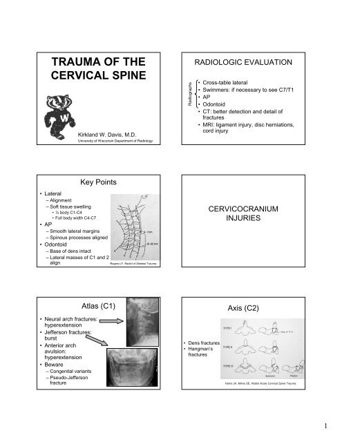
<strong>TRAUMA</strong> <strong>OF</strong> <strong>THE</strong><br />
<strong>CERVICAL</strong> <strong>SPINE</strong><br />
Kirkland W. Davis, M.D.<br />
University <strong>of</strong> Wisconsin <strong>Department</strong> <strong>of</strong> <strong>Radiology</strong><br />
Radiographs<br />
RADIOLOGIC EVALUATION<br />
• Cross-table lateral<br />
• Swimmers: if necessary to see C7/T1<br />
•AP<br />
• Odontoid<br />
• CT: better detection and detail <strong>of</strong><br />
fractures<br />
• MRI: ligament injury, disc herniations,<br />
cord injury<br />
Key Points<br />
• Lateral<br />
– Alignment<br />
– S<strong>of</strong>t tissue swelling<br />
•½body C1-C4<br />
• Full body width C4-C7<br />
•AP<br />
– Smooth lateral margins<br />
– Spinous processes aligned<br />
• Odontoid<br />
– Base <strong>of</strong> dens intact<br />
– Lateral masses <strong>of</strong> C1 and 2<br />
align<br />
Rogers LF. Radiol <strong>of</strong> Skeletal Trauma.<br />
CERVICOCRANIUM<br />
INJURIES<br />
• Neural arch fractures:<br />
hyperextension<br />
• Jefferson fractures:<br />
burst<br />
• Anterior arch<br />
avulsion:<br />
hyperextension<br />
•Beware<br />
– Congenital variants<br />
– Pseudo-Jefferson<br />
fracture<br />
Atlas (C1)<br />
• Dens fractures<br />
• Hangman’s<br />
fractures<br />
Axis (C2)<br />
Harris JH, Mirvis SE. Radiol Acute Cervical Spine Trauma.<br />
1
Mechanism: Hyperflexion<br />
INJURIES <strong>OF</strong> <strong>THE</strong><br />
LOWER<br />
<strong>CERVICAL</strong> <strong>SPINE</strong> (C3-7)<br />
• Anterior subluxation (hyperflexion sprain)<br />
• Bilateral interfacetal dislocation<br />
• Simple wedge compression fracture<br />
• Clay shoveler’s fracture<br />
• Flexion teardrop fracture<br />
Anterior Subluxation<br />
(Hyperflexion Sprain)<br />
Bilateral Interfacetal Dislocation<br />
(Facet Jump)<br />
•Unstable<br />
• Predominantly s<strong>of</strong>t<br />
tissue disruptions<br />
• Wide<br />
posteriorly/kyphosis<br />
• > 50% anterior<br />
translation<br />
• Both facets jumped<br />
• Severe neurologic injury<br />
common<br />
Rogers LF. Radiol <strong>of</strong> Skeletal Trauma.<br />
Clay Shoveler’s Fracture<br />
• Spinous process<br />
avulsion<br />
• C7 > C6 > T1<br />
• No neurologic<br />
sequelae<br />
Flexion Teardrop Fracture<br />
• Swimming pool injury<br />
• Combo flexion and axial<br />
load<br />
• Characteristic<br />
“teardrop” fragment<br />
anterior inferior corner<br />
• Kyphotic angulation<br />
• Neurologically<br />
devastating<br />
2
Mechanism: Simultaneous<br />
Flexion and Rotation<br />
• Unilateral interfacetal<br />
dislocation<br />
– < 50% anterior translation<br />
– Spinous processes<br />
deviate<br />
– Bow-tie/bat-wing/butterfly<br />
appearance <strong>of</strong> facets<br />
• Unilateral interfacetal<br />
fracture-dislocation<br />
Mechanism: Hyperextension<br />
• Hyperextension dislocation<br />
• Avulsion fracture <strong>of</strong> the anterior arch <strong>of</strong><br />
the atlas<br />
• Fracture <strong>of</strong> the posterior arch <strong>of</strong> the<br />
atlas<br />
Mechanism: Hyperextension<br />
• Extension teardrop fracture<br />
• Laminar fracture<br />
• Hangman’s fracture<br />
• Hyperextension fracture-dislocation<br />
Hyperextension Dislocation<br />
• Often elderly<br />
• Widened anteriorly<br />
• S<strong>of</strong>t-tissue swelling<br />
universal, but usually<br />
obscured by<br />
intubation<br />
• Neuro problems<br />
common: central cord<br />
syndrome<br />
Extension Teardrop Fracture<br />
Mechanism: Hyperextension<br />
and Rotation<br />
•C2<br />
• Taller than it is wide<br />
• Pillar fracture<br />
• Pedicolaminar<br />
fracture-separation<br />
3
Mechanism:<br />
Vertical Compression<br />
Mechanism:<br />
Lateral Flexion<br />
• Jefferson<br />
fracture<br />
• Burst fracture<br />
This burst fracture<br />
involves the posterior<br />
wall <strong>of</strong> the vertebra<br />
and the laminae,<br />
which is typical.<br />
• Unilateral occipital condyle fracture<br />
• Unilateral lateral mass fracture, C1<br />
• Uncinate process fracture<br />
• Transverse process fracture<br />
REASONS TO OBTAIN MRI<br />
• Neurologic impairment (MR shows cord<br />
damage)<br />
• Suspect ligament injury (CT doesn’t<br />
show s<strong>of</strong>t tissue damage)<br />
• Suspect herniated disc (MR superior to<br />
CT)<br />
NOTE: CT is the study <strong>of</strong> choice to evaluate for fracture!<br />
EVALUATION <strong>OF</strong><br />
PELVIC RING AND<br />
ACETABULAR FRACTURES<br />
Kirkland W. Davis, M.D.<br />
University <strong>of</strong> Wisconsin <strong>Department</strong> <strong>of</strong> <strong>Radiology</strong><br />
Radiologic Evaluation<br />
• AP radiograph<br />
• CT<br />
– If have known fractures<br />
– If radiographs uncertain<br />
• Inlet/outlet (45<br />
degrees)<br />
• Judet (45 degrees)<br />
As needed for treatment<br />
and follow-up<br />
Key Areas to Look<br />
• Femoral neck/intertrochanteric<br />
• Obturator ring<br />
• Pubic symphysis (< 6mm)<br />
• Iliopubic and ilioischial<br />
lines<br />
• Posterior wall acetabulum<br />
• SI joints (< 4mm)<br />
• Sacral foramina<br />
• L4 and L5 transverse<br />
processes<br />
4


