

CT Protocols: (Brain, ENT, Spine, Vascular) - Department of Radiology
CT Protocols: (Brain, ENT, Spine, Vascular) - Department of Radiology
CT Protocols: (Brain, ENT, Spine, Vascular) - Department of Radiology

You also want an ePaper? Increase the reach of your titles
YUMPU automatically turns print PDFs into web optimized ePapers that Google loves.
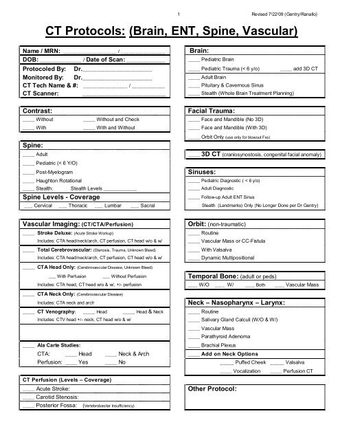
Name / MRN: ___________________ / ________________<br />
DOB: _______________ / Date <strong>of</strong> Scan: ______________ _____ Pediatric <strong>Brain</strong><br />
1 Revised 7/22/09 (Gentry/Ranallo)<br />
<strong>CT</strong> <strong>Protocols</strong>: (<strong>Brain</strong>, <strong>ENT</strong>, <strong>Spine</strong>, <strong>Vascular</strong>)<br />
<strong>Brain</strong>:<br />
Protocoled By: Dr. _________________________ _____ Pediatric Trauma (< 6 y/o) _____ add 3D <strong>CT</strong><br />
Monitored By: Dr. _________________________<br />
_____ Adult <strong>Brain</strong><br />
<strong>CT</strong> Tech Name & #: ________________ / ____________ _____ Pituitary & Cavernous Sinus<br />
<strong>CT</strong> Scanner: ______________________________ _____ Stealth (Whole <strong>Brain</strong> Treatment Planning)<br />
Contrast:<br />
Facial Trauma:<br />
_____ Without _____ Without and Check _____ Face and Mandible (No 3D)<br />
_____ With _____ With and Without _____ Face and Mandible (With 3D)<br />
_____ Orbit Only (use only for blowout Fxs)<br />
<strong>Spine</strong>:<br />
_____ Adult _____ (craniosynostosis, congenital facial anomaly)<br />
_____ Pediatric (< 6 Y/O)<br />
_____ Post-Myelogram<br />
_____ Haughton Rotational<br />
3D <strong>CT</strong><br />
Sinuses:<br />
_____ Pediatric Diagnostic ( < 6 y/o)<br />
_____ Stealth: Stealth Levels ____________ _____ Adult Diagnostic<br />
<strong>Spine</strong> Levels - Coverage<br />
_____ Follow-up Adult <strong>ENT</strong> Sinus<br />
___ Cervical ___ Thoracic ___ Lumbar ___ Sacral Stealth (Landmarks) Only (No Longer Done per Dr Gentry)<br />
<strong>Vascular</strong> Imaging: (<strong>CT</strong>/<strong>CT</strong>A/Perfusion)<br />
Orbit: (non-traumatic)<br />
_____ Stroke Deluxe: (Acute Stroke Workup) _____ Routine<br />
Includes: <strong>CT</strong>A head/neck/arch, <strong>CT</strong> perfusion, <strong>CT</strong> head w/o & w/<br />
_____ <strong>Vascular</strong> Mass or CC-Fistula<br />
_____ Total Cerebrovascular: (Stenosis, Trauma, Unknown Bleed) _____ With Valsalva<br />
Includes: <strong>CT</strong>A head/neck/arch, <strong>CT</strong> perfusion, <strong>CT</strong> head w/o & w/<br />
_____ Dynamic Multipositional<br />
_____<br />
_____<br />
<strong>CT</strong>A Head Only: (Cerebrovascular Disease, Unknown Bleed)<br />
___ With Perfusion ___ Without Perfusion Temporal Bone: (adult or peds)<br />
Includes: <strong>CT</strong>A head, <strong>CT</strong> head w/o & w/, +/- perfusion ____ W/O ____ W/ ____ Both ____ <strong>Vascular</strong> Mass<br />
<strong>CT</strong>A Neck Only: (Cerebrovascular Disease)<br />
Includes: <strong>CT</strong>A neck and arch<br />
_____ <strong>CT</strong> Venography: _____ Head _____ Head & Neck _____ Routine<br />
Includes: <strong>CT</strong>V head +/- neck, <strong>CT</strong> head w/o & w/<br />
_____ Salivary Gland Calculi (W/O & W/)<br />
_____ Aneurysm (Hi-Res COW): (Nontraumatic SAH, Known Aneurysm) _____ <strong>Vascular</strong> Mass<br />
Includes: <strong>CT</strong>A head, <strong>CT</strong> head w/o & w/, no perfusion<br />
_____ Parathyroid Adenoma<br />
_____ Ala Carte Studies:<br />
_____ Brachial Plexus<br />
<strong>CT</strong>A: _____ Head _____ Neck & Arch _____ Add on Neck Options<br />
Perfusion: _____ Yes _____ No _____ Puffed Cheek _____ Valsalva<br />
_____ Vocalization _____ Perfusion <strong>CT</strong><br />
<strong>CT</strong> Perfusion (Levels – Coverage)<br />
_____ Acute Stroke:<br />
_____ Carotid Stenosis:<br />
_____ Posterior Fossa:<br />
(Vertebrobasilar Insufficiency)<br />
Neck – Nasopharynx – Larynx:<br />
Other Protocol:
Table <strong>of</strong> Contents:<br />
2 Revised 7/22/09 (Gentry/Ranallo)<br />
Page # Protocol Exam<br />
5 1.1 Adult Head – Routine Helical<br />
8 1.2 Adult Head – Helical Scan with Angled Axial Reformations<br />
11 1.3 Adult Head – Axial<br />
13 11.1 & 11.2 Pediatric Head – Routine Helical<br />
17 11.3 & 11.4 Pediatric Head – Helical Scan with Angled Axial Reformations<br />
21 11.5 & 11.6 Pediatric Head – Axial<br />
24 11.7 & 11.8 Pediatric Head – Trauma<br />
27 2.1 Orbit – Routine<br />
33 2.2 Orbit – With and Without Valsalva<br />
34 2.3 Orbit – <strong>Vascular</strong> Mass or Carotid-Cavernous Fistula<br />
36 2.4 Orbit – Dynamic – EOM Movements<br />
27 12.1 &12.2 Pediatric Orbit – Routine<br />
33 12.3 & 12.4 Pediatric Orbit – With and Without Valsalva<br />
34 12.5 & 12.6 Pediatric Orbit – <strong>Vascular</strong> Mass or Carotid-Cavernous Fistula<br />
36 12.7 & 12.8 Pediatric Orbit – Dynamic – EOM Movements<br />
37 2.5a Maxill<strong>of</strong>acial Trauma – Routine<br />
43 2.5b Maxill<strong>of</strong>acial Trauma – Routine plus 3D<br />
37 12.9 & 12.10 Pediatric Maxill<strong>of</strong>acial Trauma – Routine<br />
43 12.9 & 12.10 Pediatric Maxill<strong>of</strong>acial Trauma – Routine plus 3D<br />
44 1.5 3D <strong>CT</strong> – Craniosynostosis, Congenital Facial Anomaly<br />
44 11.9 & 11.10 Pediatric 3D <strong>CT</strong> – Craniosynostosis, Congenital Facial Anomaly<br />
48 2.6 Pituitary Gland and Cavernous Sinus<br />
48 12.11 & 12.12 Pediatric Pituitary Gland and Cavernous Sinus<br />
52 1.10 Stealth – Stereotactic Head (Whole <strong>Brain</strong> Treatment Planning)<br />
52 11.11 & 11.12 Pediatric Stealth – Stereotactic Head (Whole <strong>Brain</strong> Treatment Planning)<br />
53 2.7 Sinuses – Diagnostic<br />
53 12.13 & 12.14 Pediatric Sinuses – Diagnostic<br />
59 2.8 Sinuses – Follow-up Adult <strong>ENT</strong> Sinus<br />
61 2.9 Sinuses – Conbined Diagnostic and Landmark<br />
61 12.16 & 12.17 Pediatric Sinuses – Conbined Diagnostic and Landmark<br />
62 2.10 Temporal Bone and Posterior Fossa (W/O Contrast)<br />
62 12.18 & 12.19 Pediatric Temporal Bone and Posterior Fossa (W/O Contrast)<br />
66 2.11 Temporal Bone and Posterior Fossa (W/O and W Contrast)<br />
66 12.20 & 12.21 Pediatric Temporal Bone and Posterior Fossa (W/O and W Contrast)<br />
70 2.12 Temporal Bone – <strong>Vascular</strong> Mass<br />
70 12.22 & 12.23 Pediatric Temporal Bone – <strong>Vascular</strong> Mass<br />
75 3.1 Neck – Routine<br />
75 3.2 Neck – Feet First<br />
81 3.1 Neck – Salivary Gland<br />
82 3.3 Neck – <strong>Vascular</strong> Mass
Table <strong>of</strong> Contents (continued):<br />
3 Revised 7/22/09 (Gentry/Ranallo)<br />
Page # Protocol Exam<br />
84 3.5 Neck – Add on Options<br />
86 3.4 Neck – Parathyroid Adenoma<br />
75 13.1 Pediatric Neck – Routine<br />
75 13.2 Pediatric Neck – Feet First<br />
81 13.1 Pediatric Neck – Salivary Gland<br />
82 13.3 Pediatric Neck – <strong>Vascular</strong> Mass<br />
86 3.4 Pediatric Neck – Parathyroid Adenoma<br />
84 13.5 Pediatric Neck – Add on Options<br />
90 3.1 Brachial Plexus – Adult<br />
90 13.1 Brachial Plexus –Pediatric<br />
91 3.5 Cervical <strong>Spine</strong> – Adult<br />
93 7.5 Thoracic <strong>Spine</strong> – Feet First – Adult<br />
94 7.6 Thoracic <strong>Spine</strong> – Head First – Adult<br />
95 7.1 Lumbar <strong>Spine</strong> – Feet First – Adult<br />
97 13.5 Cervical <strong>Spine</strong> – Pediatric<br />
100 17.5 Thoracic <strong>Spine</strong> – Pediatric<br />
103 17.1 Lumbar <strong>Spine</strong> – Pediatric<br />
106 7.2 Stealth (Stereotactic) <strong>Spine</strong><br />
107 1.6 & 3.7 <strong>Vascular</strong> Imaging: Stroke Deluxe (Acute Stroke Workup)<br />
111 1.6a <strong>Vascular</strong> Imaging: Total Cerebrovascular<br />
112 1.7 <strong>Vascular</strong> Imaging: <strong>CT</strong>A Head Only (Stenosis, Unknown Bleed)<br />
116 1.8 <strong>Vascular</strong> Imaging: Aneurysm (Hi-Res COW) (Non-traumatic SAH, Known Aneurysm)<br />
120 3.8 <strong>Vascular</strong> Imaging: <strong>CT</strong>A Neck Only (Cerebrovascular Disease)<br />
124 1.9 & 3.9 <strong>Vascular</strong> Imaging: <strong>CT</strong> Venography<br />
107 11.16 & 11.17 Pediatric <strong>Vascular</strong> Imaging: Stroke Deluxe (Acute Stroke Workup)<br />
111 11.16 & 11.17 Pediatric <strong>Vascular</strong> Imaging: Total Cerebrovascular<br />
112 11.18 & 11.19 Pediatric <strong>Vascular</strong> Imaging: <strong>CT</strong>A Head Only (Stenosis, Unknown Bleed)<br />
116 11.20 & 11.21 Pediatric <strong>Vascular</strong> Imaging: Aneurysm(Hi-Res COW)(Non-trauma SAH, Known Aneurysm)<br />
120 11.22 & 11.23 Pediatric <strong>Vascular</strong> Imaging: <strong>CT</strong>A Neck Only (Cerebrovascular Disease)<br />
124 11.24 & 11.25 Pediatric <strong>Vascular</strong> Imaging: <strong>CT</strong> Venography<br />
128 Appendix #1 <strong>CT</strong>A Head: 2D Thin and Thick Slab Reformations<br />
129 Appendix #2: <strong>CT</strong>A Neck: 2D-Reformations<br />
130 Appendix #3: <strong>CT</strong> Perfusion Protocol<br />
131 Appendix #4: <strong>CT</strong> Perfusion Coverage<br />
132 Appendix #5: <strong>CT</strong> Perfusion Analysis Instructions<br />
138 Appendix #6: Neck <strong>CT</strong> Contrast Timing for Routine Neck <strong>CT</strong><br />
139 Appendix #7: 64 Slice Scanner Prioity<br />
140 Appendix #8: <strong>CT</strong> Scanner Type<br />
141 Appendix #9: <strong>CT</strong> Scanner Limits<br />
142 Appendix #10: Direction and Naming <strong>of</strong> 2D-Reformations<br />
143 Appendix: #11 Combined Neuro and Body Contrast Studies
4 Revised 7/22/09 (Gentry/Ranallo)<br />
Scanner Nomenclature:<br />
Scanner -<br />
Location<br />
# <strong>of</strong><br />
Slices<br />
Maximum mA<br />
at 120 kV<br />
Scanner Name<br />
Naming Convention<br />
in this Protocol Book<br />
<strong>CT</strong>I – 1 16 800 Lightspeed Xtra<br />
<strong>CT</strong>I – 2 4 440 LightSpeed 16<br />
<strong>CT</strong>I – 3 16 800 LightSpeed 16 Pro<br />
<strong>CT</strong>I - 4 64 800 LightSpeed V<strong>CT</strong> 64<br />
ER 64 800 LightSpeed V<strong>CT</strong> 64<br />
<strong>CT</strong>-RP 8 440 LightSpeed 8<br />
East 8 440 LightSpeed 8<br />
LS Xtra<br />
LS 16<br />
LS 16 Pro<br />
LS V<strong>CT</strong> 64<br />
LS V<strong>CT</strong> 64<br />
LS 8<br />
LS 8
5 Revised 7/22/09 (Gentry/Ranallo)<br />
Adult Head: Routine (Helical Mode) (Protocol # 1.1)<br />
Billing: 1. <strong>CT</strong> Head without, or with, or without and with<br />
2. Contrast if used<br />
Setup: 1. Supine, AP and lateral scouts, no gantry angle<br />
2. Helical mode should be used routinely for adult head <strong>CT</strong> scans. Only use axial mode<br />
when you cannot move the patient’s head into proper position (trauma, cervical<br />
collar, rigid neck).<br />
3. Patient Positioning: Tilt the patients head so that a line connecting the lateral<br />
canthus <strong>of</strong> the eye and the EAC is perpendicular to the <strong>CT</strong> tabletop (see below). Use<br />
axial mode and angle the gantry if you cannot place the patient’s head within 15<br />
degrees <strong>of</strong> the proper setup angle.<br />
4. Start scans at the bottom <strong>of</strong> C1 and scan through the top <strong>of</strong> the head<br />
DFOV: Preferred 20 cm (Range 18-22)<br />
Contrast: 1. 150 ml <strong>of</strong> 240 mg/dl non-ionic contrast @ 0.6 ml/sec (4.2 minutes)<br />
2. Begin scanning as soon as contrast injection is finished<br />
Other Info:
6 Revised 7/22/09 (Gentry/Ranallo)<br />
Head: Helical<br />
Adult<br />
Non-Contrast<br />
<strong>CT</strong> 1 <strong>CT</strong> 2 <strong>CT</strong> 3 <strong>CT</strong> 4 & ER <strong>CT</strong> East & RP <strong>CT</strong><br />
Scanner GE LS Xtra GE LS 16 GE LS 16 Pro GE LS V<strong>CT</strong> 64 GE LS 8<br />
Scan Type Helical Helical Helical Helical Helical<br />
Rotation Time (sec) 0.5 0.6 0.4 0.4 0.7<br />
Detector Coverage (mm)<br />
Beam Collimation (mm)<br />
10 10 10 20 10<br />
Detector Rows 16 16 16 16 8<br />
Pitch 0.562 0.562 0.562 0.531 0.625<br />
Speed (mm/rot) 5.625 5.625 5.625 10.62 6.25<br />
Detector Configuration 16 x 0.625 16 x 0.625 16 x 0.625 64 x 0.625 8 x 1.25<br />
Slice Thickness (mm) 5 5 5 5 5<br />
Interval (mm) 2.5 2.5 2.5 2.5 2.5<br />
Scan FOV Head Head Head Head Head<br />
kV 120 120 120 120 120<br />
Smart mA/ Auto mA Range 200-660 130-440 200-660 190-620 130-440<br />
Noise Index 2.8 2.8 2.8 2.8 2.8<br />
(Manual mA) 530 350 530 500 340<br />
Recon 1:<br />
DFOV 22 22 22 22 22<br />
Recon Type S<strong>of</strong>t S<strong>of</strong>t S<strong>of</strong>t S<strong>of</strong>t S<strong>of</strong>t<br />
WW/ WL 80/25 80/25 80/25 80/25 80/25<br />
Recon Option Full Full Full Full Full<br />
Recon 2:<br />
DFOV 22 22 22 22 22<br />
Recon Type Bone Plus Bone Plus Bone Plus Bone Plus Bone Plus<br />
WW/ WL 3000/300 3000/300 3000/300 3000/300 3000/300<br />
Recon Option Full Full Full Full Full<br />
Slice Thickness (mm) 2.5 2.5 2.5 2.5 2.5<br />
Interval (mm) 1.25 1.25 1.25 1.25 1.25
7 Revised 7/22/09 (Gentry/Ranallo)<br />
Head: Helical<br />
Adult<br />
Contrast<br />
<strong>CT</strong> 1 <strong>CT</strong> 2 <strong>CT</strong> 3 <strong>CT</strong> 4 & ER <strong>CT</strong> East & RP <strong>CT</strong><br />
Scanner GE LS Xtra GE LS 16 GE LS 16 Pro GE LS V<strong>CT</strong> 64 GE LS 8<br />
Scan Type Helical Helical Helical Helical Helical<br />
Rotation Time (sec) 0.6 0.9 0.5 0.5 1.0<br />
Detector Coverage (mm)<br />
Beam Collimation (mm)<br />
10 10 10 20 10<br />
Detector Rows 16 16 16 16 8<br />
Pitch 0.562 0.562 0.562 0.531 0.625<br />
Speed (mm/rot) 5.625 5.625 5.625 10.62 6.25<br />
Detector Configuration 16 x 0.625 16 x 0.625 16 x 0.625 64 x 0.625 8 x 1.25<br />
Slice Thickness (mm) 5 5 5 5 5<br />
Interval (mm) 2.5 2.5 2.5 2.5 2.5<br />
Scan FOV Head Head Head Head Head<br />
kV 100 100 100 100 100<br />
Smart mA/ Auto mA Range 230-750 130-420 230-750 210-700 130-420<br />
Noise Index 3.3 3.3 3.3 3.3 3.3<br />
(Manual mA) 620 330 600 570 340<br />
Recon 1:<br />
DFOV 22 22 22 22 22<br />
Recon Type S<strong>of</strong>t S<strong>of</strong>t S<strong>of</strong>t S<strong>of</strong>t S<strong>of</strong>t<br />
WW/ WL 90/30 90/30 90/30 90/30 90/30<br />
Recon Option Full Full Full Full Full<br />
Recon 2:<br />
DFOV 22 22 22 22 22<br />
Recon Type Bone Plus Bone Plus Bone Plus Bone Plus Bone Plus<br />
WW/ WL 3500/350 3500/350 3500/350 3500/350 3500/350<br />
Recon Option Full Full Full Full Full<br />
Slice Thickness (mm) 2.5 2.5 2.5 2.5 2.5<br />
Interval (mm) 1.25 1.25 1.25 1.25 1.25
8 Revised 7/22/09 (Gentry/Ranallo)<br />
Adult Head: Helical Scan with Angled Axial Reformations (Protocol # 1.2)<br />
Billing: 1. <strong>CT</strong> Head without, or with, or without and with<br />
2. Contrast if used<br />
Setup: 1. Use this protocol when the head cannot be properly positioned for a routine helical<br />
head scan. Example: when you cannot move the patient’s head into proper position<br />
(trauma, cervical collar, rigid neck).<br />
2. Supine, AP and lateral scouts, no gantry angle<br />
3. Start the scans at C2 and scan through the top <strong>of</strong> the head<br />
4. Do not send the source images to PACS (Only send the 2D-reformations)<br />
5. Obtain 2D-reformations parallel to a line connecting the infraorbital rim with the<br />
opisthion (see below). Use a sagittal view on Imageworks slightly <strong>of</strong>f midline to<br />
choose proper angle <strong>of</strong> reconstruction. Start reformations at the bottom <strong>of</strong> C1 and<br />
go to the top <strong>of</strong> the head using a 20 cm DFOV.<br />
6. Important: Be certain that dental filling artifact does not extend across the brain on<br />
the helical raw data. If it does, then use the axial mode head protocol instead.<br />
DFOV: Preferred 20 cm (Range 18-22)<br />
Contrast: 1. 150 ml <strong>of</strong> 240 mg/dl non-ionic contrast @ 0.6 ml/sec (4.2 minutes)<br />
2. Begin scanning as soon as contrast injection is finished<br />
Other Info: 1. 2D-Reformations<br />
a. Axial S<strong>of</strong>t Tissue: 5 mm thick with an interval <strong>of</strong> 2.5 mm<br />
b. Axial Bone: 2.5 mm thick with an interval <strong>of</strong> 1.25 mm
9 Revised 7/22/09 (Gentry/Ranallo)<br />
Head: Helical Scan with Angled Axial<br />
Reformations - Adult<br />
Scanner GE LS Xtra GE LS 16 GE LS 16 Pro GE LS V<strong>CT</strong> 64 GE LS 8<br />
Scan Type Helical Helical Helical Helical Helical<br />
Rotation Time (sec) 0.6 0.6 0.5 0.5 0.7<br />
Detector Coverage (mm)<br />
Beam Collimation (mm)<br />
10 10 10 20 10<br />
Detector Rows 16 16 16 16 8<br />
Pitch 0.562 0.562 0.562 0.531 0.625<br />
Speed (mm/rot) 5.625 5.625 5.625 10.62 6.25<br />
Detector Configuration 16 x 0.625 16 x 0.625 16 x 0.625 64 x 0.625 8 x 1.25<br />
Slice Thickness (mm) 1.25 1.25 1.25 1.25 1.25<br />
Interval (mm) 0.65 0.65 0.65 0.65 0.65<br />
Scan FOV Head Head Head Head Head<br />
kV 120 120 120 120 120<br />
Smart mA/ Auto mA Range 170-550 130-440 160-530 150-500 130-440<br />
Noise Index 5.6 5.6 5.6 5.6 5.6<br />
(Manual mA) 440 350 420 400 340<br />
Recon 1:<br />
DFOV 22 22 22 22 22<br />
Recon Type S<strong>of</strong>t S<strong>of</strong>t S<strong>of</strong>t S<strong>of</strong>t S<strong>of</strong>t<br />
WW/ WL 80/25 80/25 80/25 80/25 80/25<br />
Recon Option Plus Plus Plus Full Plus<br />
Recon Option<br />
Recon 2:<br />
IQ Enhance<br />
DFOV 22 22 22 22 22<br />
Recon Type Bone Plus Bone Plus Bone Plus Bone Plus Bone Plus<br />
WW/ WL 3000/300 3000/300 3000/300 3000/300 3000/300<br />
Recon Option Plus Plus Plus Full Plus<br />
Recon Option<br />
Non-Contrast<br />
<strong>CT</strong> 1 <strong>CT</strong> 2 <strong>CT</strong> 3 <strong>CT</strong> 4 & ER <strong>CT</strong> East & RP <strong>CT</strong><br />
IQ Enhance<br />
Slice Thickness (mm) 0.625 0.625 0.625 0.625 1.25<br />
Interval (mm) 0.375 0.375 0.375 0.312 0.75
10 Revised 7/22/09 (Gentry/Ranallo)<br />
Head: Helical Scan with Angled Axial<br />
Reformations - Adult<br />
Scanner GE LS Xtra GE LS 16 GE LS 16 Pro GE LS V<strong>CT</strong> 64 GE LS 8<br />
Scan Type Helical Helical Helical Helical Helical<br />
Rotation Time (sec) 0.6 0.9 0.5 0.5 1.0<br />
Detector Coverage (mm)<br />
Beam Collimation (mm)<br />
10 10 10 20 10<br />
Detector Rows 16 16 16 16 8<br />
Pitch 0.562 0.562 0.562 0.531 0.625<br />
Speed (mm/rot) 5.625 5.625 5.625 10.62 6.25<br />
Detector Configuration 16 x 0.625 16 x 0.625 16 x 0.625 64 x 0.625 8 x 1.25<br />
Slice Thickness (mm) 1.25 1.25 1.25 1.25 1.25<br />
Interval (mm) 0.65 0.65 0.65 0.65 0.65<br />
Scan FOV Head Head Head Head Head<br />
kV 100 100 100 100 100<br />
Smart mA/ Auto mA Range 240-750 130-420 230-750 210-700 130-420<br />
Noise Index 6.6 6.6 6.6 6.6 6.6<br />
(Manual mA) 630 330 600 570 340<br />
Recon 1:<br />
DFOV 22 22 22 22 22<br />
Recon Type S<strong>of</strong>t S<strong>of</strong>t S<strong>of</strong>t S<strong>of</strong>t S<strong>of</strong>t<br />
WW/ WL 90/30 90/30 90/30 90/30 90/30<br />
Recon Option Plus Plus Plus Full Plus<br />
Recon Option<br />
Recon 2:<br />
IQ Enhance<br />
DFOV 22 22 22 22 22<br />
Recon Type Bone Plus Bone Plus Bone Plus Bone Plus Bone Plus<br />
WW/ WL 3500/350 3500/350 3500/350 3500/350 3500/350<br />
Recon Option Plus Plus Plus Full Plus<br />
Recon Option<br />
Contrast<br />
<strong>CT</strong> 1 <strong>CT</strong> 2 <strong>CT</strong> 3 <strong>CT</strong> 4 & ER <strong>CT</strong> East & RP <strong>CT</strong><br />
IQ Enhance<br />
Slice Thickness (mm) 0.625 0.625 0.625 0.625 1.25<br />
Interval (mm) 0.375 0.375 0.375 0.312 0.75
Adult Head: (Axial Mode) (Protocol # 1.3)<br />
11 Revised 7/22/09 (Gentry/Ranallo)<br />
Billing: 1. <strong>CT</strong> Head without, or with, or without and with<br />
2. Contrast if used<br />
Setup: 1. Supine, AP and lateral scouts<br />
2. Helical mode should be used routinely `for adult head <strong>CT</strong> scans. Only use axial<br />
mode when you cannot move the patient’s head into proper position (trauma,<br />
cervical collar, rigid neck). This mode can also be used in unstable patients in the<br />
emergency department when the <strong>CT</strong> scan time must be expedited.<br />
3. Patient Positioning: Using the lateral scout image, angle the gantry so that it is<br />
parallel to a line connecting the infraorbital rim with the opisthion (see below).<br />
4. Start scans at the bottom <strong>of</strong> C1 and scan through the top <strong>of</strong> the head<br />
DFOV: Preferred 20 cm (Range 18-22)<br />
Contrast: 1. 150 ml <strong>of</strong> 240 mg/dl non-ionic contrast @ 0.6 ml/sec (4.2 minutes)<br />
2. Begin scanning as soon as contrast injection is finished<br />
Other Info:
12 Revised 7/22/09 (Gentry/Ranallo)<br />
Head: Axial<br />
Adult<br />
Non-Contrast<br />
<strong>CT</strong> 1 <strong>CT</strong> 2 <strong>CT</strong> 3 <strong>CT</strong> 4 & ER <strong>CT</strong> East & RP <strong>CT</strong><br />
Scanner GE LS Xtra GE LS 16 GE LS 16 Pro GE LS V<strong>CT</strong> 64 GE LS 8<br />
Scan Type Axial Axial Axial Axial Axial<br />
Rotation Time (sec) 0.7 0.9 0.6 0.6 0.9<br />
Detector Coverage (mm)<br />
Beam Collimation (mm)<br />
10 10 10 10 10<br />
Detector Rows 16 16 16 16 8<br />
Number <strong>of</strong> Images per<br />
Rotation<br />
2i 2i 2i 2i 2i<br />
Detector Configuration 16 x 0.625 16 x 0.625 16 x 0.625 16 x 0.625 8 x 1.25<br />
Slice Thickness (mm) 5 5 5 5 5<br />
Scan FOV Head Head Head Head Head<br />
kV 120 120 120 120 120<br />
mA 670 420 630 630 420<br />
Recon 1:<br />
DFOV 22 22 22 22 22<br />
Recon Type S<strong>of</strong>t S<strong>of</strong>t S<strong>of</strong>t S<strong>of</strong>t S<strong>of</strong>t<br />
WW/ WL 80/25 80/25 80/25 80/25 80/25<br />
Recon 2:<br />
DFOV 22 22 22 22 22<br />
Recon Type Bone Plus Bone Plus Bone Plus Bone Plus Bone Plus<br />
WW/ WL 3000/300 3000/300 3000/300 3000/300 3000/300<br />
Slice Thickness (mm) 2.5 2.5 2.5 2.5 2.5<br />
Head: Axial<br />
Adult<br />
Contrast<br />
<strong>CT</strong> 1 <strong>CT</strong> 2 <strong>CT</strong> 3 <strong>CT</strong> 4 & ER <strong>CT</strong> East & RP <strong>CT</strong><br />
Scanner GE LS Xtra GE LS 16 GE LS 16 Pro GE LS V<strong>CT</strong> 64 GE LS 8<br />
Scan Type Axial Axial Axial Axial Axial<br />
Rotation Time (sec) 1.0 0.9 0.8 0.8 0.9<br />
Detector Coverage (mm)<br />
Beam Collimation (mm)<br />
10 10 10 10 10<br />
Detector Rows 16 16 16 16 8<br />
Number <strong>of</strong> Images per<br />
Rotation<br />
2i 2i 2i 2i 2i<br />
Detector Configuration 16 x 0.625 16 x 0.625 16 x 0.625 16 x 0.625 8 x 1.25<br />
Slice Thickness (mm) 5 5 5 5 5<br />
Scan FOV Head Head Head Head Head<br />
kV 100 120 100 100 120<br />
mA 670 400 670 670 400<br />
Recon 1:<br />
DFOV 22 22 22 22 22<br />
Recon Type S<strong>of</strong>t S<strong>of</strong>t S<strong>of</strong>t S<strong>of</strong>t S<strong>of</strong>t<br />
WW/ WL 90/30 90/30 90/30 90/30 90/30<br />
Recon 2:<br />
DFOV 22 22 22 22 22<br />
Recon Type Bone Plus Bone Plus Bone Plus Bone Plus Bone Plus<br />
WW/ WL 3500/350 3500/350 3500/350 3500/350 3500/350<br />
Slice Thickness (mm) 2.5 2.5 2.5 2.5 2.5
13 Revised 7/22/09 (Gentry/Ranallo)<br />
Pediatric Head: Routine (Helical Mode) (< 6 years <strong>of</strong> age)<br />
(Protocol # 11.1 & 11.2)<br />
Billing: 1. <strong>CT</strong> Head without, or with, or without and with<br />
2. Contrast if used<br />
Setup: 1. Supine, AP and lateral scouts, no gantry angle<br />
2. Helical mode should be used routinely for pediatric head <strong>CT</strong> scans. Only use axial<br />
mode when you cannot move the patient’s head into proper position (trauma,<br />
cervical collar, rigid neck).<br />
3. Patient Positioning: Tilt the patients head so that a line connecting the lateral<br />
canthus <strong>of</strong> the eye and the EAC is perpendicular to the <strong>CT</strong> tabletop (see below). Use<br />
axial mode and angle the gantry if you cannot place the patient’s head within 15<br />
degrees <strong>of</strong> the proper setup angle.<br />
4. Start scans at the bottom <strong>of</strong> C1 and scan through the top <strong>of</strong> the head<br />
Preferred:<br />
Preferred 16 cm (Range 14-18 cm)<br />
Contrast: 1. 1 ml / pound (2 ml/kg) <strong>of</strong> 240 non-ionic contrast @ 0.6 ml/sec<br />
2. Begin scanning as soon as contrast injection is finished<br />
Patient Age:<br />
Choose the <strong>CT</strong> scan factors on the scanner for the proper age range <strong>of</strong> the patient<br />
1. Child: (3 – 6 years)<br />
2. Infant: (0 – 3 years)<br />
Other Info:
14 Revised 7/22/09 (Gentry/Ranallo)<br />
Head: Helical<br />
Pediatric Child (3 – 6 yr)<br />
Non-Contrast<br />
<strong>CT</strong> 1 <strong>CT</strong> 2 <strong>CT</strong> 3 <strong>CT</strong> 4 & ER <strong>CT</strong> East & RP <strong>CT</strong><br />
Scanner GE LS Xtra GE LS 16 GE LS 16 Pro GE LS V<strong>CT</strong> 64 GE LS 8<br />
Scan Type Helical Helical Helical Helical Helical<br />
Rotation Time (sec) 0.5 0.6 0.4 0.4 0.7<br />
Detector Coverage (mm)<br />
Beam Collimation (mm)<br />
10 10 10 20 10<br />
Detector Rows 16 16 16 16 8<br />
Pitch 0.562 0.562 0.562 0.531 0.625<br />
Speed (mm/rot) 5.625 5.625 5.625 10.62 6.25<br />
Detector Configuration 16 x 0.625 16 x 0.625 16 x 0.625 64 x 0.625 8 x 1.25<br />
Slice Thickness (mm) 5 5 5 5 5<br />
Interval (mm) 2.5 2.5 2.5 2.5 2.5<br />
Scan FOV Head Head Head Small Head Head<br />
kV 100 100 100 100 100<br />
Smart mA/ Auto mA Range 170-680 110-400 170-680 160-640 110-420<br />
Noise Index 2.6 2.6 2.6 2.6 2.6<br />
(Manual mA) 510 340 510 480 330<br />
Recon 1:<br />
DFOV 22 22 22 22 22<br />
Recon Type S<strong>of</strong>t S<strong>of</strong>t S<strong>of</strong>t S<strong>of</strong>t S<strong>of</strong>t<br />
WW/ WL 80/25 80/25 80/25 80/25 80/25<br />
Recon Option Full Full Full Full Full<br />
Recon 2:<br />
DFOV 22 22 22 22 22<br />
Recon Type Bone Plus Bone Plus Bone Plus Bone Plus Bone Plus<br />
WW/ WL 3000/300 3000/300 3000/300 3000/300 3000/300<br />
Recon Option Full Full Full Full Full<br />
Slice Thickness (mm) 2.5 2.5 2.5 2.5 2.5<br />
Interval (mm) 1.25 1.25 1.25 1.25 1.25
15 Revised 7/22/09 (Gentry/Ranallo)<br />
Head: Helical<br />
Pediatric Child (3 – 6 yr)<br />
Contrast<br />
<strong>CT</strong> 1 <strong>CT</strong> 2 <strong>CT</strong> 3 <strong>CT</strong> 4 & ER <strong>CT</strong> East & RP <strong>CT</strong><br />
Scanner GE LS Xtra GE LS 16 GE LS 16 Pro GE LS V<strong>CT</strong> 64 GE LS 8<br />
Scan Type Helical Helical Helical Helical Helical<br />
Rotation Time (sec) 0.6 0.8 0.5 0.5 1.0<br />
Detector Coverage (mm)<br />
Beam Collimation (mm)<br />
10 10 10 20 10<br />
Detector Rows 16 16 16 16 8<br />
Pitch 0.562 0.562 0.562 0.531 0.625<br />
Speed (mm/rot) 5.625 5.625 5.625 10.62 6.25<br />
Detector Configuration 16 x 0.625 16 x 0.625 16 x 0.625 64 x 0.625 8 x 1.25<br />
Slice Thickness (mm) 5 5 5 5 5<br />
Interval (mm) 2.5 2.5 2.5 2.5 2.5<br />
Scan FOV Head Head Head Small Head Head<br />
kV 80 80 80 80 80<br />
Smart mA/ Auto mA Range 190-675 110-400 180-675 170-675 100-400<br />
Noise Index 3.0 3.0 3.0 3.0 3.0<br />
(Manual mA) 560 340 540 510 310<br />
Recon 1:<br />
DFOV 22 22 22 22 22<br />
Recon Type S<strong>of</strong>t S<strong>of</strong>t S<strong>of</strong>t S<strong>of</strong>t S<strong>of</strong>t<br />
WW/ WL 80/25 80/25 80/25 80/25 80/25<br />
Recon Option Full Full Full Full Full<br />
Recon 2:<br />
DFOV 22 22 22 22 22<br />
Recon Type Bone Plus Bone Plus Bone Plus Bone Plus Bone Plus<br />
WW/ WL 3000/300 3000/300 3000/300 3000/300 3000/300<br />
Recon Option Full Full Full Full Full<br />
Slice Thickness (mm) 2.5 2.5 2.5 2.5 2.5<br />
Interval (mm) 1.25 1.25 1.25 1.25 1.25
16 Revised 7/22/09 (Gentry/Ranallo)<br />
Head: Helical<br />
Pediatric Infant (0– 3 yr)<br />
Non-Contrast or Contrast<br />
<strong>CT</strong> 1 <strong>CT</strong> 2 <strong>CT</strong> 3 <strong>CT</strong> 4 & ER <strong>CT</strong> East & RP <strong>CT</strong><br />
Scanner GE LS Xtra GE LS 16 GE LS 16 Pro GE LS V<strong>CT</strong> 64 GE LS 8<br />
Scan Type Helical Helical Helical Helical Helical<br />
Rotation Time (sec) 0.5 0.7 0.4 0.4 0.8<br />
Detector Coverage (mm)<br />
Beam Collimation (mm)<br />
10 10 10 20 10<br />
Detector Rows 16 16 16 16 8<br />
Pitch 0.562 0.562 0.562 0.531 0.625<br />
Speed (mm/rot) 5.625 5.625 5.625 10.62 6.25<br />
Detector Configuration 16 x 0.625 16 x 0.625 16 x 0.625 64 x 0.625 8 x 1.25<br />
Slice Thickness (mm) 2.5 2.5 2.5 2.5 2.5<br />
Interval (mm) 1.5 1.5 1.5 1.5 1.5<br />
Scan FOV Head Head Head Small Head Head<br />
kV 80 80 80 80 80<br />
Smart mA/ Auto mA Range 140-675 80-390 140-675 130-640 80-390<br />
Noise Index 3.3 3.3 3.3 3.3 3.3<br />
(Manual mA) 460 260 460 430 260<br />
Recon 1:<br />
DFOV 20 20 20 20 20<br />
Recon Type Standard Standard Standard Standard Standard<br />
WW/ WL 80/25 80/25 80/25 80/25 80/25<br />
Recon Option Plus Plus Plus Plus Plus<br />
Recon 2:<br />
DFOV 20 20 20 20 20<br />
Recon Type Bone Plus Bone Plus Bone Plus Bone Plus Bone Plus<br />
WW/ WL 3000/300 3000/300 3000/300 3000/300 3000/300<br />
Recon Option Full Full Full Full Full<br />
Slice Thickness (mm) 2.5 2.5 2.5 2.5 2.5<br />
Interval (mm) 1.25 1.25 1.25 1.25 1.25
17 Revised 7/22/09 (Gentry/Ranallo)<br />
Pediatric Head: Helical Scan with Angled Axial Reformations (< 6 years <strong>of</strong> age)<br />
(Protocol # 11.3 & 11.4)<br />
Billing: 1. <strong>CT</strong> Head without, or with, or without and with<br />
2. Contrast if used<br />
Setup: 1. Use this protocol when the head cannot be properly positioned for a routine helical<br />
head scan. Example: when you cannot move the patient’s head into proper position<br />
(trauma, cervical collar, rigid neck)<br />
2. Supine, AP and lateral scouts, no gantry angle<br />
3. Start the scans at C2 and scan through the top <strong>of</strong> the head<br />
4. Do not send the source data to PACS (Only send the 2D-reformations)<br />
5. Obtain 2D-reformations parallel to a line connecting the infraorbital rim with the<br />
opisthion (see below). Start reformations at the bottom <strong>of</strong> C1 and go to the top <strong>of</strong> the<br />
head using a 20 cm DFOV.<br />
6. Important: Be certain that dental filling artifact does not extend across the brain on<br />
the helical raw data. If it does, then use the axial mode head protocol instead.<br />
DFOV:<br />
Preferred 16 cm (Range 14-18 cm)<br />
Contrast: 1. 1 ml / pound (2 ml/kg) <strong>of</strong> 240 non-ionic contrast @ 0.6 ml/sec<br />
2. Begin scanning as soon as contrast injection is finished<br />
Patient Age:<br />
Choose the <strong>CT</strong> scan factors on the scanner for the proper age range <strong>of</strong> the patient<br />
1. Child: (3 – 6 years)<br />
2. Infant: (0 – 3 years)<br />
Other Info: 1. 2D-Reformations<br />
a. Axial S<strong>of</strong>t Tissue: 5 mm thick with an interval <strong>of</strong> 2.5 mm<br />
b. Axial Bone: 2.5 mm with an interval <strong>of</strong> 1.25 mm
18 Revised 7/22/09 (Gentry/Ranallo)<br />
Head: Helical Scan with Angled Axial<br />
Reformations - Pediatric Child (3 – 6 yr)<br />
Scanner GE LS Xtra GE LS 16 GE LS 16 Pro GE LS V<strong>CT</strong> 64 GE LS 8<br />
Scan Type Helical Helical Helical Helical Helical<br />
Rotation Time (sec) 0.6 0.6 0.5 0.5 0.7<br />
Detector Coverage (mm)<br />
Beam Collimation (mm)<br />
10 10 10 20 10<br />
Detector Rows 16 16 16 16 8<br />
Pitch 0.562 0.562 0.562 0.531 0.625<br />
Speed (mm/rot) 5.625 5.625 5.625 10.62 6.25<br />
Detector Configuration 16 x 0.625 16 x 0.625 16 x 0.625 64 x 0.625 8 x 1.25<br />
Slice Thickness (mm) 0.625 0.625 0.625 0.625 1.25<br />
Interval (mm) 0.375 0.375 0.375 0.312 0.75<br />
Scan FOV Head Head Head Small Head Head<br />
kV 100 100 100 100 100<br />
Smart mA/ Auto mA Range 130-560 110-420 130-540 120-510 110-420<br />
Noise Index 5.2 5.2 5.2 5.2 5.2<br />
(Manual mA) 420 340 400 380 330<br />
Recon 1:<br />
DFOV 22 22 22 22 22<br />
Recon Type S<strong>of</strong>t S<strong>of</strong>t S<strong>of</strong>t S<strong>of</strong>t S<strong>of</strong>t<br />
WW/ WL 80/25 80/25 80/25 80/25 80/25<br />
Recon Option Plus Plus Plus Full Plus<br />
Recon Option<br />
Recon 2:<br />
IQ Enhance<br />
DFOV 22 22 22 22 22<br />
Recon Type Bone Plus Bone Plus Bone Plus Bone Plus Bone Plus<br />
WW/ WL 3000/300 3000/300 3000/300 3000/300 3000/300<br />
Recon Option Plus Plus Plus Full Plus<br />
Recon Option<br />
Non-Contrast<br />
<strong>CT</strong> 1 <strong>CT</strong> 2 <strong>CT</strong> 3 <strong>CT</strong> 4 & ER <strong>CT</strong> East & RP <strong>CT</strong><br />
IQ Enhance<br />
Slice Thickness (mm) 0.625 0.625 0.625 0.625 1.25<br />
Interval (mm) 0.375 0.375 0.375 0.312 0.75
19 Revised 7/22/09 (Gentry/Ranallo)<br />
Head: Helical Scan with Angled Axial<br />
Reformations - Pediatric Child (3 – 6 yr)<br />
Scanner GE LS Xtra GE LS 16 GE LS 16 Pro GE LS V<strong>CT</strong> 64 GE LS 8<br />
Scan Type Helical Helical Helical Helical Helical<br />
Rotation Time (sec) 0.6 0.8 0.5 0.5 1.0<br />
Detector Coverage (mm)<br />
Beam Collimation (mm)<br />
10 10 10 20 10<br />
Detector Rows 16 16 16 16 8<br />
Pitch 0.562 0.562 0.562 0.531 0.625<br />
Speed (mm/rot) 5.625 5.625 5.625 10.62 6.25<br />
Detector Configuration 16 x 0.625 16 x 0.625 16 x 0.625 64 x 0.625 8 x 1.25<br />
Slice Thickness (mm) 0.625 0.625 0.625 0.625 1.25<br />
Interval (mm) 0.375 0.375 0.375 0.312 0.75<br />
Scan FOV Head Head Head Small Head Head<br />
kV 80 80 80 80 80<br />
Smart mA/ Auto mA Range 190-675 110-400 180-675 170-675 100-400<br />
Noise Index 6.0 6.0 6.0 6.0 6.0<br />
(Manual mA) 560 340 540 510 310<br />
Recon 1:<br />
DFOV 22 22 22 22 22<br />
Recon Type S<strong>of</strong>t S<strong>of</strong>t S<strong>of</strong>t S<strong>of</strong>t S<strong>of</strong>t<br />
WW/ WL 80/25 80/25 80/25 80/25 80/25<br />
Recon Option Plus Plus Plus Full Plus<br />
Recon Option<br />
Recon 2:<br />
IQ Enhance<br />
DFOV 22 22 22 22 22<br />
Recon Type Bone Plus Bone Plus Bone Plus Bone Plus Bone Plus<br />
WW/ WL 3000/300 3000/300 3000/300 3000/300 3000/300<br />
Recon Option Plus Plus Plus Full Plus<br />
Recon Option<br />
Contrast<br />
<strong>CT</strong> 1 <strong>CT</strong> 2 <strong>CT</strong> 3 <strong>CT</strong> 4 & ER <strong>CT</strong> East & RP <strong>CT</strong><br />
IQ Enhance<br />
Slice Thickness (mm) 0.625 0.625 0.625 0.625 1.25<br />
Interval (mm) 0.375 0.375 0.375 0.312 0.75
20 Revised 7/22/09 (Gentry/Ranallo)<br />
Head: Helical Scan with Angled Axial<br />
Reformations - Pediatric Infant (0– 3 yr)<br />
Scanner GE LS Xtra GE LS 16 GE LS 16 Pro GE LS V<strong>CT</strong> 64 GE LS 8<br />
Scan Type Helical Helical Helical Helical Helical<br />
Rotation Time (sec) 0.6 0.7 0.5 0.5 0.8<br />
Detector Coverage (mm)<br />
Beam Collimation (mm)<br />
10 10 10 20 10<br />
Detector Rows 16 16 16 16 8<br />
Pitch 0.562 0.562 0.562 0.531 0.625<br />
Speed (mm/rot) 5.625 5.625 5.625 10.62 6.25<br />
Detector Configuration 16 x 0.625 16 x 0.625 16 x 0.625 64 x 0.625 8 x 1.25<br />
Slice Thickness (mm) 0.625 0.625 0.625 0.625 1.25<br />
Interval (mm) 0.375 0.375 0.375 0.312 0.75<br />
Scan FOV Head Head Head Small Head Head<br />
kV 80 80 80 80 80<br />
Smart mA/ Auto mA Range 110-570 80-390 110-550 100-520 80-390<br />
Noise Index 6.6 6.6 6.6 6.6 6.6<br />
(Manual mA) 370 260 360 340 260<br />
Recon 1:<br />
DFOV 20 20 20 20 20<br />
Recon Type Standard Standard Standard Standard Standard<br />
WW/ WL 80/25 80/25 80/25 80/25 80/25<br />
Recon Option Plus Plus Plus Full Plus<br />
Recon Option<br />
Recon 2:<br />
IQ Enhance<br />
DFOV 20 20 20 20 20<br />
Recon Type Bone Plus Bone Plus Bone Plus Bone Plus Bone Plus<br />
WW/ WL 3000/300 3000/300 3000/300 3000/300 3000/300<br />
Recon Option Plus Plus Plus Full Plus<br />
Recon Option<br />
Non-Contrast or Contrast<br />
<strong>CT</strong> 1 <strong>CT</strong> 2 <strong>CT</strong> 3 <strong>CT</strong> 4 & ER <strong>CT</strong> East & RP <strong>CT</strong><br />
IQ Enhance<br />
Slice Thickness (mm) 0.625 0.625 0.625 0.625 1.25<br />
Interval (mm) 0.375 0.375 0.375 0.312 0.75
21 Revised 7/22/09 (Gentry/Ranallo)<br />
Pediatric Head: (Axial Mode) (Less than 6 years <strong>of</strong> age) (Protocol # 11.5 & 11.6)<br />
Billing: 1. <strong>CT</strong> Head without, or with, or without and with<br />
2. Contrast if used<br />
Setup: 1. Supine, AP and lateral scouts<br />
2. Helical mode should be used routinely for pediatric head <strong>CT</strong> scans. Only use axial<br />
mode when you cannot move the patient’s head into proper position (trauma,<br />
cervical collar, rigid neck). This mode can also be used in unstable patients in the<br />
emergency department when the <strong>CT</strong> scan time must be expedited.<br />
3. Patient Positioning: Using the lateral scout image, angle the gantry so that it is<br />
parallel to a line connecting the infraorbital rim with the opisthion (see below).<br />
4. Start scans at the bottom <strong>of</strong> C1 and scan through the top <strong>of</strong> the head<br />
DFOV:<br />
Preferred 16 cm (Range 14-18 cm)<br />
Contrast: 1. 1 ml / pound (2 ml/kg) <strong>of</strong> 240 non-ionic contrast @ 0.6 ml/sec<br />
2. Begin scanning as soon as contrast injection is finished<br />
Patient Age:<br />
Other Info:<br />
Choose the <strong>CT</strong> scan factors on the scanner for the proper age range <strong>of</strong> the patient<br />
1. Child: (3 – 6 years)<br />
2. Infant: (0 – 3 years)
22 Revised 7/22/09 (Gentry/Ranallo)<br />
Head: Axial<br />
Pediatric Child (3 – 6 yr)<br />
Non-Contrast<br />
<strong>CT</strong> 1 <strong>CT</strong> 2 <strong>CT</strong> 3 <strong>CT</strong> 4 & ER <strong>CT</strong> East & RP <strong>CT</strong><br />
Scanner GE LS Xtra GE LS 16 GE LS 16 Pro GE LS V<strong>CT</strong> 64 GE LS 8<br />
Scan Type Axial Axial Axial Axial Axial<br />
Rotation Time (sec) 0.7 0.8 0.6 0.6 0.8<br />
Detector Coverage (mm)<br />
Beam Collimation (mm)<br />
10 10 10 10 10<br />
Detector Rows 16 16 16 16 8<br />
Number <strong>of</strong> Images per<br />
Rotation<br />
2i 2i 2i 2i 2i<br />
Detector Configuration 16 x 0.625 16 x 0.625 16 x 0.625 16 x 0.625 8 x 1.25<br />
Slice Thickness (mm) 5 5 5 5 5<br />
Scan FOV Head Head Head Small Head Head<br />
kV 100 100 100 100 100<br />
mA 640 420 600 600 420<br />
Recon 1:<br />
DFOV 22 22 22 22 22<br />
Recon Type S<strong>of</strong>t S<strong>of</strong>t S<strong>of</strong>t S<strong>of</strong>t S<strong>of</strong>t<br />
WW/ WL 80/25 80/25 80/25 80/25 80/25<br />
Recon 2:<br />
DFOV 22 22 22 22 22<br />
Recon Type Bone Plus Bone Plus Bone Plus Bone Plus Bone Plus<br />
WW/ WL 3000/300 3000/300 3000/300 3000/300 3000/300<br />
Slice Thickness (mm) 2.5 2.5 2.5 2.5 2.5<br />
Head: Axial<br />
Pediatric Child (3 – 6 yr)<br />
Contrast<br />
<strong>CT</strong> 1 <strong>CT</strong> 2 <strong>CT</strong> 3 <strong>CT</strong> 4 & ER <strong>CT</strong> East & RP <strong>CT</strong><br />
Scanner GE LS Xtra GE LS 16 GE LS 16 Pro GE LS V<strong>CT</strong> 64 GE LS 8<br />
Scan Type Axial Axial Axial Axial Axial<br />
Rotation Time (sec) 0.9 0.8 0.8 0.8 0.8<br />
Detector Coverage (mm)<br />
Beam Collimation (mm)<br />
10 10 10 10 10<br />
Detector Rows 16 16 16 16 8<br />
Number <strong>of</strong> Images per<br />
Rotation<br />
2i 2i 2i 2i 2i<br />
Detector Configuration 16 x 0.625 16 x 0.625 16 x 0.625 16 x 0.625 8 x 1.25<br />
Slice Thickness (mm) 5 5 5 5 5<br />
Scan FOV Head Head Head Small Head Head<br />
kV 80 100 80 80 100<br />
mA 670 420 600 600 420<br />
Recon 1:<br />
DFOV 22 22 22 22 22<br />
Recon Type S<strong>of</strong>t S<strong>of</strong>t S<strong>of</strong>t S<strong>of</strong>t S<strong>of</strong>t<br />
WW/ WL 80/25 80/25 80/25 80/25 80/25<br />
Recon 2:<br />
DFOV 22 22 22 22 22<br />
Recon Type Bone Plus Bone Plus Bone Plus Bone Plus Bone Plus<br />
WW/ WL 3000/300 3000/300 3000/300 3000/300 3000/300<br />
Slice Thickness (mm) 2.5 2.5 2.5 2.5 2.5
23 Revised 7/22/09 (Gentry/Ranallo)<br />
Head: Axial<br />
Pediatric Infant (0– 3 yr)<br />
Non-Contrast or Contrast<br />
<strong>CT</strong> 1 <strong>CT</strong> 2 <strong>CT</strong> 3 <strong>CT</strong> 4 & ER <strong>CT</strong> East & RP <strong>CT</strong><br />
Scanner GE LS Xtra GE LS 16 GE LS 16 Pro GE LS V<strong>CT</strong> 64 GE LS 8<br />
Scan Type Axial Axial Axial Axial Axial<br />
Rotation Time (sec) 0.6 0.8 0.5 0.5 0.8<br />
Detector Coverage (mm)<br />
Beam Collimation (mm)<br />
10 10 10 10 10<br />
Detector Rows 16 16 16 16 8<br />
Number <strong>of</strong> Images per<br />
Rotation<br />
2i 2i 2i 2i 2i<br />
Detector Configuration 16 x 0.625 16 x 0.625 16 x 0.625 16 x 0.625 8 x 1.25<br />
Slice Thickness (mm) 5 5 5 5 5<br />
Scan FOV Head Head Head Small Head Head<br />
kV 80 80 80 80 80<br />
mA 660 400 640 640 400<br />
Recon 1:<br />
DFOV 20 20 20 20 20<br />
Recon Type Standard Standard Standard Standard Standard<br />
WW/ WL 80/25 80/25 80/25 80/25 80/25<br />
Recon 2:<br />
DFOV 20 20 20 20 20<br />
Recon Type Bone Plus Bone Plus Bone Plus Bone Plus Bone Plus<br />
WW/ WL 3000/300 3000/300 3000/300 3000/300 3000/300<br />
Slice Thickness (mm) 2.5 2.5 2.5 2.5 2.5
24 Revised 7/22/09 (Gentry/Ranallo)<br />
Pediatric Head: (Trauma) (< 6 y/o) (with or without 3D <strong>CT</strong>) (Protocol # 11.7 & 11.8)<br />
Billing: 1. <strong>CT</strong> Head without<br />
2. 3D <strong>CT</strong> Head if done<br />
Setup: 1. Patient Supine, AP and lateral scouts, no gantry angle<br />
3. Remove all metallic and high-density objects from the scanning area<br />
3. Patient Positioning: Tilt the patient’s head so that a line connecting the lateral<br />
canthus <strong>of</strong> the eye and the EAC is perpendicular to the <strong>CT</strong> tabletop (see below).<br />
4. Start scans at the bottom <strong>of</strong> C1 and scan completely through the top <strong>of</strong> the head<br />
5. Must be done with helical mode.<br />
DFOV:<br />
Patient Age:<br />
Preferred 16 cm (Range 14-18 cm)<br />
Choose the <strong>CT</strong> scan factors on the scanner for the proper age range <strong>of</strong> the patient<br />
1. Child: (3 – 6 years)<br />
2. Infant: (0 – 3 years)<br />
Contrast: 1. None<br />
Other Info: 1. See Maxill<strong>of</strong>acial Trauma Protocol for more information on 3D reconstructions.<br />
2. 3D Exam: Please perform three 360 0 rotations at 10 0 intervals as follows<br />
a. From a right lateral view = rotate the head horizontally for 360 0<br />
b. From an AP view = rotate vertically for 360 0<br />
c. From a Water’s type view = rotate horizontally for 360 0 (note: to get a Water’s type<br />
projection rotate the patient’s nose upward about 20 0 )<br />
3. Networking 3D images to ALI Store<br />
4. Place the 3D request in the 3D slot on the wall in the E3/3 control room!
25 Revised 7/22/09 (Gentry/Ranallo)<br />
Head Trauma: Helical<br />
Pediatric Child (3 – 6 yr)<br />
Scanner GE LS Xtra GE LS 16 GE LS 16 Pro GE LS V<strong>CT</strong> 64 GE LS 8<br />
Scan Type Helical Helical Helical Helical Helical<br />
Rotation Time (sec) 0.5 0.6 0.4 0.4 0.7<br />
Detector Coverage (mm)<br />
Beam Collimation (mm)<br />
10 10 10 20 10<br />
Detector Rows 16 16 16 16 8<br />
Pitch 0.562 0.562 0.562 0.531 0.625<br />
Speed (mm/rot) 5.625 5.625 5.625 10.62 6.25<br />
Detector Configuration 16 x 0.625 16 x 0.625 16 x 0.625 64 x 0.625 8 x 1.25<br />
Slice Thickness (mm) 5 5 5 5 5<br />
Interval (mm) 2.5 2.5 2.5 2.5 2.5<br />
Scan FOV Head Head Head Small Head Head<br />
kV 100 100 100 100 100<br />
Smart mA/ Auto mA Range 170-680 110-420 170-680 160-640 110-420<br />
Noise Index 2.6 2.6 2.6 2.6 2.6<br />
(Manual mA) 510 340 510 480 330<br />
Recon 1:<br />
DFOV 22 22 22 22 22<br />
Recon Type S<strong>of</strong>t S<strong>of</strong>t S<strong>of</strong>t S<strong>of</strong>t S<strong>of</strong>t<br />
WW/ WL 80/25 80/25 80/25 80/25 80/25<br />
Recon Option Full Full Full Full Full<br />
Recon 2:<br />
DFOV 22 22 22 22 22<br />
Recon Type Bone Plus Bone Plus Bone Plus Bone Plus Bone Plus<br />
WW/ WL 3000/300 3000/300 3000/300 3000/300 3000/300<br />
Recon Option Plus Plus Plus Full Plus<br />
Recon Option<br />
Non-Contrast<br />
<strong>CT</strong> 1 <strong>CT</strong> 2 <strong>CT</strong> 3 <strong>CT</strong> 4 & ER <strong>CT</strong> East & RP <strong>CT</strong><br />
IQ Enhance<br />
Slice Thickness (mm) 1.25 1.25 1.25 1.25 1.25<br />
Interval (mm) 0.75 0.75 0.75 0.625 0.75
26 Revised 7/22/09 (Gentry/Ranallo)<br />
Head Trauma: Helical<br />
Pediatric Infant (0 – 3 yr)<br />
Scanner GE LS Xtra GE LS 16 GE LS 16 Pro GE LS V<strong>CT</strong> 64 GE LS 8<br />
Scan Type Helical Helical Helical Helical Helical<br />
Rotation Time (sec) 0.5 0.7 0.4 0.4 0.8<br />
Detector Coverage (mm)<br />
Beam Collimation (mm)<br />
10 10 10 20 10<br />
Detector Rows 16 16 16 16 8<br />
Pitch 0.562 0.562 0.562 0.531 0.625<br />
Speed (mm/rot) 5.625 5.625 5.625 10.62 6.25<br />
Detector Configuration 16 x 0.625 16 x 0.625 16 x 0.625 64 x 0.625 8 x 1.25<br />
Slice Thickness (mm) 2.5 2.5 2.5 2.5 2.5<br />
Interval (mm) 1.5 1.5 1.5 1.5 1.5<br />
Scan FOV Head Head Head Small Head Head<br />
kV 80 80 80 80 80<br />
Smart mA/ Auto mA Range 140-680 80-390 140-680 130-640 80-390<br />
Noise Index 3.3 3.3 3.3 3.3 3.3<br />
(Manual mA) 460 260 460 430 260<br />
Recon 1:<br />
DFOV 20 20 20 20 20<br />
Recon Type Standard Standard Standard Standard Standard<br />
WW/ WL 80/25 80/25 80/25 80/25 80/25<br />
Recon Option Plus Plus Plus Plus Plus<br />
Recon 2:<br />
DFOV 20 20 20 20 20<br />
Recon Type Bone Plus Bone Plus Bone Plus Bone Plus Bone Plus<br />
WW/ WL 3000/300 3000/300 3000/300 3000/300 3000/300<br />
Recon Option Plus Plus Plus Full Plus<br />
Recon Option<br />
Non-Contrast<br />
<strong>CT</strong> 1 <strong>CT</strong> 2 <strong>CT</strong> 3 <strong>CT</strong> 4 & ER <strong>CT</strong> East & RP <strong>CT</strong><br />
IQ Enhance<br />
Slice Thickness (mm) 1.25 1.25 1.25 1.25 1.25<br />
Interval (mm) 0.75 0.75 0.75 0.625 0.75
27 Revised 7/22/09 (Gentry/Ranallo)<br />
Orbit: (Routine) (Protocol – Adult: # 2.1 – Pediatric: # 12.1 & 12.2)<br />
Billing:<br />
Setup:<br />
DFOV:<br />
Contrast:<br />
Patient Age:<br />
1. <strong>CT</strong> Orbit without, or with, or with and without<br />
2. Contrast if used<br />
1. Patient Supine, AP and lateral scouts, no gantry angle.<br />
2. Extend the scouts to include aortic arch for smart prep.<br />
3. Patient Positioning: Tilt the patients head so that a line connecting the lateral canthus<br />
<strong>of</strong> the eye and the EAC is perpendicular to the <strong>CT</strong> tabletop (see head <strong>CT</strong> protocol).<br />
You may need to put a foam pad under the occiput to get the head in this position.<br />
4. Ask the patient to look straight ahead and hold their eyes in a very still position.<br />
5. Start the scans at the infraorbital rim and scan through the top <strong>of</strong> the orbit<br />
Preferred 14 cm (Range 14-16 cm)<br />
1. Adults: 75 ml <strong>of</strong> 240 mg/ml nonionic contrast media (use 150ml <strong>of</strong> 240mg/ml if a <strong>CT</strong><br />
<strong>of</strong> the head will also be obtained)<br />
2. Pediatrics: 1 ml / pound (2 ml/kg) <strong>of</strong> 240 non-ionic contrast media<br />
3. Injection Rate: Adults: 3 ml/sec; Pediatric: 2 ml/sec<br />
4. Smart prep over the cavernous sinus (adults) or aortic arch (peds). Begin scanning<br />
10 seconds (adults) or 6 seconds (pediatric) after arrival <strong>of</strong> contrast.<br />
Choose the <strong>CT</strong> scan factors on the scanner for the proper age range <strong>of</strong> the patient<br />
1. Child: (3 – 6 years)<br />
2. Infant: (0 – 3 years)<br />
Recons & Reformats:<br />
1. All 2-D reformats described below are to be done as 2 mm x 2 mm reformats. Do them in<br />
the coronal and bilateral oblique sagittal planes as shown in the image below.<br />
2. If this is an exam solely with contrast or solely without contrast: Do 2D-reformats using<br />
both the standard 1.25 mm images (Recon 1) AND the bone 0.625 mm images (Recon 3)<br />
3. If this is a “with & without” contrast study: Do not do Recons 2 and 3 on the contrast scan.<br />
Do 2D-reformats using the 1.25 mm standard algorithm images (Recon 1) only from the<br />
contrast series AND also do 2 mm x 2 mm reformats using the bone 0.625 mm images<br />
from the non-contrast series (Recon 3).<br />
4. Do not send the 0.625 mm (Recon 3) bone images to PACS.
28 Revised 7/22/09 (Gentry/Ranallo)<br />
Orbit: Routine<br />
Adult<br />
Scanner GE LS Xtra GE LS 16 GE LS 16 Pro GE LS V<strong>CT</strong> 64 GE LS 8<br />
Scan Type Helical Helical Helical Helical Helical<br />
Rotation Time (sec) 0.6 0.6 0.5 0.5 0.7<br />
Detector Coverage (mm)<br />
Beam Collimation (mm)<br />
10 10 10 20 10<br />
Detector Rows 16 16 16 16 8<br />
Pitch 0.562 0.562 0.562 0.531 0.625<br />
Speed (mm/rot) 5.625 5.625 5.625 10.62 6.25<br />
Detector Configuration 16 x 0.625 16 x 0.625 16 x 0.625 64 x 0.625 8 x 1.25<br />
Slice Thickness (mm) 1.25 1.25 1.25 1.25 1.25<br />
Interval (mm) 0.75 0.75 0.75 0.625 0.75<br />
Scan FOV Head Head Head Head Head<br />
kV 120 120 120 120 120<br />
Smart mA/ Auto mA Range 160-550 130-440 160-530 150-500 130-440<br />
Noise Index 5.6 5.6 5.6 5.6 5.6<br />
(Manual mA) 440 350 420 400 340<br />
Recon 1:<br />
DFOV 18 18 18 18 18<br />
Recon Type Standard Standard Standard Standard Standard<br />
WW/ WL 300/0 300/0 300/0 300/0 300/0<br />
Recon Option Plus Plus Plus Full Plus<br />
Recon Option<br />
Recon 2:<br />
IQ Enhance<br />
DFOV 18 18 18 18 18<br />
Recon Type Bone Plus Bone Plus Bone Plus Bone Plus Bone Plus<br />
WW/ WL 3000/300 3000/300 3000/300 3000/300 3000/300<br />
Recon Option Plus Plus Plus Full Plus<br />
Recon Option<br />
IQ Enhance<br />
Slice Thickness (mm) 1.25 1.25 1.25 1.25 1.25<br />
Interval (mm) 0.75 0.75 0.75 0.625 0.75<br />
Recon 3:<br />
DFOV 18 18 18 18<br />
Recon Type Bone Plus Bone Plus Bone Plus Bone Plus<br />
WW/ WL 3000/300 3000/300 3000/300 3000/300<br />
Recon Option Plus Plus Plus Full<br />
Recon Option<br />
Non-Contrast<br />
<strong>CT</strong> 1 <strong>CT</strong> 2 <strong>CT</strong> 3 <strong>CT</strong> 4 & ER <strong>CT</strong> East & RP <strong>CT</strong><br />
IQ Enhance<br />
Slice Thickness (mm) 0.625 0.625 0.625 0.625<br />
Interval (mm) 0.375 0.375 0.375 0.312
29 Revised 7/22/09 (Gentry/Ranallo)<br />
Smart Prep<br />
Prep Over<br />
mA<br />
Monitoring Delay<br />
(sec)<br />
Monitoring<br />
ISD (sec)<br />
Enhancement<br />
Threshold<br />
Diagnostic Delay<br />
(sec)<br />
Cavernous Sinus 80 10.0 2.0 50 12.0<br />
Orbit: Routine<br />
Adult<br />
Scanner GE LS Xtra GE LS 16 GE LS 16 Pro GE LS V<strong>CT</strong> 64 GE LS 8<br />
Scan Type Helical Helical Helical Helical Helical<br />
Rotation Time (sec) 0.6 0.9 0.5 0.5 1.0<br />
Detector Coverage (mm)<br />
Beam Collimation (mm)<br />
10 10 10 20 10<br />
Detector Rows 16 16 16 16 8<br />
Pitch 0.562 0.562 0.562 0.531 0.625<br />
Speed (mm/rot) 5.625 5.625 5.625 10.62 6.25<br />
Detector Configuration 16 x 0.625 16 x 0.625 16 x 0.625 64 x 0.625 8 x 1.25<br />
Slice Thickness (mm) 1.25 1.25 1.25 1.25 1.25<br />
Interval (mm) 0.75 0.75 0.75 0.625 0.75<br />
Scan FOV Head Head Head Head Head<br />
kV 100 100 100 100 100<br />
Smart mA/ Auto mA Range 240-750 120-420 230-750 210-700 130-420<br />
Noise Index 6.6 6.6 6.6 6.6 6.6<br />
(Manual mA) 620 330 600 570 340<br />
Recon 1:<br />
DFOV 18 18 18 18 18<br />
Recon Type Standard Standard Standard Standard Standard<br />
WW/ WL 350/20 350/20 350/20 350/20 350/20<br />
Recon Option Plus Plus Plus Full Plus<br />
Recon Option<br />
Recon 2:<br />
IQ Enhance<br />
DFOV 18 18 18 18 18<br />
Recon Type Bone Plus Bone Plus Bone Plus Bone Plus Bone Plus<br />
WW/ WL 3500/350 3500/350 3500/350 3500/350 3500/350<br />
Recon Option Plus Plus Plus Full Plus<br />
Recon Option<br />
IQ Enhance<br />
Slice Thickness (mm) 1.25 1.25 1.25 1.25 1.25<br />
Interval (mm) 0.75 0.75 0.75 0.625 0.75<br />
Recon 3:<br />
DFOV 18 18 18 18<br />
Recon Type Bone Plus Bone Plus Bone Plus Bone Plus<br />
WW/ WL 3500/350 3500/350 3500/350 3500/350<br />
Recon Option Plus Plus Plus Full<br />
Recon Option<br />
Contrast<br />
<strong>CT</strong> 1 <strong>CT</strong> 2 <strong>CT</strong> 3 <strong>CT</strong> 4 & ER <strong>CT</strong> East & RP <strong>CT</strong><br />
IQ Enhance<br />
Slice Thickness (mm) 0.625 0.625 0.625 0.625<br />
Interval (mm) 0.375 0.375 0.375 0.312
30 Revised 7/22/09 (Gentry/Ranallo)<br />
Orbit: Routine<br />
Pediatric Child (3 – 6 yr)<br />
Scanner GE LS Xtra GE LS 16 GE LS 16 Pro GE LS V<strong>CT</strong> 64 GE LS 8<br />
Scan Type Helical Helical Helical Helical Helical<br />
Rotation Time (sec) 0.6 0.7 0.5 0.5 0.8<br />
Detector Coverage (mm)<br />
Beam Collimation (mm)<br />
10 10 10 20 10<br />
Detector Rows 16 16 16 16 8<br />
Pitch 0.562 0.562 0.562 0.531 0.625<br />
Speed (mm/rot) 5.625 5.625 5.625 10.62 6.25<br />
Detector Configuration 16 x 0.625 16 x 0.625 16 x 0.625 64 x 0.625 8 x 1.25<br />
Slice Thickness (mm) 1.25 1.25 1.25 1.25 1.25<br />
Interval (mm) 0.75 0.75 0.75 0.625 0.75<br />
Scan FOV Head Head Head Small Head Head<br />
kV 100 100 100 100 100<br />
Smart mA/ Auto mA Range 140-560 100-390 130-540 120-510 100-380<br />
Noise Index 5.2 5.2 5.2 5.2 5.2<br />
(Manual mA) 420 290 400 380 290<br />
Recon 1:<br />
DFOV 18 18 18 18 18<br />
Recon Type Standard Standard Standard Standard Standard<br />
WW/ WL 300/0 300/0 300/0 300/0 300/0<br />
Recon Option Plus Plus Plus Full Plus<br />
Recon Option<br />
Recon 2:<br />
IQ Enhance<br />
DFOV 18 18 18 18 18<br />
Recon Type Bone Plus Bone Plus Bone Plus Bone Plus Bone Plus<br />
WW/ WL 3000/300 3000/300 3000/300 3000/300 3000/300<br />
Recon Option Plus Plus Plus Full Plus<br />
Recon Option<br />
IQ Enhance<br />
Slice Thickness (mm) 1.25 1.25 1.25 1.25 1.25<br />
Interval (mm) 0.75 0.75 0.75 0.625 0.75<br />
Recon 3:<br />
DFOV 18 18 18 18<br />
Recon Type Bone Plus Bone Plus Bone Plus Bone Plus<br />
WW/ WL 3000/300 3000/300 3000/300 3000/300<br />
Recon Option Plus Plus Plus Full<br />
Recon Option<br />
Non-Contrast<br />
<strong>CT</strong> 1 <strong>CT</strong> 2 <strong>CT</strong> 3 <strong>CT</strong> 4 & ER <strong>CT</strong> East & RP <strong>CT</strong><br />
IQ Enhance<br />
Slice Thickness (mm) 0.625 0.625 0.625 0.625<br />
Interval (mm) 0.375 0.375 0.375 0.312
31 Revised 7/22/09 (Gentry/Ranallo)<br />
Smart Prep<br />
Prep Over<br />
mA<br />
Monitoring Delay<br />
(sec)<br />
Monitoring<br />
ISD (sec)<br />
Enhancement<br />
Threshold<br />
Diagnostic Delay<br />
(sec)<br />
Aortic Arch 40 10.0 2.0 50 6.0<br />
Orbit: Routine<br />
Pediatric Child (3 – 6 yr)<br />
Scanner GE LS Xtra GE LS 16 GE LS 16 Pro GE LS V<strong>CT</strong> 64 GE LS 8<br />
Scan Type Helical Helical Helical Helical Helical<br />
Rotation Time (sec) 0.7 0.8 0.6 0.5 0.9<br />
Detector Coverage (mm)<br />
Beam Collimation (mm)<br />
10 10 10 20 10<br />
Detector Rows 16 16 16 16 8<br />
Pitch 0.562 0.562 0.562 0.531 0.625<br />
Speed (mm/rot) 5.625 5.625 5.625 10.62 6.25<br />
Detector Configuration 16 x 0.625 16 x 0.625 16 x 0.625 64 x 0.625 8 x 1.25<br />
Slice Thickness (mm) 1.25 1.25 1.25 1.25 1.25<br />
Interval (mm) 0.75 0.75 0.75 0.625 0.75<br />
Scan FOV Head Head Head Small Head Head<br />
kV 80 80 80 80 80<br />
Smart mA/ Auto mA Range 160-650 110-400 150-610 170-675 110-400<br />
Noise Index 6.0 6.0 6.0 6.0 6.0<br />
(Manual mA) 510 340 460 510 340<br />
Recon 1:<br />
DFOV 18 18 18 18 18<br />
Recon Type Standard Standard Standard Standard Standard<br />
WW/ WL 300/0 300/0 300/0 300/0 300/0<br />
Recon Option Plus Plus Plus Full Plus<br />
Recon Option<br />
Recon 2:<br />
IQ Enhance<br />
DFOV 18 18 18 18 18<br />
Recon Type Bone Plus Bone Plus Bone Plus Bone Plus Bone Plus<br />
WW/ WL 3000/300 3000/300 3000/300 3000/300 3000/300<br />
Recon Option Plus Plus Plus Full Plus<br />
Recon Option<br />
IQ Enhance<br />
Slice Thickness (mm) 1.25 1.25 1.25 1.25 1.25<br />
Interval (mm) 0.75 0.75 0.75 0.625 0.75<br />
Recon 3:<br />
DFOV 18 18 18 18<br />
Recon Type Bone Plus Bone Plus Bone Plus Bone Plus<br />
WW/ WL 3000/300 3000/300 3000/300 3000/300<br />
Recon Option Plus Plus Plus Full<br />
Recon Option<br />
Contrast<br />
<strong>CT</strong> 1 <strong>CT</strong> 2 <strong>CT</strong> 3 <strong>CT</strong> 4 & ER <strong>CT</strong> East & RP <strong>CT</strong><br />
IQ Enhance<br />
Slice Thickness (mm) 0.625 0.625 0.625 0.625<br />
Interval (mm) 0.375 0.375 0.375 0.312
32 Revised 7/22/09 (Gentry/Ranallo)<br />
Smart Prep<br />
Prep Over<br />
mA<br />
Monitoring Delay<br />
(sec)<br />
Monitoring<br />
ISD (sec)<br />
Enhancement<br />
Threshold<br />
Diagnostic Delay<br />
(sec)<br />
Aortic Arch 40 10.0 2.0 50 6.0<br />
Orbit: Routine<br />
Pediatric Infant (0 – 3 yr)<br />
Scanner GE LS Xtra GE LS 16 GE LS 16 Pro GE LS V<strong>CT</strong> 64 GE LS 8<br />
Scan Type Helical Helical Helical Helical Helical<br />
Rotation Time (sec) 0.6 0.7 0.5 0.5 0.8<br />
Detector Coverage (mm)<br />
Beam Collimation (mm)<br />
10 10 10 20 10<br />
Detector Rows 16 16 16 16 8<br />
Pitch 0.562 0.562 0.562 0.531 0.625<br />
Speed (mm/rot) 5.625 5.625 5.625 10.62 6.25<br />
Detector Configuration 16 x 0.625 16 x 0.625 16 x 0.625 64 x 0.625 8 x 1.25<br />
Slice Thickness (mm) 1.25 1.25 1.25 1.25 1.25<br />
Interval (mm) 0.75 0.75 0.75 0.625 0.75<br />
Scan FOV Head Head Head Small Head Head<br />
kV 80 80 80 80 80<br />
Smart mA/ Auto mA Range 110-570 80-390 110-550 100-520 80-390<br />
Noise Index 5.1 5.1 5.1 5.1 5.1<br />
(Manual mA) 370 260 360 340 260<br />
Recon 1:<br />
DFOV 15 15 15 15 15<br />
Recon Type Standard Standard Standard Standard Standard<br />
WW/ WL 300/0 300/0 300/0 300/0 300/0<br />
Recon Option Plus Plus Plus Full Plus<br />
Recon Option<br />
Recon 2:<br />
IQ Enhance<br />
DFOV 15 15 15 15 15<br />
Recon Type Bone Plus Bone Plus Bone Plus Bone Plus Bone Plus<br />
WW/ WL 3000/300 3000/300 3000/300 3000/300 3000/300<br />
Recon Option Plus Plus Plus Full Plus<br />
Recon Option<br />
IQ Enhance<br />
Slice Thickness (mm) 1.25 1.25 1.25 1.25 1.25<br />
Interval (mm) 0.75 0.75 0.75 0.625 0.75<br />
Recon 3:<br />
DFOV 15 15 15 15<br />
Recon Type Bone Plus Bone Plus Bone Plus Bone Plus<br />
WW/ WL 3000/300 3000/300 3000/300 3000/300<br />
Recon Option Plus Plus Plus Full<br />
Recon Option<br />
Non-Contrast or Contrast<br />
<strong>CT</strong> 1 <strong>CT</strong> 2 <strong>CT</strong> 3 <strong>CT</strong> 4 & ER <strong>CT</strong> East & RP <strong>CT</strong><br />
IQ Enhance<br />
Slice Thickness (mm) 0.625 0.625 0.625 0.625<br />
Interval (mm) 0.375 0.375 0.375 0.312
33 Revised 7/22/09 (Gentry/Ranallo)<br />
Orbit: (With and Without Valsalva) (Protocol – Adult: # 2.2 – Pediatric: # 12.3 & 12.4)<br />
Billing:<br />
Setup Info:<br />
1. <strong>CT</strong> Orbit with or with and without<br />
2. Contrast<br />
1. Monitored Exam: <strong>ENT</strong> attending or fellow<br />
2. Patient Supine, AP and lateral scouts, no gantry angle<br />
3. Extend the scouts to include aortic arch for smart prep.<br />
4. Patient Positioning: Tilt the patients head so that a line connecting the lateral<br />
canthus <strong>of</strong> the eye and the EAC is perpendicular to the <strong>CT</strong> tabletop (see head<br />
<strong>CT</strong> protocol). You may need to put a foam pad under the occiput to get the head<br />
in this position.<br />
5. Ask the patient to look straight ahead and hold their eyes in a very still position.<br />
6. Start the scans at the infraorbital rim and scan through the top <strong>of</strong> the orbit<br />
7. Always done following either a routine orbit <strong>CT</strong> with contrast or a vascular mass<br />
<strong>CT</strong> <strong>of</strong> the orbit with and without contrast<br />
Exam:<br />
Part 1:<br />
Routine Orbit <strong>CT</strong> with contrast or Orbit <strong>Vascular</strong> Mass <strong>CT</strong> without and with contrast<br />
Part 2: Valsalva Protocol (as below)<br />
- Use routine orbit scan factors (but only Recon 1 – No bone images)<br />
- To be monitored by <strong>ENT</strong> attending or <strong>ENT</strong> fellow if possible<br />
- If there is any indication <strong>of</strong> increased intra-orbital or intra-ocular pressure, the<br />
radiologist should 1st clear this procedure with the ordering ophthalmologist<br />
- First rehearse the maneuver with the patient<br />
- Ask the patient to take a deep breath and Valsalva for 15 seconds (do not do<br />
<strong>CT</strong> scans at this point)<br />
- Have the patient take another deep breath, then<br />
- Do Valsalva maneuver again.<br />
- Begin 1.25 mm helical scans through entire orbit 5 seconds after the start <strong>of</strong><br />
the 2 nd Valsalva maneuver<br />
- Acquire 1.25 mm helical images in axial plane using just the standard<br />
algorithm (no bone algorithm)<br />
DFOV:<br />
Reformats:<br />
Preferred 14 cm (Range 14-16 cm)<br />
1. Do the routine 2D orbital reformations for Part 1.<br />
2. Do additional 2 x 2 mm 2D-reformations (s<strong>of</strong>t tissue only) from the post Valsalva<br />
images in Part 2 in the coronal and bilateral oblique sagittal planes as shown below.
Orbit: (<strong>Vascular</strong> Mass or Carotid-Cavernous Fistula)<br />
(Protocol – Adult: # 2.3 – Pediatric: # 12.5 & 12.6)<br />
Billing:<br />
Setup:<br />
DFOV:<br />
Exam:<br />
Reformats:<br />
Part 1:<br />
Part 2:<br />
Part 3:<br />
1. <strong>CT</strong> Orbit (with and without)<br />
2. Contrast<br />
34 Revised 7/22/09 (Gentry/Ranallo)<br />
1. Patient Supine, AP and lateral scouts, no gantry angle<br />
2. Extend the scouts to include aortic arch and superior vena cava for smart prep.<br />
3. Always do exam with and without contrast<br />
4. Patient Positioning: Tilt the patients head so that a line connecting the lateral<br />
canthus <strong>of</strong> the eye and the EAC is perpendicular to the <strong>CT</strong> tabletop (see head<br />
<strong>CT</strong> protocol). You may need to put a foam pad under the occiput to get the head<br />
in this position.<br />
5. Ask the patient to look straight ahead and hold their eyes in a very still position.<br />
6. Start the scans at the infraorbital rim and scan through the top <strong>of</strong> the orbit<br />
14 cm (14-16 cm)<br />
Routine Orbit <strong>CT</strong> without contrast<br />
- Standard algorithm (Recon 1) only (no bone algorithm images & no reformats)<br />
Dynamic <strong>CT</strong> scans through lesion (as described below and on next page)<br />
- Choose eight 2.5 mm slices through the mass for dynamic scans<br />
- Smart prep over the superior vena cava<br />
- To be monitored by <strong>ENT</strong> attending or <strong>ENT</strong> fellow if possible<br />
- Adults: Inject 100 ml <strong>of</strong> 240mg/ml at 3.5 ml per sec (Injection time = 28.5 sec)<br />
(note: use 150ml <strong>of</strong> 240mg/ml if a <strong>CT</strong> <strong>of</strong> the head will also be obtained)<br />
- Pediatrics: Inject 1 ml/lb (2 ml/kg) <strong>of</strong> 240 mg/ml contrast at 2 ml per sec<br />
- Begin scanning with the arrival <strong>of</strong> contrast in the superior vena cava<br />
- 1 image per sec X 5 (Using Cine Mode), then 1 image every 3 sec. X 3, then 1<br />
image every 10 seconds X 2 (5 groups using Axial Mode with a Prep Group<br />
Delay <strong>of</strong> 2 sec and 9 sec respectively) (scan time = 34 sec).<br />
- Use 100 kV; 300 mA for adults, 150 mA for peds less than 6 y/o.<br />
- Allow all <strong>of</strong> the contrast to be injected then do Part 3<br />
- no reformats or bone algorithm with Part 2, only axial standard 2.5 mm images<br />
Post-contrast Orbit <strong>CT</strong><br />
- Scan as in routine orbit “with contrast” protocol performing all 3 Recons:<br />
standard, bone and, thin bone<br />
1. Only do 2D-Reconstructions from the Part 3 contrast-enhanced images<br />
2. Do 2 mm x 2 mm 2D-reformations using both the standard 1.25 mm images<br />
(Recon 1) AND the bone 0.625 mm images (Recon 3) in the coronal and bilateral<br />
oblique sagittal planes as outlined below.<br />
3. Do not send the 0.625 mm (Recon 3) bone images to PACS
35 Revised 7/22/09 (Gentry/Ranallo)<br />
Scan Factors for Part 2 only: (Adult and Pediatric)<br />
Smart Prep<br />
Prep Over<br />
Superior Vena<br />
Cava<br />
mA<br />
40 (Adult)<br />
20 (Peds)<br />
Monitoring Delay<br />
(sec)<br />
Monitoring<br />
ISD (sec)<br />
Enhancement<br />
Threshold<br />
Diagnostic Delay<br />
(sec)<br />
10.0 2.0 50 3.0<br />
Orbit: (<strong>Vascular</strong> Mass / Carotid-Cavernous Fistula)<br />
Adult and Pediatric<br />
Contrast (Part 2 Only)<br />
<strong>CT</strong> 1 <strong>CT</strong> 2 <strong>CT</strong> 3 <strong>CT</strong> 4 & ER <strong>CT</strong> East & RP <strong>CT</strong><br />
Scanner GE LS Xtra GE LS 16 GE LS 16 Pro GE LS V<strong>CT</strong> 64 GE LS 8<br />
Scan Type Cine then Axial Cine then Axial Cine then Axial Cine then Axial Cine then Axial<br />
Rotation Time (sec) 1.0 1.0 1.0 1.0 1.0<br />
Detector Coverage (mm) 20 20 20 20 20<br />
Slice Thickness (mm) 2.5 2.5 2.5 2.5 2.5<br />
Scan FOV Head Head Head Head Head<br />
kV 100 100 100 100 100<br />
mA Adults 380 300 300 300 300<br />
mA Peds < 6 y/o 190 150 150 150 150<br />
Recon 1:<br />
DFOV 18 18 18 18 18<br />
Recon Type Standard Standard Standard Standard Standard<br />
WW/ WL 350/20 350/20 350/20 350/20 350/20
36 Revised 7/22/09 (Gentry/Ranallo)<br />
Orbit: (Dynamic (EOM Movements) (Protocol – Adult: # 2.4 – Pediatric: # 12.7 & 12.8)<br />
Billing:<br />
Setup Info:<br />
Part 1:<br />
Part 2:<br />
DFOV:<br />
Reformats:<br />
1. <strong>CT</strong> Orbit without<br />
1. Monitored Exam: (Only scheduled and done when an <strong>ENT</strong> attending is available)<br />
2. Always obtained following a Routine Orbit <strong>CT</strong> without contrast<br />
3. Do only on 64 slice scanner<br />
4. Patient Supine, AP and lateral scouts, no gantry angle<br />
5. Patient Positioning: Tilt the patients head so that a line connecting the lateral<br />
canthus <strong>of</strong> the eye and the EAC is perpendicular to the <strong>CT</strong> tabletop (see head<br />
<strong>CT</strong> protocol). You may need to put a foam pad under the occiput to get the head<br />
in this position.<br />
6. Use a head restraint to help the patient hold their head still.<br />
7. Ask the patient to look straight ahead and hold their eyes in a very still position.<br />
8. Start the scans at the infraorbital rim and scan through the top <strong>of</strong> the orbit<br />
Routine Orbit <strong>CT</strong> without contrast<br />
- The routine orbit protocol is performed as usual<br />
- The patient must be instructed to look straight ahead prior to the scan<br />
Dynamic Maneuver<br />
- Use the routine noncontrast orbit <strong>CT</strong> scan factors<br />
- Helical 1.25 mm scans will be obtained using standard algorithm only<br />
(no bone algorithm)<br />
- Ask the patient to hold their head still and only move their eyes (practice this<br />
with the patient ahead <strong>of</strong> time)<br />
- Do the following scans in these directions <strong>of</strong> gaze – IN THIS ORDER:<br />
- Left gaze<br />
- Right gaze<br />
- Up gaze<br />
- Down gaze<br />
Preferred 14 cm (Range 14-16 cm)<br />
1. Do the standard 2 x 2 mm 2D-reformations for the routine orbit exam (Part 1)<br />
2. Do additional 2 x 2 mm 2D-reformations from Part 2 the as described below.<br />
- Neutral gaze: coronal and bilateral oblique sagittal recons (Part 1)<br />
- Left gaze: No 2D recons<br />
- Right gaze: No 2D recons<br />
- Up gaze: coronal and bilateral oblique sagittal recons (Part 2)<br />
- Down gaze: coronal and bilateral oblique sagittal recons (Part 2)
37 Revised 7/22/09 (Gentry/Ranallo)<br />
Maxill<strong>of</strong>acial Trauma: (Routine)<br />
(Protocol – Adult: # 2.5a – Pediatric: # 12.9 & 12.10)<br />
Billing:<br />
Setup:<br />
1. <strong>CT</strong> Maxi-face without, or with, or with and without<br />
2. Contrast if used<br />
1. Patient Supine, AP and lateral scouts, no gantry angle<br />
2. Extend the scouts to include aortic arch for smart prep.<br />
3. Remove all metallic and high-density objects from the scanning area.<br />
4. Patient Positioning: Tilt the patient’s head so that a line connecting the lateral<br />
canthus and the EAC is perpendicular to the <strong>CT</strong> tabletop (see head <strong>CT</strong> protocol).<br />
5. Scanning begins just below the genu <strong>of</strong> the mandible to the top <strong>of</strong> the frontal sinuses<br />
Contrast:<br />
Patient Age<br />
1. Routine: w/o contrast. Use the following injection parameters if done w/ contrast.<br />
2. Adults: 100 ml <strong>of</strong> 240 mg/ml nonionic contrast (use 150ml <strong>of</strong> 240mg/ml if a <strong>CT</strong> head<br />
will also be obtained); Pediatrics: 1 ml/lb (2 ml/kg) <strong>of</strong> 240 non-ionic contrast.<br />
3. Injection Rate: Adults: 3.5 ml/sec; Pediatric: 2 ml/sec<br />
4. Smart prep over the aortic arch and begin scanning 15 seconds (adults) or 8<br />
seconds (pediatrics) after arrival <strong>of</strong> contrast in the arch<br />
: Choose the <strong>CT</strong> scan factors on the scanner for the proper age range <strong>of</strong> the patient<br />
1. Child: (3 – 6 years)<br />
2. Infant: (0 – 3 years)<br />
DFOV:<br />
Preferred 17.2 cm (Range 15-18 cm)<br />
Recons & Reformats:<br />
1. All 2-D reformats described below are to be done as 2 mm x 2 mm reformats as shown in<br />
the image below<br />
Oblique Sagittal: Through each orbit parallel to the optic nerves.<br />
Sagittal:<br />
Coronal:<br />
Through both orbits. Be sure to include both TMJs.<br />
From the anterior aspect <strong>of</strong> the superior orbital rim through the<br />
sella. Be sure to include all <strong>of</strong> the TMJ.<br />
2. If this is an exam solely with contrast or solely without contrast: Do 2D-reformats using<br />
both the standard 1.25 mm images (Recon 1) AND the bone images (Recon 2)<br />
3. If this is a “with & without” contrast study: Do not do Recon 2 on the contrast scan. Do<br />
2D-reformats using the standard 1.25 mm images (Recon 1) only from the contrast series<br />
AND also do 2mm x 2mm reformats using the bone images (Recon 2) from the noncontrast<br />
series.
38 Revised 7/22/09 (Gentry/Ranallo)<br />
Maxill<strong>of</strong>acial Trauma<br />
Adult<br />
Scanner GE LS Xtra GE LS 16 GE LS 16 Pro GE LS V<strong>CT</strong> 64 GE LS 8<br />
Scan Type Helical Helical Helical Helical Helical<br />
Rotation Time (sec) 0.6 0.6 0.5 0.5 0.7<br />
Detector Coverage (mm)<br />
Beam Collimation (mm)<br />
10 10 10 20 10<br />
Detector Rows 16 16 16 16 8<br />
Pitch 0.562 0.562 0.562 0.531 0.625<br />
Speed (mm/rot) 5.625 5.625 5.625 10.62 6.25<br />
Detector Configuration 16 x 0.625 16 x 0.625 16 x 0.625 64 x 0.625 8 x 1.25<br />
Slice Thickness (mm) 1.25 1.25 1.25 1.25 1.25<br />
Interval (mm) 0.75 0.75 0.75 0.625 0.75<br />
Scan FOV Head Head Head Head Head<br />
kV 120 120 120 120 120<br />
Smart mA/ Auto mA Range 170-550 130-440 160-530 150-500 130-440<br />
Noise Index 5.6 5.6 5.6 5.6 5.6<br />
(Manual mA) 440 350 420 400 340<br />
Recon 1:<br />
DFOV 17.2 17.2 17.2 17.2 17.2<br />
Recon Type Standard Standard Standard Standard Standard<br />
WW/ WL 400/50 400/50 400/50 400/50 400/50<br />
Recon Option Plus Plus Plus Full Plus<br />
Recon Option<br />
Recon 2:<br />
IQ Enhance<br />
DFOV 17.2 17.2 17.2 17.2 17.2<br />
Recon Type Bone Plus Bone Plus Bone Plus Bone Plus Bone Plus<br />
WW/ WL 3000/300 3000/300 3000/300 3000/300 3000/300<br />
Recon Option Plus Plus Plus Full Plus<br />
Recon Option<br />
Non-Contrast<br />
<strong>CT</strong> 1 <strong>CT</strong> 2 <strong>CT</strong> 3 <strong>CT</strong> 4 & ER <strong>CT</strong> East & RP <strong>CT</strong><br />
IQ Enhance<br />
Slice Thickness (mm) 0.625 0.625 0.625 0.625 1.25<br />
Interval (mm) 0.625 0.625 0.625 0.625 0.75
39 Revised 7/22/09 (Gentry/Ranallo)<br />
Smart Prep<br />
Prep Over<br />
mA<br />
Monitoring Delay<br />
(sec)<br />
Monitoring<br />
ISD (sec)<br />
Enhancement<br />
Threshold<br />
Diagnostic Delay<br />
(sec)<br />
Aortic Arch 80 10.0 2.5 50 15.0<br />
Maxill<strong>of</strong>acial Trauma<br />
Adult<br />
Scanner GE LS Xtra GE LS 16 GE LS 16 Pro GE LS V<strong>CT</strong> 64 GE LS 8<br />
Scan Type Helical Helical Helical Helical Helical<br />
Rotation Time (sec) 0.6 0.9 0.5 0.5 1.0<br />
Detector Coverage (mm)<br />
Beam Collimation (mm)<br />
10 10 10 20 10<br />
Detector Rows 16 16 16 16 8<br />
Pitch 0.562 0.562 0.562 0.531 0.625<br />
Speed (mm/rot) 5.625 5.625 5.625 10.62 6.25<br />
Detector Configuration 16 x 0.625 16 x 0.625 16 x 0.625 64 x 0.625 8 x 1.25<br />
Slice Thickness (mm) 1.25 1.25 1.25 1.25 1.25<br />
Interval (mm) 0.75 0.75 0.75 0.625 0.75<br />
Scan FOV Head Head Head Head Head<br />
kV 100 100 100 100 100<br />
Smart mA/ Auto mA Range 240-750 130-420 230-750 210-700 130-420<br />
Noise Index 6.6 6.6 6.6 6.6 6.6<br />
(Manual mA) 620 330 600 570 340<br />
Recon 1:<br />
DFOV 17.2 17.2 17.2 17.2 17.2<br />
Recon Type Standard Standard Standard Standard Standard<br />
WW/ WL 450/70 450/70 450/70 450/70 450/70<br />
Recon Option Plus Plus Plus Full Plus<br />
Recon Option<br />
Recon 2:<br />
IQ Enhance<br />
DFOV 17.2 17.2 17.2 17.2 17.2<br />
Recon Type Bone Plus Bone Plus Bone Plus Bone Plus Bone Plus<br />
WW/ WL 3500/350 3500/350 3500/350 3500/350 3500/350<br />
Recon Option Plus Plus Plus Full Plus<br />
Recon Option<br />
Contrast<br />
<strong>CT</strong> 1 <strong>CT</strong> 2 <strong>CT</strong> 3 <strong>CT</strong> 4 & ER <strong>CT</strong> East & RP <strong>CT</strong><br />
IQ Enhance<br />
Slice Thickness (mm) 0.625 0.625 0.625 0.625 1.25<br />
Interval (mm) 0.625 0.625 0.625 0.625 0.75
40 Revised 7/22/09 (Gentry/Ranallo)<br />
Maxill<strong>of</strong>acial Trauma:<br />
Pediatric Child (3 – 6 yr)<br />
Scanner GE LS Xtra GE LS 16 GE LS 16 Pro GE LS V<strong>CT</strong> 64 GE LS 8<br />
Scan Type Helical Helical Helical Helical Helical<br />
Rotation Time (sec) 0.6 0.7 0.5 0.5 0.8<br />
Detector Coverage (mm)<br />
Beam Collimation (mm)<br />
10 10 10 20 10<br />
Detector Rows 16 16 16 16 8<br />
Pitch 0.562 0.562 0.562 0.531 0.625<br />
Speed (mm/rot) 5.625 5.625 5.625 10.62 6.25<br />
Detector Configuration 16 x 0.625 16 x 0.625 16 x 0.625 64 x 0.625 8 x 1.25<br />
Slice Thickness (mm) 1.25 1.25 1.25 1.25 1.25<br />
Interval (mm) 0.75 0.75 0.75 0.625 0.75<br />
Scan FOV Head Head Head Small Head Head<br />
kV 100 100 100 100 100<br />
Smart mA/ Auto mA Range 130-560 100-390 130-540 120-510 100-380<br />
Noise Index 5.2 5.2 5.2 5.2 5.2<br />
(Manual mA) 420 290 400 380 290<br />
Recon 1:<br />
DFOV 17.2 17.2 17.2 17.2 17.2<br />
Recon Type Standard Standard Standard Standard Standard<br />
WW/ WL 400/50 400/50 400/50 400/50 400/50<br />
Recon Option Plus Plus Plus Full Plus<br />
Recon Option<br />
Recon 2:<br />
IQ Enhance<br />
DFOV 17.2 17.2 17.2 17.2 17.2<br />
Recon Type Bone Plus Bone Plus Bone Plus Bone Plus Bone Plus<br />
WW/ WL 3000/300 3000/300 3000/300 3000/300 3000/300<br />
Recon Option Plus Plus Plus Full Plus<br />
Recon Option<br />
Non-Contrast<br />
<strong>CT</strong> 1 <strong>CT</strong> 2 <strong>CT</strong> 3 <strong>CT</strong> 4 & ER <strong>CT</strong> East & RP <strong>CT</strong><br />
IQ Enhance<br />
Slice Thickness (mm) 0.625 0.625 0.625 0.625 1.25<br />
Interval (mm) 0.625 0.625 0.625 0.625 0.75
41 Revised 7/22/09 (Gentry/Ranallo)<br />
Smart Prep<br />
Prep Over<br />
mA<br />
Monitoring Delay<br />
(sec)<br />
Monitoring<br />
ISD (sec)<br />
Enhancement<br />
Threshold<br />
Diagnostic Delay<br />
(sec)<br />
Aortic Arch 40 10.0 2.5 50 8.0<br />
Maxill<strong>of</strong>acial Trauma:<br />
Pediatric Child (3 – 6 yr)<br />
Scanner GE LS Xtra GE LS 16 GE LS 16 Pro GE LS V<strong>CT</strong> 64 GE LS 8<br />
Scan Type Helical Helical Helical Helical Helical<br />
Rotation Time (sec) 0.7 0.8 0.6 0.5 0.9<br />
Detector Coverage (mm)<br />
Beam Collimation (mm)<br />
10 10 10 20 10<br />
Detector Rows 16 16 16 16 8<br />
Pitch 0.562 0.562 0.562 0.531 0.625<br />
Speed (mm/rot) 5.625 5.625 5.625 10.62 6.25<br />
Detector Configuration 16 x 0.625 16 x 0.625 16 x 0.625 64 x 0.625 8 x 1.25<br />
Slice Thickness (mm) 1.25 1.25 1.25 1.25 1.25<br />
Interval (mm) 0.75 0.75 0.75 0.625 0.75<br />
Scan FOV Head Head Head Small Head Head<br />
kV 80 80 80 80 80<br />
Smart mA/ Auto mA Range 160-650 110-400 150-610 170-680 110-400<br />
Noise Index 6.0 6.0 6.0 6.0 6.0<br />
(Manual mA) 490 340 460 510 340<br />
Recon 1:<br />
DFOV 17.2 17.2 17.2 17.2 17.2<br />
Recon Type Standard Standard Standard Standard Standard<br />
WW/ WL 400/50 400/50 400/50 400/50 400/50<br />
Recon Option Plus Plus Plus Full Plus<br />
Recon Option<br />
Recon 2:<br />
IQ Enhance<br />
DFOV 17.2 17.2 17.2 17.2 17.2<br />
Recon Type Bone Plus Bone Plus Bone Plus Bone Plus Bone Plus<br />
WW/ WL 3000/300 3000/300 3000/300 3000/300 3000/300<br />
Recon Option Plus Plus Plus Full Plus<br />
Recon Option<br />
Contrast<br />
<strong>CT</strong> 1 <strong>CT</strong> 2 <strong>CT</strong> 3 <strong>CT</strong> 4 & ER <strong>CT</strong> East & RP <strong>CT</strong><br />
IQ Enhance<br />
Slice Thickness (mm) 0.625 0.625 0.625 0.625 1.25<br />
Interval (mm) 0.625 0.625 0.625 0.625 0.75
42 Revised 7/22/09 (Gentry/Ranallo)<br />
Smart Prep<br />
Prep Over<br />
mA<br />
Monitoring Delay<br />
(sec)<br />
Monitoring<br />
ISD (sec)<br />
Enhancement<br />
Threshold<br />
Diagnostic Delay<br />
(sec)<br />
Aortic Arch 40 10.0 2.5 50 8.0<br />
Maxill<strong>of</strong>acial Trauma:<br />
Pediatric Infant (0 – 3 yr)<br />
Scanner GE LS Xtra GE LS 16 GE LS 16 Pro GE LS V<strong>CT</strong> 64 GE LS 8<br />
Scan Type Helical Helical Helical Helical Helical<br />
Rotation Time (sec) 0.6 0.7 0.5 0.5 0.8<br />
Detector Coverage (mm)<br />
Beam Collimation (mm)<br />
10 10 10 20 10<br />
Detector Rows 16 16 16 16 8<br />
Pitch 0.562 0.562 0.562 0.531 0.625<br />
Speed (mm/rot) 5.625 5.625 5.625 10.62 6.25<br />
Detector Configuration 16 x 0.625 16 x 0.625 16 x 0.625 64 x 0.625 8 x 1.25<br />
Slice Thickness (mm) 1.25 1.25 1.25 1.25 1.25<br />
Interval (mm) 0.75 0.75 0.75 0.625 0.75<br />
Scan FOV Head Head Head Small Head Head<br />
kV 80 80 80 80 80<br />
Smart mA/ Auto mA Range 110-570 80-390 110-550 100-520 80-390<br />
Noise Index 5.1 5.1 5.1 5.1 5.1<br />
(Manual mA) 370 260 360 340 260<br />
Recon 1:<br />
DFOV 17.2 17.2 17.2 17.2 17.2<br />
Recon Type Standard Standard Standard Standard Standard<br />
WW/ WL 400/50 400/50 400/50 400/50 400/50<br />
Recon Option Plus Plus Plus Full Plus<br />
Recon Option<br />
Recon 2:<br />
IQ Enhance<br />
DFOV 17.2 17.2 17.2 17.2 17.2<br />
Recon Type Bone Plus Bone Plus Bone Plus Bone Plus Bone Plus<br />
WW/ WL 3000/300 3000/300 3000/300 3000/300 3000/300<br />
Recon Option Plus Plus Plus Full Plus<br />
Recon Option<br />
Non-Contrast or Contrast<br />
<strong>CT</strong> 1 <strong>CT</strong> 2 <strong>CT</strong> 3 <strong>CT</strong> 4 & ER <strong>CT</strong> East & RP <strong>CT</strong><br />
IQ Enhance<br />
Slice Thickness (mm) 0.625 0.625 0.625 0.625 1.25<br />
Interval (mm) 0.625 0.625 0.625 0.625 0.75
43 Revised 7/22/09 (Gentry/Ranallo)<br />
Maxill<strong>of</strong>acial Trauma: (Routine plus 3D)<br />
(Protocol – Adult: # 2.5b – Pediatric: # 12.9 & 12.10)<br />
Billing:<br />
Setup:<br />
Exam:<br />
1. <strong>CT</strong> Maxi-face without<br />
2. 3D <strong>CT</strong> charge<br />
1. Patient Supine, AP and lateral scouts, no gantry angle<br />
2. Remove all metallic and high-density objects from the scanning area.<br />
3. Patient Positioning: Tilt the patient’s head so that a line connecting the lateral<br />
canthus and the EAC is perpendicular to the <strong>CT</strong> tabletop (see head <strong>CT</strong> protocol).<br />
4. Scanning begins just below the genu <strong>of</strong> the mandible to the top <strong>of</strong> the frontal sinuses<br />
Part 1: Routine Maxiface Trauma Protocol<br />
1. Do a routine maxill<strong>of</strong>acial trauma protocol<br />
2. Scanning begins at the bottom <strong>of</strong> the mandible to the top <strong>of</strong> the frontal sinus (get<br />
one air scan below chin to allow the 3D to show the entire mandible without<br />
artifacts)<br />
Part 2: 3D Exam Post-Processing:<br />
1. Cut away as much <strong>of</strong> the c-spine as possible<br />
2. Please perform three 360 degree rotations at 10 degree intervals as follows<br />
a. From a right lateral view = rotate horizontally for 360 degrees<br />
b. From an AP view = rotate vertically for 360 degrees<br />
c. From a Water’s type view = rotate horizontally for 360 degrees (note: to get a<br />
Water’s type projection rotate the patient’s nose upward about 20 degrees)<br />
Other Info:<br />
1. Do 2 mm x 2 mm 2D-Reformations as in the routine maxifacial protocol<br />
2. Network the 2D and 3D images to ALI Store<br />
3. Place the 3D request in the 3D slot on the wall in the E3/3 control room!
3D <strong>CT</strong>: (Craniosynostosis, Congenital Facial Anomaly)<br />
(Protocol – Adult: # 1.5 – Pediatric: # 11.9 & 11.10)<br />
44 Revised 7/22/09 (Gentry/Ranallo)<br />
Billing:<br />
Setup:<br />
DFOV:<br />
Contrast:<br />
Patient Age:<br />
Other Info:<br />
1. <strong>CT</strong> Head without contrast<br />
2. 3D <strong>CT</strong> Charge<br />
1. Patient Supine, AP and lateral scouts, no gantry angle<br />
2. Use 64 slice scanners if possible<br />
3. Remove all metallic and high-density objects from the scanning area.<br />
4. Patient Positioning: Tilt the patient’s head so that a line connecting the lateral<br />
canthus and the EAC is perpendicular to the <strong>CT</strong> tabletop. (see head <strong>CT</strong> protocol)<br />
5. Scanning begins below the genu <strong>of</strong> the mandible all the way through the top <strong>of</strong> the<br />
head (Get 1-2 air scans above the head and below the chin to allow the 3D program to show<br />
the entire head and mandible without artifacts)<br />
Preferred 20 cm (Range 18-22 cm) Must include the entire head, face, and mandible<br />
1. Only done without contrast.<br />
Choose the <strong>CT</strong> scan factors on the scanner for the proper age range <strong>of</strong> the patient<br />
1. Child: (3 – 6 years)<br />
2. Infant: (0 – 3 years)<br />
1. Send the following source images to PACS: Recon 1: 5 mm slice thickness<br />
standard images (2.5 mm Plus recon for infants), and Recon 2: 1.25 mm slice<br />
thickness bone images.<br />
2. Do not send the 0.625 mm (Recon 3) bone images to PACS.<br />
3D Reconstructions:<br />
1. Do 3D Reconstructions only from the Recon 3 thin bone images<br />
2. Cut away as much <strong>of</strong> the c-spine as possible<br />
3. Please perform three 360 degree rotations at 10 degree intervals as follows<br />
a. From a right lateral view = rotate horizontally for 360 degrees<br />
b. From an AP view = rotate vertically for 360 degrees<br />
c. From a Water’s type view = rotate horizontally for 360 degrees (note: to get a<br />
Water’s type projection rotate the patient’s nose upward about 20 degrees)<br />
4. Networking all 3D images to ALI Store<br />
5. Make sure the 3D request is placed in the 3D slot on the wall in the E3/3 control<br />
room!
45 Revised 7/22/09 (Gentry/Ranallo)<br />
3D <strong>CT</strong>: (Craniosynostosis, Congenital Facial Anomaly)<br />
Adult<br />
Scanner GE LS Xtra GE LS 16 GE LS 16 Pro GE LS V<strong>CT</strong> 64 GE LS 8<br />
Scan Type Helical Helical Helical Helical Helical<br />
Rotation Time (sec) 0.6 0.6 0.5 0.5 0.7<br />
Detector Coverage (mm)<br />
Beam Collimation (mm)<br />
10 10 10 20 10<br />
Detector Rows 16 16 16 16 8<br />
Pitch 0.562 0.562 0.562 0.531 0.625<br />
Speed (mm/rot) 5.625 5.625 5.625 10.62 6.25<br />
Detector Configuration 16 x 0.625 16 x 0.625 16 x 0.625 64 x 0.625 8 x 1.25<br />
Slice Thickness (mm) 5 5 5 5 5<br />
Interval (mm) 2.5 2.5 2.5 2.5 2.5<br />
Scan FOV Head Head Head Head Head<br />
kV 120 120 120 120 120<br />
Smart mA/ Auto mA Range 170-550 130-440 160-530 150-500 130-440<br />
Noise Index 2.8 2.8 2.8 2.8 2.8<br />
(Manual mA) 440 350 420 400 340<br />
Recon 1:<br />
DFOV 22 22 22 22 22<br />
Recon Type Standard Standard Standard Standard Standard<br />
WW/ WL 80/25 80/25 80/25 80/25 80/25<br />
Recon Option Full Full Full Full Full<br />
Recon 2:<br />
DFOV 22 22 22 22 22<br />
Recon Type Bone Plus Bone Plus Bone Plus Bone Plus Bone Plus<br />
WW/ WL 3000/300 3000/300 3000/300 3000/300 3000/300<br />
Recon Option Plus Plus Plus Full Plus<br />
Recon Option<br />
IQ Enhance<br />
Slice Thickness (mm) 1.25 1.25 1.25 1.25 1.25<br />
Interval (mm) 0.75 0.75 0.75 0.625 0.75<br />
Recon 3:<br />
DFOV 22 22 22 22<br />
Recon Type Bone Plus Bone Plus Bone Plus Bone Plus<br />
WW/ WL 3000/300 3000/300 3000/300 3000/300<br />
Recon Option Plus Plus Plus Full<br />
Recon Option<br />
Non-Contrast<br />
<strong>CT</strong> 1 <strong>CT</strong> 2 <strong>CT</strong> 3 <strong>CT</strong> 4 & ER <strong>CT</strong> East & RP <strong>CT</strong><br />
IQ Enhance<br />
Slice Thickness (mm) 0.625 0.625 0.625 0.625<br />
Interval (mm) 0.375 0.375 0.375 0.312
46 Revised 7/22/09 (Gentry/Ranallo)<br />
3D <strong>CT</strong>: (Craniosynostosis, Congenital Facial Anomaly)<br />
Pediatric Child (3 – 6 yr)<br />
Scanner GE LS Xtra GE LS 16 GE LS 16 Pro GE LS V<strong>CT</strong> 64 GE LS 8<br />
Scan Type Helical Helical Helical Helical Helical<br />
Rotation Time (sec) 0.6 0.7 0.5 0.5 0.8<br />
Detector Coverage (mm)<br />
Beam Collimation (mm)<br />
10 10 10 20 10<br />
Detector Rows 16 16 16 16 8<br />
Pitch 0.562 0.562 0.562 0.531 0.625<br />
Speed (mm/rot) 5.625 5.625 5.625 10.62 6.25<br />
Detector Configuration 16 x 0.625 16 x 0.625 16 x 0.625 64 x 0.625 8 x 1.25<br />
Slice Thickness (mm) 5 5 5 5 5<br />
Interval (mm) 2.5 2.5 2.5 2.5 2.5<br />
Scan FOV Head Head Head Small Head Head<br />
kV 100 100 100 100 100<br />
Smart mA/ Auto mA Range 140-560 100-390 130-540 120-510 100-380<br />
Noise Index 2.6 2.6 2.6 2.6 2.6<br />
(Manual mA) 420 290 400 380 290<br />
Recon 1:<br />
DFOV 22 22 22 22 22<br />
Recon Type Standard Standard Standard Standard Standard<br />
WW/ WL 80/25 80/25 80/25 80/25 80/25<br />
Recon Option Full Full Full Full Full<br />
Recon 2:<br />
DFOV 22 22 22 22 22<br />
Recon Type Bone Plus Bone Plus Bone Plus Bone Plus Bone Plus<br />
WW/ WL 3000/300 3000/300 3000/300 3000/300 3000/300<br />
Recon Option Plus Plus Plus Full Plus<br />
Recon Option<br />
IQ Enhance<br />
Slice Thickness (mm) 1.25 1.25 1.25 1.25 1.25<br />
Interval (mm) 0.75 0.75 0.75 0.625 0.75<br />
Recon 3:<br />
DFOV 22 22 22 22<br />
Recon Type Bone Plus Bone Plus Bone Plus Bone Plus<br />
WW/ WL 3000/300 3000/300 3000/300 3000/300<br />
Recon Option Plus Plus Plus Full<br />
Recon Option<br />
Non-Contrast<br />
<strong>CT</strong> 1 <strong>CT</strong> 2 <strong>CT</strong> 3 <strong>CT</strong> 4 & ER <strong>CT</strong> East & RP <strong>CT</strong><br />
IQ Enhance<br />
Slice Thickness (mm) 0.625 0.625 0.625 0.625<br />
Interval (mm) 0.375 0.375 0.375 0.312
47 Revised 7/22/09 (Gentry/Ranallo)<br />
3D <strong>CT</strong>: (Craniosynostosis, Congenital Facial Anomaly)<br />
Pediatric Infant (0 – 3 yr)<br />
Scanner GE LS Xtra GE LS 16 GE LS 16 Pro GE LS V<strong>CT</strong> 64 GE LS 8<br />
Scan Type Helical Helical Helical Helical Helical<br />
Rotation Time (sec) 0.6 0.7 0.5 0.5 0.8<br />
Detector Coverage (mm)<br />
Beam Collimation (mm)<br />
10 10 10 20 10<br />
Detector Rows 16 16 16 16 8<br />
Pitch 0.562 0.562 0.562 0.531 0.625<br />
Speed (mm/rot) 5.625 5.625 5.625 10.62 6.25<br />
Detector Configuration 16 x 0.625 16 x 0.625 16 x 0.625 64 x 0.625 8 x 1.25<br />
Slice Thickness (mm) 2.5 2.5 2.5 2.5 2.5<br />
Interval (mm) 1.5 1.5 1.5 1.5 1.5<br />
Scan FOV Head Head Head Small Head Head<br />
kV 80 80 80 80 80<br />
Smart mA/ Auto mA Range 110-570 80-390 110-550 100-520 80-390<br />
Noise Index 3.3 3.3 3.3 3.3 3.3<br />
(Manual mA) 370 260 360 340 260<br />
Recon 1:<br />
DFOV 20 20 20 20 20<br />
Recon Type Standard Standard Standard Standard Standard<br />
WW/ WL 80/25 80/25 80/25 80/25 80/25<br />
Recon Option Full Full Full Full Full<br />
Recon 2:<br />
DFOV 20 20 20 20 20<br />
Recon Type Bone Plus Bone Plus Bone Plus Bone Plus Bone Plus<br />
WW/ WL 3000/300 3000/300 3000/300 3000/300 3000/300<br />
Recon Option Plus Plus Plus Full Plus<br />
Recon Option<br />
IQ Enhance<br />
Slice Thickness (mm) 1.25 1.25 1.25 1.25 1.25<br />
Interval (mm) 0.75 0.75 0.75 0.625 0.75<br />
Recon 3:<br />
DFOV 20 20 20 20<br />
Recon Type Bone Plus Bone Plus Bone Plus Bone Plus<br />
WW/ WL 3000/300 3000/300 3000/300 3000/300<br />
Recon Option Plus Plus Plus Full<br />
Recon Option<br />
Non-Contrast<br />
<strong>CT</strong> 1 <strong>CT</strong> 2 <strong>CT</strong> 3 <strong>CT</strong> 4 & ER <strong>CT</strong> East & RP <strong>CT</strong><br />
IQ Enhance<br />
Slice Thickness (mm) 0.625 0.625 0.625 0.625<br />
Interval (mm) 0.375 0.375 0.375 0.312
Pituitary Gland and Cavernous Sinus:<br />
(Protocol – Adult: # 2.6 – Pediatric: # 12.11 & 12.12)<br />
48 Revised 7/22/09 (Gentry/Ranallo)<br />
Billing:<br />
Setup:<br />
Exam:<br />
DFOV:<br />
For Part 2:<br />
1. <strong>CT</strong> Head with and without contrast; <strong>CT</strong> Sella with contrast<br />
2. Contrast<br />
1. Patient Supine, AP and lateral scouts, no gantry angle<br />
2. Extend the scouts to include aortic arch for smart prep.<br />
3. Use 64 slice scanners if possible<br />
4. Patient Positioning: Tilt the patient’s head so that a line connecting the lateral<br />
canthus <strong>of</strong> the eye and the EAC is perpendicular to the <strong>CT</strong> tabletop (see below).<br />
Part 1: <strong>CT</strong> Head Without Contrast (do as in routine <strong>CT</strong> head)<br />
Part 2: Pituitary <strong>CT</strong> with Contrast (as outlined below)<br />
- Use a DFOV <strong>of</strong> approximately 14 cm (include the orbit and the sella<br />
- Perform helical axial scans between yellow lines as diagramed at bottom <strong>of</strong> page<br />
- Use a bolus contrast injection as listed below:<br />
1. Adults: 150 ml <strong>of</strong> 240 mg/ml nonionic contrast at 3.5 ml/sec<br />
2. Pediatrics: 1 ml/lb (2 ml/kg) <strong>of</strong> 240 nonionic at 2.0 to 2.5 ml/sec<br />
- Smart prep over the aortic arch and begin scanning 15 seconds (adults) or 8<br />
seconds (pediatrics) after arrival <strong>of</strong> contrast in the arch<br />
- Begin Part 3 when all <strong>of</strong> contrast is in.<br />
Part 3: <strong>CT</strong> Head With Contrast (do as in routine <strong>CT</strong> head)<br />
Head:<br />
Pituitary:<br />
Preferred 20 cm (Range 18-22 cm)<br />
14 cm<br />
1. Only send the Standard 1.25 mm (Recon 1) images to PACS. Do not send the<br />
Recon 2 bone images to PACS.<br />
2. Do 2 mm x 2 mm 2D-reformats using both the standard 1.25 mm (Recon 1) images<br />
AND the bone 0.625 mm (Recon 2) images as outlined below.
49 Revised 7/22/09 (Gentry/Ranallo)<br />
Scan Factors for Part 2 only:<br />
Smart Prep<br />
Prep Over<br />
mA<br />
Monitoring Delay<br />
(sec)<br />
Monitoring<br />
ISD (sec)<br />
Enhancement<br />
Threshold<br />
Diagnostic Delay<br />
(sec)<br />
Aortic Arch 80 10.0 2.5 50 15.0<br />
Pituitary Gland and Cavernous Sinus<br />
Adult<br />
Scanner GE LS Xtra GE LS 16 GE LS 16 Pro GE LS V<strong>CT</strong> 64 GE LS 8<br />
Scan Type Helical Helical Helical Helical Helical<br />
Rotation Time (sec) 0.6 0.8 0.5 0.5 0.9<br />
Detector Coverage (mm)<br />
Beam Collimation (mm)<br />
10 10 10 20 10<br />
Detector Rows 16 16 16 16 8<br />
Pitch 0.562 0.562 0.562 0.531 0.625<br />
Speed (mm/rot) 5.625 5.625 5.625 10.62 6.25<br />
Detector Configuration 16 x 0.625 16 x 0.625 16 x 0.625 64 x 0.625 8 x 1.25<br />
Slice Thickness (mm) 1.25 1.25 1.25 1.25 1.25<br />
Interval (mm) 0.75 0.75 0.75 0.625 0.75<br />
Scan FOV Head Head Head Head Head<br />
kV 100 100 100 100 100<br />
Smart mA/ Auto mA Range 240-750 140-420 230-750 210-700 140-420<br />
Noise Index 6.6 6.6 6.6 6.6 6.6<br />
(Manual mA) 620 370 600 570 370<br />
Recon 1:<br />
DFOV 14 14 14 14 14<br />
Recon Type Standard Standard Standard Standard Standard<br />
WW/ WL 350/20 350/20 350/20 350/20 350/20<br />
Recon Option Plus Plus Plus Full Plus<br />
Recon Option<br />
Recon 2:<br />
IQ Enhance<br />
DFOV 14 14 14 14 14<br />
Recon Type Bone Plus Bone Plus Bone Plus Bone Plus Bone Plus<br />
WW/ WL 3500/350 3500/350 3500/350 3500/350 3500/350<br />
Recon Option Plus Plus Plus Full Plus<br />
Recon Option<br />
Contrast<br />
<strong>CT</strong> 1 <strong>CT</strong> 2 <strong>CT</strong> 3 <strong>CT</strong> 4 & ER <strong>CT</strong> East & RP <strong>CT</strong><br />
IQ Enhance<br />
Slice Thickness (mm) 0.625 0.625 0.625 0.625 1.25<br />
Interval (mm) 0.375 0.375 0.375 0.312 0.75
50 Revised 7/22/09 (Gentry/Ranallo)<br />
Scan Factors for Part 2 only:<br />
Smart Prep<br />
Prep Over<br />
mA<br />
Monitoring Delay<br />
(sec)<br />
Monitoring<br />
ISD (sec)<br />
Enhancement<br />
Threshold<br />
Diagnostic Delay<br />
(sec)<br />
Aortic Arch 40 10.0 2.5 50 8.0<br />
Pituitary Gland and Cavernous Sinus<br />
Pediatric Child (3 – 6 yr)<br />
Scanner GE LS Xtra GE LS 16 GE LS 16 Pro GE LS V<strong>CT</strong> 64 GE LS 8<br />
Scan Type Helical Helical Helical Helical Helical<br />
Rotation Time (sec) 0.6 0.8 0.5 0.5 0.9<br />
Detector Coverage (mm)<br />
Beam Collimation (mm)<br />
10 10 10 20 10<br />
Detector Rows 16 16 16 16 8<br />
Pitch 0.562 0.562 0.562 0.531 0.625<br />
Speed (mm/rot) 5.625 5.625 5.625 10.62 6.25<br />
Detector Configuration 16 x 0.625 16 x 0.625 16 x 0.625 64 x 0.625 8 x 1.25<br />
Slice Thickness (mm) 1.25 1.25 1.25 1.25 1.25<br />
Interval (mm) 0.75 0.75 0.75 0.625 0.75<br />
Scan FOV Head Head Head Small Head Head<br />
kV 80 80 80 80 80<br />
Smart mA/ Auto mA Range 190-675 110-400 180-675 170-675 110-400<br />
Noise Index 6.0 6.0 6.0 6.0 6.0<br />
(Manual mA) 560 340 540 510 340<br />
Recon 1:<br />
DFOV 14 14 14 14 14<br />
Recon Type Standard Standard Standard Standard Standard<br />
WW/ WL 300/0 300/0 300/0 300/0 300/0<br />
Recon Option Plus Plus Plus Full Plus<br />
Recon Option<br />
Recon 2:<br />
IQ Enhance<br />
DFOV 14 14 14 14 14<br />
Recon Type Bone Plus Bone Plus Bone Plus Bone Plus Bone Plus<br />
WW/ WL 3000/300 3000/300 3000/300 3000/300 3000/300<br />
Recon Option Plus Plus Plus Full Plus<br />
Recon Option<br />
Contrast<br />
<strong>CT</strong> 1 <strong>CT</strong> 2 <strong>CT</strong> 3 <strong>CT</strong> 4 & ER <strong>CT</strong> East & RP <strong>CT</strong><br />
IQ Enhance<br />
Slice Thickness (mm) 0.625 0.625 0.625 0.625 1.25<br />
Interval (mm) 0.375 0.375 0.375 0.312 0.75
51 Revised 7/22/09 (Gentry/Ranallo)<br />
Scan Factors for Part 2 only:<br />
Smart Prep<br />
Prep Over<br />
mA<br />
Monitoring Delay<br />
(sec)<br />
Monitoring<br />
ISD (sec)<br />
Enhancement<br />
Threshold<br />
Diagnostic Delay<br />
(sec)<br />
Aortic Arch 40 10.0 2.5 50 8.0<br />
Pituitary Gland and Cavernous Sinus<br />
Pediatric Infant (0 – 3 yr)<br />
Scanner GE LS Xtra GE LS 16 GE LS 16 Pro GE LS V<strong>CT</strong> 64 GE LS 8<br />
Scan Type Helical Helical Helical Helical Helical<br />
Rotation Time (sec) 0.6 0.6 0.5 0.5 0.7<br />
Detector Coverage (mm)<br />
Beam Collimation (mm)<br />
10 10 10 20 10<br />
Detector Rows 16 16 16 16 8<br />
Pitch 0.562 0.562 0.562 0.531 0.625<br />
Speed (mm/rot) 5.625 5.625 5.625 10.62 6.25<br />
Detector Configuration 16 x 0.625 16 x 0.625 16 x 0.625 64 x 0.625 8 x 1.25<br />
Slice Thickness (mm) 1.25 1.25 1.25 1.25 1.25<br />
Interval (mm) 0.75 0.75 0.75 0.625 0.75<br />
Scan FOV Head Head Head Small Head Head<br />
kV 80 80 80 80 80<br />
Smart mA/ Auto mA Range 110-570 90-400 110-550 100-520 90-400<br />
Noise Index 5.1 5.1 5.1 5.1 5.1<br />
(Manual mA) 370 300 360 340 290<br />
Recon 1:<br />
DFOV 12 12 12 12 12<br />
Recon Type Standard Standard Standard Standard Standard<br />
WW/ WL 300/0 300/0 300/0 300/0 300/0<br />
Recon Option Plus Plus Plus Full Plus<br />
Recon Option<br />
Recon 2:<br />
IQ Enhance<br />
DFOV 12 12 12 12 12<br />
Recon Type Bone Plus Bone Plus Bone Plus Bone Plus Bone Plus<br />
WW/ WL 3000/300 3000/300 3000/300 3000/300 3000/300<br />
Recon Option Plus Plus Plus Full Plus<br />
Recon Option<br />
Contrast<br />
<strong>CT</strong> 1 <strong>CT</strong> 2 <strong>CT</strong> 3 <strong>CT</strong> 4 & ER <strong>CT</strong> East & RP <strong>CT</strong><br />
IQ Enhance<br />
Slice Thickness (mm) 0.625 0.625 0.625 0.625 1.25<br />
Interval (mm) 0.375 0.375 0.375 0.312 0.75
52 Revised 7/22/09 (Gentry/Ranallo)<br />
Stealth - Stereotactic Head: (Whole <strong>Brain</strong> Treatment Planning)<br />
(Protocol – Adult: # 1.10 – Pediatric: # 11.11 & 11.12)<br />
Billing:<br />
Setup:<br />
Contrast:<br />
Scan Factors:<br />
General Notes:<br />
1. <strong>CT</strong> Stereotactic<br />
2. Contrast if used<br />
1. Patient Supine on table top<br />
2. AP and lateral scouts<br />
3. No gantry angle<br />
4. Using the lasers, line up patient so that the canthomeatal line is perpendicular to<br />
the <strong>CT</strong> table. This may require you to tilt the patients head either up on down<br />
slightly.<br />
5. Scan from the hard palate to the top <strong>of</strong> the head<br />
1. Adult: 150 ml <strong>of</strong> 240 mg/dl non-ionic contrast @ 0.6 ml/sec (4.2 minutes)<br />
2. Peds: 1 ml / pound (2 ml/kg) <strong>of</strong> 240 non-ionic contrast @ 0.6 ml/sec<br />
3. Begin scanning as soon as contrast injection is finished<br />
Mode Axial<br />
Time 1 sec<br />
Mode 1.25 @ 8i - Interval 10.0<br />
KVP 120<br />
mA 250<br />
Algorithm Standard<br />
SFOV Head<br />
DFOV 22 to 25<br />
1. If there is ANY patient motion, start the scan over.<br />
2. All images should be networked to ALI Store.<br />
3. Archive only the axial images to the MOD that goes to the surgeon.<br />
4. Technical support is available 24 hours and day, 7 days a week at 800-595-9709.
Sinuses (Diagnostic): (Adult and Pediatric)<br />
(Protocol – Adult: # 2.7 – Pediatric: # 12.13 &12.14)<br />
Billing:<br />
Setup:<br />
Contrast:<br />
Patient Age:<br />
DFOV:<br />
1. <strong>CT</strong> Sinus without, or with, or with and without<br />
2. Contrast if used<br />
53 Revised 7/22/09 (Gentry/Ranallo)<br />
1. Patient Supine, AP and lateral scouts, no gantry angle<br />
2. Extend the scouts to include aortic arch for smart prep if the exam is to be done with<br />
contrast.<br />
3. Patient Positioning: Tilt the patient’s head so that a line connecting the lateral<br />
canthus <strong>of</strong> the eye and the EAC is perpendicular to the <strong>CT</strong> tabletop (see below).<br />
The tip <strong>of</strong> the nose and the both zygomatic bones must be on the scan.<br />
4. Scan from the bottom <strong>of</strong> maxillary teeth to the top <strong>of</strong> the frontal sinuses (see below)<br />
5. Note: If the scan is protocoled as a “with contrast” scan, start the scans at the genu<br />
<strong>of</strong> mandible<br />
6. Use axial <strong>CT</strong> scan factors if a coronal plane scan is ordered.<br />
1. Adults: 100 ml <strong>of</strong> 240 mg/ml nonionic contrast media; Pediatrics: 1 ml/lb (2 ml/kg) <strong>of</strong><br />
240 nonionic contrast media.<br />
2. Injection Rate: Adults: 3.5 ml/sec; Pediatric: 0.6 to 2.5 ml/sec<br />
3. Smart prep over the aortic arch (use cavernous sinus if arch cannot be seen) and<br />
begin scanning 15 seconds (adults) or 8 seconds (pediatrics) after arrival <strong>of</strong> contrast<br />
in the arch<br />
Choose the <strong>CT</strong> scan factors on the scanner for the proper age range <strong>of</strong> the patient<br />
1. Child: (3 – 6 years)<br />
2. Infant: (0 – 3 years)<br />
Preferred 14 cm (Range 14-16 cm)<br />
Recons & Reformats:<br />
1. All 2-D reformats described below are to be done as 2 mm x 2 mm reformats (see below)<br />
Sagittal: Through both orbits. Be sure to include both TMJs.<br />
Coronal: From the anterior aspect <strong>of</strong> the superior orbital rim through the sella. Be<br />
sure to include all <strong>of</strong> the TMJ.<br />
2. If this is an exam solely with contrast or solely without contrast: Do 2D reformats using both<br />
standard 1.25 mm images (Recon 1) and the bone images (Recon 2)<br />
3. If this is a “with & without” contrast study: Do not do Recon 2 on the contrast scan. Do 2Dreformats<br />
using the standard 1.25 mm images (Recon 1) only from the contrast series and<br />
also do 2 mm x 2 mm reformats using bone images (Recon 2) from the non-contrast series.
54 Revised 7/22/09 (Gentry/Ranallo)<br />
Sinuses (Diagnostic)<br />
Adult<br />
Scanner GE LS Xtra GE LS 16 GE LS 16 Pro GE LS V<strong>CT</strong> 64 GE LS 8<br />
Scan Type Helical Helical Helical Helical Helical<br />
Rotation Time (sec) 0.5 0.5 0.5 0.5 0.5<br />
Detector Coverage (mm)<br />
Beam Collimation (mm)<br />
10 10 10 20 10<br />
Detector Rows 16 16 16 16 8<br />
Pitch 0.562 0.562 0.562 0.531 0.625<br />
Speed (mm/rot) 5.625 5.625 5.625 10.62 6.25<br />
Detector Configuration 16 x 0.625 16 x 0.625 16 x 0.625 64 x 0.625 8 x 1.25<br />
Slice Thickness (mm) 1.25 1.25 1.25 1.25 1.25<br />
Interval (mm) 0.75 0.75 0.75 0.625 0.75<br />
Scan FOV Head Head Head Head Head<br />
kV 120 120 120 120 120<br />
Smart mA/ Auto mA Range 110-400 90-320 90-320 90-300 100-360<br />
Noise Index 7.2 7.2 7.2 7.2 7.2<br />
(Manual mA) 310 250 250 240 290<br />
Recon 1:<br />
DFOV 14 14 14 14 14<br />
Recon Type Standard Standard Standard Standard Standard<br />
WW/ WL 400/30 400/30 400/30 400/30 400/30<br />
Recon Option Plus Plus Plus Full Plus<br />
Recon Option<br />
Recon 2:<br />
IQ Enhance<br />
DFOV 14 14 14 14 14<br />
Recon Type Bone Plus Bone Plus Bone Plus Bone Plus Bone Plus<br />
WW/ WL 3000/300 3000/300 3000/300 3000/300 3000/300<br />
Recon Option Plus Plus Plus Full Plus<br />
Recon Option<br />
Non-Contrast<br />
<strong>CT</strong> 1 <strong>CT</strong> 2 <strong>CT</strong> 3 <strong>CT</strong> 4 & ER <strong>CT</strong> East & RP <strong>CT</strong><br />
IQ Enhance<br />
Slice Thickness (mm) 0.625 0.625 0.625 0.625 1.25<br />
Interval (mm) 0.625 0.625 0.625 0.625 0.75
55 Revised 7/22/09 (Gentry/Ranallo)<br />
Smart Prep<br />
Prep Over<br />
mA<br />
Monitoring Delay<br />
(sec)<br />
Monitoring<br />
ISD (sec)<br />
Enhancement<br />
Threshold<br />
Diagnostic Delay<br />
(sec)<br />
Aortic Arch 80 10.0 2.5 50 15.0<br />
Sinuses (Diagnostic)<br />
Adult<br />
Scanner GE LS Xtra GE LS 16 GE LS 16 Pro GE LS V<strong>CT</strong> 64 GE LS 8<br />
Scan Type Helical Helical Helical Helical Helical<br />
Rotation Time (sec) 0.5 0.5 0.5 0.5 0.6<br />
Detector Coverage (mm)<br />
Beam Collimation (mm)<br />
10 10 10 20 10<br />
Detector Rows 16 16 16 16 8<br />
Pitch 0.562 0.562 0.562 0.531 0.625<br />
Speed (mm/rot) 5.625 5.625 5.625 10.62 6.25<br />
Detector Configuration 16 x 0.625 16 x 0.625 16 x 0.625 64 x 0.625 8 x 1.25<br />
Slice Thickness (mm) 1.25 1.25 1.25 1.25 1.25<br />
Interval (mm) 0.75 0.75 0.75 0.625 0.75<br />
Scan FOV Head Head Head Head Head<br />
kV 100 100 100 100 100<br />
Smart mA/ Auto mA Range 160-560 130-420 130-450 120-430 120-420<br />
Noise Index 8.5 8.5 8.5 8.5 8.5<br />
(Manual mA) 450 360 360 340 340<br />
Recon 1:<br />
DFOV 14 14 14 14 14<br />
Recon Type Standard Standard Standard Standard Standard<br />
WW/ WL 450/50 450/50 450/50 450/50 450/50<br />
Recon Option Plus Plus Plus Full Plus<br />
Recon Option<br />
Recon 2:<br />
IQ Enhance<br />
DFOV 14 14 14 14 14<br />
Recon Type Bone Plus Bone Plus Bone Plus Bone Plus Bone Plus<br />
WW/ WL 3500/350 3500/350 3500/350 3500/350 3500/350<br />
Recon Option Plus Plus Plus Full Plus<br />
Recon Option<br />
Contrast<br />
<strong>CT</strong> 1 <strong>CT</strong> 2 <strong>CT</strong> 3 <strong>CT</strong> 4 & ER <strong>CT</strong> East & RP <strong>CT</strong><br />
IQ Enhance<br />
Slice Thickness (mm) 0.625 0.625 0.625 0.625 1.25<br />
Interval (mm) 0.625 0.625 0.625 0.625 0.75
56 Revised 7/22/09 (Gentry/Ranallo)<br />
Sinuses (Diagnostic)<br />
Pediatric Child (3 – 6 yr)<br />
Scanner GE LS Xtra GE LS 16 GE LS 16 Pro GE LS V<strong>CT</strong> 64 GE LS 8<br />
Scan Type Helical Helical Helical Helical Helical<br />
Rotation Time (sec) 0.5 0.5 0.5 0.5 0.5<br />
Detector Coverage (mm)<br />
Beam Collimation (mm)<br />
10 10 10 20 10<br />
Detector Rows 16 16 16 16 8<br />
Pitch 0.562 0.562 0.562 0.531 0.625<br />
Speed (mm/rot) 5.625 5.625 5.625 10.62 6.25<br />
Detector Configuration 16 x 0.625 16 x 0.625 16 x 0.625 64 x 0.625 8 x 1.25<br />
Slice Thickness (mm) 1.25 1.25 1.25 1.25 1.25<br />
Interval (mm) 0.75 0.75 0.75 0.625 0.75<br />
Scan FOV Head Head Head Small Head Head<br />
kV 100 100 100 100 100<br />
Smart mA/ Auto mA Range 100-410 80-330 80-330 80-310 90-370<br />
Noise Index 6.8 6.8 6.8 6.8 6.8<br />
(Manual mA) 300 240 240 230 280<br />
Recon 1:<br />
DFOV 14 14 14 14 14<br />
Recon Type Standard Standard Standard Standard Standard<br />
WW/ WL 400/30 400/30 400/30 400/30 400/30<br />
Recon Option Plus Plus Plus Full Plus<br />
Recon Option<br />
Recon 2:<br />
IQ Enhance<br />
DFOV 14 14 14 14 14<br />
Recon Type Bone Plus Bone Plus Bone Plus Bone Plus Bone Plus<br />
WW/ WL 3000/300 3000/300 3000/300 3000/300 3000/300<br />
Recon Option Plus Plus Plus Full Plus<br />
Recon Option<br />
Non-Contrast<br />
<strong>CT</strong> 1 <strong>CT</strong> 2 <strong>CT</strong> 3 <strong>CT</strong> 4 & ER <strong>CT</strong> East & RP <strong>CT</strong><br />
IQ Enhance<br />
Slice Thickness (mm) 0.625 0.625 0.625 0.625 1.25<br />
Interval (mm) 0.625 0.625 0.625 0.625 0.75
57 Revised 7/22/09 (Gentry/Ranallo)<br />
Smart Prep<br />
Prep Over<br />
mA<br />
Monitoring Delay<br />
(sec)<br />
Monitoring<br />
ISD (sec)<br />
Enhancement<br />
Threshold<br />
Diagnostic Delay<br />
(sec)<br />
Aortic Arch 40 10.0 2.5 50 8.0<br />
Sinuses (Diagnostic)<br />
Pediatric Child (3 – 6 yr)<br />
Scanner GE LS Xtra GE LS 16 GE LS 16 Pro GE LS V<strong>CT</strong> 64 GE LS 8<br />
Scan Type Helical Helical Helical Helical Helical<br />
Rotation Time (sec) 0.5 0.5 0.5 0.5 0.6<br />
Detector Coverage (mm)<br />
Beam Collimation (mm)<br />
10 10 10 20 10<br />
Detector Rows 16 16 16 16 8<br />
Pitch 0.562 0.562 0.562 0.531 0.625<br />
Speed (mm/rot) 5.625 5.625 5.625 10.62 6.25<br />
Detector Configuration 16 x 0.625 16 x 0.625 16 x 0.625 64 x 0.625 8 x 1.25<br />
Slice Thickness (mm) 1.25 1.25 1.25 1.25 1.25<br />
Interval (mm) 0.75 0.75 0.75 0.625 0.75<br />
Scan FOV Head Head Head Small Head Head<br />
kV 80 80 80 80 80<br />
Smart mA/ Auto mA Range 140-550 110-400 110-440 100-420 100-400<br />
Noise Index 7.7 7.7 7.7 7.7 7.7<br />
(Manual mA) 410 330 330 310 310<br />
Recon 1:<br />
DFOV 14 14 14 14 14<br />
Recon Type Standard Standard Standard Standard Standard<br />
WW/ WL 400/30 400/30 400/30 400/30 400/30<br />
Recon Option Plus Plus Plus Full Plus<br />
Recon Option<br />
Recon 2:<br />
IQ Enhance<br />
DFOV 14 14 14 14 14<br />
Recon Type Bone Plus Bone Plus Bone Plus Bone Plus Bone Plus<br />
WW/ WL 3000/300 3000/300 3000/300 3000/300 3000/300<br />
Recon Option Plus Plus Plus Full Plus<br />
Recon Option<br />
Contrast<br />
<strong>CT</strong> 1 <strong>CT</strong> 2 <strong>CT</strong> 3 <strong>CT</strong> 4 & ER <strong>CT</strong> East & RP <strong>CT</strong><br />
IQ Enhance<br />
Slice Thickness (mm) 0.625 0.625 0.625 0.625 1.25<br />
Interval (mm) 0.625 0.625 0.625 0.625 0.75
58 Revised 7/22/09 (Gentry/Ranallo)<br />
Smart Prep<br />
Prep Over<br />
mA<br />
Monitoring Delay<br />
(sec)<br />
Monitoring<br />
ISD (sec)<br />
Enhancement<br />
Threshold<br />
Diagnostic Delay<br />
(sec)<br />
Aortic Arch 40 10.0 2.5 50 8.0<br />
Sinuses (Diagnostic)<br />
Pediatric Infant (0 – 3 yr)<br />
Scanner GE LS Xtra GE LS 16 GE LS 16 Pro GE LS V<strong>CT</strong> 64 GE LS 8<br />
Scan Type Helical Helical Helical Helical Helical<br />
Rotation Time (sec) 0.5 0.5 0.5 0.5 0.5<br />
Detector Coverage (mm)<br />
Beam Collimation (mm)<br />
10 10 10 20 10<br />
Detector Rows 16 16 16 16 8<br />
Pitch 0.562 0.562 0.562 0.531 0.625<br />
Speed (mm/rot) 5.625 5.625 5.625 10.62 6.25<br />
Detector Configuration 16 x 0.625 16 x 0.625 16 x 0.625 64 x 0.625 8 x 1.25<br />
Slice Thickness (mm) 1.25 1.25 1.25 1.25 1.25<br />
Interval (mm) 0.75 0.75 0.75 0.625 0.75<br />
Scan FOV Head Head Head Small Head Head<br />
kV 80 80 80 80 80<br />
Smart mA/ Auto mA Range 90-410 70-330 70-330 60-310 70-370<br />
Noise Index 6.6 6.6 6.6 6.6 6.6<br />
(Manual mA) 280 220 220 210 250<br />
Recon 1:<br />
DFOV 14 14 14 14 14<br />
Recon Type Standard Standard Standard Standard Standard<br />
WW/ WL 400/30 400/30 400/30 400/30 400/30<br />
Recon Option Plus Plus Plus Full Plus<br />
Recon Option<br />
Recon 2:<br />
IQ Enhance<br />
DFOV 14 14 14 14 14<br />
Recon Type Bone Plus Bone Plus Bone Plus Bone Plus Bone Plus<br />
WW/ WL 3000/300 3000/300 3000/300 3000/300 3000/300<br />
Recon Option Plus Plus Plus Full Plus<br />
Recon Option<br />
Non-Contrast or Contrast<br />
<strong>CT</strong> 1 <strong>CT</strong> 2 <strong>CT</strong> 3 <strong>CT</strong> 4 & ER <strong>CT</strong> East & RP <strong>CT</strong><br />
IQ Enhance<br />
Slice Thickness (mm) 0.625 0.625 0.625 0.625 1.25<br />
Interval (mm) 0.625 0.625 0.625 0.625 0.75
59 Revised 7/22/09 (Gentry/Ranallo)<br />
Sinuses (Follow-up Adult <strong>ENT</strong> Sinus): (Protocol: # 2.7)<br />
Billing:<br />
Setup:<br />
DFOV:<br />
Contrast:<br />
Reformats:<br />
1. <strong>CT</strong> Sinus without only (never use this protocol when giving contrast)<br />
1. Patient Supine, AP and lateral scouts, no gantry angle<br />
2. Only use this protocol for <strong>ENT</strong> and allergy patients with multiple prior diagnostic<br />
sinus <strong>CT</strong> scans<br />
3. Patient Positioning: Tilt the patient’s head so that a line connecting the lateral<br />
canthus <strong>of</strong> the eye and the EAC is perpendicular to the <strong>CT</strong> tabletop (see below).<br />
4. Scan from the bottom <strong>of</strong> maxillary teeth to the top <strong>of</strong> the frontal sinuses (see below)<br />
Preferred 14 cm (Range 14-16 cm)<br />
1. Never give contrast with this protocol<br />
1. Do 2 mm x 2 mm 2D-reformations using both the standard 1.25 mm images AND the<br />
bone 0.625 mm images as shown below.
60 Revised 7/22/09 (Gentry/Ranallo)<br />
Sinuses: (Follow-up <strong>ENT</strong>)<br />
Adult<br />
Scanner GE LS Xtra GE LS 16 GE LS 16 Pro GE LS V<strong>CT</strong> 64 GE LS 8<br />
Scan Type Helical Helical Helical Helical Helical<br />
Rotation Time (sec) 0.5 0.5 0.5 0.5 0.5<br />
Detector Coverage (mm)<br />
Beam Collimation (mm)<br />
10 10 10 20 10<br />
Detector Rows 16 16 16 16 8<br />
Pitch 0.562 0.562 0.562 0.531 0.625<br />
Speed (mm/rot) 5.625 5.625 5.625 10.62 6.25<br />
Detector Configuration 16 x 0.625 16 x 0.625 16 x 0.625 64 x 0.625 8 x 1.25<br />
Slice Thickness (mm) 1.25 1.25 1.25 1.25 1.25<br />
Interval (mm) 0.75 0.75 0.75 0.625 0.75<br />
Scan FOV Head Head Head Head Head<br />
kV 120 120 120 120 120<br />
Smart mA/ Auto mA Range 80-260 60-210 60-210 60-200 70-240<br />
Noise Index 8.8 8.8 8.8 8.8 8.8<br />
(Manual mA) 210 170 170 160 190<br />
Recon 1:<br />
DFOV 14 14 14 14 14<br />
Recon Type Standard Standard Standard Standard Standard<br />
WW/ WL 400/30 400/30 400/30 400/30 400/30<br />
Recon Option Plus Plus Plus Full Plus<br />
Recon Option<br />
Recon 2:<br />
IQ Enhance<br />
DFOV 14 14 14 14 14<br />
Recon Type Bone Plus Bone Plus Bone Plus Bone Plus Bone Plus<br />
WW/ WL 3000/300 3000/300 3000/300 3000/300 3000/300<br />
Recon Option Plus Plus Plus Full Plus<br />
Recon Option<br />
Non-Contrast<br />
<strong>CT</strong> 1 <strong>CT</strong> 2 <strong>CT</strong> 3 <strong>CT</strong> 4 & ER <strong>CT</strong> East & RP <strong>CT</strong><br />
IQ Enhance<br />
Slice Thickness (mm) 0.625 0.625 0.625 0.625 1.25<br />
Interval (mm) 0.625 0.625 0.625 0.625 0.75
61 Revised 7/22/09 (Gentry/Ranallo)<br />
Sinuses: (Combined Diagnostic and Landmarx): (Adult and Pediatric)<br />
(Protocol – Adult: # 2.9 – Pediatric: # 12.16 & 12.17)<br />
This exam is no longer done! Do a Diagnostic Sinus <strong>CT</strong> instead. If there is any<br />
questions by referring doctors ask them to contact either Dr. Gentry or Dr. Hartman.
62 Revised 7/22/09 (Gentry/Ranallo)<br />
Temporal Bone: (W/O Contrast) (Protocol – Adult: # 2.10 – Pediatric: # 12.18 & 12.19)<br />
Billing:<br />
Setup:<br />
1. Temporal Bone without<br />
1. Patient Supine, AP and lateral scouts, no gantry angle<br />
2. Only use 16 and 64 slice scanners<br />
3. Patient Positioning: Tilt the patient’s head so that a line connecting the lateral canthus <strong>of</strong> the<br />
eye and the EAC is perpendicular to the <strong>CT</strong> tabletop (see below).<br />
4. Start scans at the mastoid tip and finish at the top <strong>of</strong> the petrous bone (see below)<br />
DFOV:<br />
Patient Age:<br />
1. Recon 1 and Retro Recons: Preferred 20 cm (Range 18-22 cm)<br />
2. Recon 2 and 3 <strong>of</strong> TB: 9.6 cm<br />
Choose the <strong>CT</strong> scan factors on the scanner for the proper age range <strong>of</strong> the patient<br />
1. Child: (3 – 6 years)<br />
2. Infant: (0 – 3 years)<br />
Recons & Reformats:<br />
1. Recon 1: 2.5 mm axial images using a bone algorithm.<br />
2. Recon 2 & 3: Obtain left and right 0.625 mm temporal bone axial images with a DFOV <strong>of</strong> 9.6 cm.<br />
3. Perform 1 additional Retro Recons to get the following axial images <strong>of</strong> the entire scan range:<br />
At 20 cm DFOV, standard algorithm, 2.5 mm slice thickness, 1.25 mm increment, (WW/WL:<br />
400/30)<br />
4. Do 1 mm by 1 mm 2D-reformats in the coronal, Stenver’s, and Pöschl planes <strong>of</strong> each temporal<br />
bone using Recon 2 & 3 as source images (below)
63 Revised 7/22/09 (Gentry/Ranallo)<br />
Temporal Bone<br />
Adult<br />
Scanner GE LS Xtra GE LS 16 GE LS 16 Pro GE LS V<strong>CT</strong> 64 GE LS 8<br />
Scan Type Helical Helical Helical Helical Helical<br />
Rotation Time (sec) 0.6 0.7 0.5 0.5<br />
Detector Coverage (mm)<br />
Beam Collimation (mm)<br />
10 10 10 20<br />
Detector Rows 16 16 16 16<br />
Pitch 0.562 0.562 0.562 0.531<br />
Speed (mm/rot) 5.625 5.625 5.625 10.62<br />
Detector Configuration 16 x 0.625 16 x 0.625 16 x 0.625 64 x 0.625<br />
Slice Thickness (mm) 2.5 2.5 2.5 2.5<br />
Interval (mm) 1.25 1.25 1.25 1.25<br />
Scan FOV Head Head Head Head<br />
kV 120 120 120 120<br />
Smart mA/ Auto mA Range 190-610 130-420 180-590 170-560<br />
Noise Index 3.7 3.7 3.7 3.7<br />
(Manual mA) 490 340 470 450<br />
Recon 1:<br />
DFOV 20 20 20 20<br />
Recon Type Bone Plus Bone Plus Bone Plus Bone Plus<br />
WW/ WL 3000/300 3000/300 3000/300 3000/300<br />
Recon Option Full Full Full Full<br />
Recon 2:<br />
DFOV 9.6 9.6 9.6 9.6<br />
Recon Type Bone Plus Bone Plus Bone Plus Bone Plus<br />
WW/ WL 3000/300 3000/300 3000/300 3000/300<br />
Recon Option Plus Plus Plus Full<br />
Recon Option<br />
IQ Enhance<br />
Slice Thickness (mm) 0.625 0.625 0.625 0.625<br />
Interval (mm) 0.375 0.375 0.375 0.312<br />
Recon 3:<br />
DFOV 9.6 9.6 9.6 9.6<br />
Recon Type Bone Plus Bone Plus Bone Plus Bone Plus<br />
WW/ WL 3000/300 3000/300 3000/300 3000/300<br />
Recon Option Plus Plus Plus Full<br />
Recon Option<br />
Non-Contrast<br />
<strong>CT</strong> 1 <strong>CT</strong> 2 <strong>CT</strong> 3 <strong>CT</strong> 4 & ER <strong>CT</strong> East & RP <strong>CT</strong><br />
IQ Enhance<br />
Slice Thickness (mm) 0.625 0.625 0.625 0.625<br />
Interval (mm) 0.375 0.375 0.375 0.312
64 Revised 7/22/09 (Gentry/Ranallo)<br />
Temporal Bone<br />
Pediatric Child (3 – 6 yr)<br />
Scanner GE LS Xtra GE LS 16 GE LS 16 Pro GE LS V<strong>CT</strong> 64 GE LS 8<br />
Scan Type Helical Helical Helical Helical Helical<br />
Rotation Time (sec) 0.6 0.7 0.5 0.5<br />
Detector Coverage (mm)<br />
Beam Collimation (mm)<br />
10 10 10 20<br />
Detector Rows 16 16 16 16<br />
Pitch 0.562 0.562 0.562 0.531<br />
Speed (mm/rot) 5.625 5.625 5.625 10.62<br />
Detector Configuration 16 x 0.625 16 x 0.625 16 x 0.625 64 x 0.625<br />
Slice Thickness (mm) 2.5 2.5 2.5 2.5<br />
Interval (mm) 1.25 1.25 1.25 1.25<br />
Scan FOV Head Head Head Small Head<br />
kV 100 100 100 100<br />
Smart mA/ Auto mA Range 160-630 110-420 150-610 140-570<br />
Noise Index 3.5 3.5 3.5 3.5<br />
(Manual mA) 480 330 460 430<br />
Recon 1:<br />
DFOV 20 20 20 20<br />
Recon Type Bone Plus Bone Plus Bone Plus Bone Plus<br />
WW/ WL 3000/300 3000/300 3000/300 3000/300<br />
Recon Option Full Full Full Full<br />
Recon Option<br />
Recon 2: 9.6 9.6 9.6 9.6<br />
DFOV Bone Plus Bone Plus Bone Plus Bone Plus<br />
Recon Type 3000/300 3000/300 3000/300 3000/300<br />
WW/ WL Plus Plus Plus Full<br />
Recon Option<br />
Non-Contrast<br />
<strong>CT</strong> 1 <strong>CT</strong> 2 <strong>CT</strong> 3 <strong>CT</strong> 4 & ER <strong>CT</strong> East & RP <strong>CT</strong><br />
IQ Enhance<br />
Slice Thickness (mm) 0.625 0.625 0.625 0.625<br />
Interval (mm) 0.375 0.375 0.375 0.312<br />
Recon 3:<br />
DFOV 9.6 9.6 9.6 9.6<br />
Recon Type Bone Plus Bone Plus Bone Plus Bone Plus<br />
WW/ WL 3000/300 3000/300 3000/300 3000/300<br />
Recon Option Plus Plus Plus Full<br />
Recon Option<br />
IQ Enhance<br />
Slice Thickness (mm) 0.625 0.625 0.625 0.625<br />
Interval (mm) 0.375 0.375 0.375 0.312
65 Revised 7/22/09 (Gentry/Ranallo)<br />
Temporal Bone<br />
Pediatric Infant (0 – 3 yr)<br />
Scanner GE LS Xtra GE LS 16 GE LS 16 Pro GE LS V<strong>CT</strong> 64 GE LS 8<br />
Scan Type Helical Helical Helical Helical Helical<br />
Rotation Time (sec) 0.6 0.7 0.5 0.5<br />
Detector Coverage (mm)<br />
Beam Collimation (mm)<br />
10 10 10 20<br />
Detector Rows 16 16 16 16<br />
Pitch 0.562 0.562 0.562 0.531<br />
Speed (mm/rot) 5.625 5.625 5.625 10.62<br />
Detector Configuration 16 x 0.625 16 x 0.625 16 x 0.625 64 x 0.625<br />
Slice Thickness (mm) 2.5 2.5 2.5 2.5<br />
Interval (mm) 1.25 1.25 1.25 1.25<br />
Scan FOV Head Head Head Small Head<br />
kV 80 80 80 80<br />
Smart mA/ Auto mA Range 120-630 90-400 120-610 120-580<br />
Noise Index 3.4 3.4 3.4 3.4<br />
(Manual mA) 430 290 410 380<br />
Recon 1:<br />
DFOV 20 20 20 20<br />
Recon Type Bone Plus Bone Plus Bone Plus Bone Plus<br />
WW/ WL 3000/300 3000/300 3000/300 3000/300<br />
Recon Option Plus Plus Plus Full<br />
Recon Option<br />
Recon 2: 9.6 9.6 9.6 9.6<br />
DFOV Bone Plus Bone Plus Bone Plus Bone Plus<br />
Recon Type 3000/300 3000/300 3000/300 3000/300<br />
WW/ WL Full Full Full Full<br />
Recon Option<br />
IQ Enhance<br />
Slice Thickness (mm) 0.625 0.625 0.625 0.625<br />
Interval (mm) 0.375 0.375 0.375 0.312<br />
Recon 3:<br />
DFOV 9.6 9.6 9.6 9.6<br />
Recon Type Bone Plus Bone Plus Bone Plus Bone Plus<br />
WW/ WL 3000/300 3000/300 3000/300 3000/300<br />
Recon Option Plus Plus Plus Full<br />
Recon Option<br />
Non-Contrast<br />
<strong>CT</strong> 1 <strong>CT</strong> 2 <strong>CT</strong> 3 <strong>CT</strong> 4 & ER <strong>CT</strong> East & RP <strong>CT</strong><br />
IQ Enhance<br />
Slice Thickness (mm) 0.625 0.625 0.625 0.625<br />
Interval (mm) 0.375 0.375 0.375 0.312
Temporal Bone: (W/O & W/ Contrast) or (W/ Contrast Only):<br />
(Protocol – Adult: # 2.11 – Pediatric: # 12.20 & 12.21)<br />
66 Revised 7/22/09 (Gentry/Ranallo)<br />
Billing:<br />
Setup:<br />
DFOV:<br />
Exam:<br />
1. <strong>CT</strong> Temporal Bone (without and with)<br />
2. Contrast<br />
1. Patient Supine, AP and lateral scouts, no gantry angle:<br />
2. Extend the scouts to include aortic arch for smart prep.<br />
3. Only use 16 and 64 slice scanners<br />
4. Patient Positioning: Tilt the patient’s head so that a line connecting the lateral canthus <strong>of</strong><br />
the eye and the EAC is perpendicular to the <strong>CT</strong> tabletop (see head <strong>CT</strong> protocol).<br />
1. Posterior Fossa: Preferred 20 cm (Range 18-22 cm)<br />
2. TB: 9.6 cm<br />
Part 1: Temporal Bone <strong>CT</strong> Without Contrast<br />
1. Use the same protocol as described in the Temporal Bone W/O Contrast protocol<br />
2. This includes axial Recon 1, 2 and 3; the additional Retro recons <strong>of</strong> s<strong>of</strong>t tissue and <strong>of</strong><br />
bone; and all the reformatted 1 x 1 cm images <strong>of</strong> the temporal bones.<br />
Part 2: Temporal Bone Examination With Contrast<br />
1. Inject contrast and then obtain standard algorithm 1.25 mm scans with a 0.75 mm<br />
interval from the bottom <strong>of</strong> C1 to 1 cm above the top <strong>of</strong> temporal bone.<br />
2. Contrast Usage: Adults: 150 ml <strong>of</strong> 240 mg/ml nonionic contrast media; Pediatrics: 1 ml/lb<br />
(2 ml/kg) <strong>of</strong> 240 nonionic contrast media.<br />
3. Injection Rate: Adults: 3.5 ml/sec; Pediatric: 2.0 to 2.5 ml/sec<br />
4. Smart prep over the aortic arch and begin scanning 30 seconds (adults) or 15 seconds<br />
(pediatrics) after arrival <strong>of</strong> contrast in the arch.<br />
5. Reconstruct the 1.25 mm axial slices into 2 mm x 2 mm images <strong>of</strong> the posterior fossa<br />
(axial, coronal, and sagittal) using an 18-22 cm DFOV with standard algorithm only.<br />
Patient Age: Choose the <strong>CT</strong> scan factors on the scanner for the proper age range <strong>of</strong> the patient<br />
1. Child: (3 – 6 years)<br />
2. Infant: (0 – 3 years)<br />
Reformats:<br />
1. Part 1: Do 1 mm by 1 mm 2D-reformats in the coronal, Stenver’s, and Pöschl planes<br />
2. Part 2: Do 2 mm by 2 mm 2D-reformats in the axial, coronal, and sagittal planes<br />
Exam:<br />
Temporal Bone <strong>CT</strong> With Contrast Only<br />
1. Use the same technique as above except do Part 2 first then do Part 1 following the<br />
contrast study.
67 Revised 7/22/09 (Gentry/Ranallo)<br />
Scan Factors: (For Part 2 Only - With Contrast)<br />
Smart Prep<br />
Prep Over<br />
mA<br />
Monitoring Delay<br />
(sec)<br />
Monitoring<br />
ISD (sec)<br />
Enhancement<br />
Threshold<br />
Diagnostic Delay<br />
(sec)<br />
Aortic Arch 80 10.0 3 50 30.0<br />
Temporal Bone: (W/O & W/ Contrast)<br />
Adult<br />
Scanner GE LS Xtra GE LS 16 GE LS 16 Pro GE LS V<strong>CT</strong> 64 GE LS 8<br />
Scan Type Helical Helical Helical Helical Helical<br />
Rotation Time (sec) 0.7 0.9 0.6 0.6<br />
Detector Coverage (mm)<br />
Beam Collimation (mm)<br />
10 10 10 20<br />
Detector Rows 16 16 16 16<br />
Pitch 0.562 0.562 0.562 0.531<br />
Speed (mm/rot) 5.625 5.625 5.625 10.62<br />
Detector Configuration 16 x 0.625 16 x 0.625 16 x 0.625 64 x 0.625<br />
Slice Thickness (mm) 1.25 1.25 1.25 1.25<br />
Interval (mm) 0.75 0.75 0.75 0.625<br />
Scan FOV Head Head Head Head<br />
kV 100 100 100 100<br />
Smart mA/ Auto mA Range 230-750 140-420 210-700 200-660<br />
Noise Index 8.8 8.8 8.8 8.8<br />
(Manual mA) 600 370 560 530<br />
Recon 1:<br />
DFOV 20 20 20 20<br />
Recon Type Standard Standard Standard Standard<br />
WW/ WL 400/40 400/40 400/40 400/40<br />
Recon Option Plus Plus Plus Full<br />
Recon Option<br />
Contrast (Part 2 Only)<br />
<strong>CT</strong> 1 <strong>CT</strong> 2 <strong>CT</strong> 3 <strong>CT</strong> 4 & ER <strong>CT</strong> East & RP <strong>CT</strong><br />
IQ Enhance
68 Revised 7/22/09 (Gentry/Ranallo)<br />
Scan Factors: (For Part 2 Only - With Contrast)<br />
Smart Prep<br />
Prep Over<br />
mA<br />
Monitoring Delay<br />
(sec)<br />
Monitoring<br />
ISD (sec)<br />
Enhancement<br />
Threshold<br />
Diagnostic Delay<br />
(sec)<br />
Aortic Arch 40 10.0 2.0 50 15.0<br />
Temporal Bone: (W/O & W/ Contrast)<br />
Pediatric Child (3 – 6 yr)<br />
Scanner GE LS Xtra GE LS 16 GE LS 16 Pro GE LS V<strong>CT</strong> 64 GE LS 8<br />
Scan Type Helical Helical Helical Helical Helical<br />
Rotation Time (sec) 0.7 0.9 0.6 0.6<br />
Detector Coverage (mm)<br />
Beam Collimation (mm)<br />
10 10 10 20<br />
Detector Rows 16 16 16 16<br />
Pitch 0.562 0.562 0.562 0.531<br />
Speed (mm/rot) 5.625 5.625 5.625 10.62<br />
Detector Configuration 16 x 0.625 16 x 0.625 16 x 0.625 64 x 0.625<br />
Slice Thickness (mm) 1.25 1.25 1.25 1.25<br />
Interval (mm) 0.75 0.75 0.75 0.625<br />
Scan FOV Head Head Head Small Head<br />
kV 80 80 80 80<br />
Smart mA/ Auto mA Range 180-675 110-400 170-675 160-640<br />
Noise Index 7.9 7.9 7.9 7.9<br />
(Manual mA) 550 340 510 480<br />
Recon 1:<br />
DFOV 20 20 20 20<br />
Recon Type Standard Standard Standard Standard<br />
WW/ WL 400/40 400/40 400/40 400/40<br />
Recon Option Plus Plus Plus Full<br />
Recon Option<br />
Contrast (Part 2 Only)<br />
<strong>CT</strong> 1 <strong>CT</strong> 2 <strong>CT</strong> 3 <strong>CT</strong> 4 & ER <strong>CT</strong> East & RP <strong>CT</strong><br />
IQ Enhance
69 Revised 7/22/09 (Gentry/Ranallo)<br />
Scan Factors: (For Part 2 Only - With Contrast)<br />
Smart Prep<br />
Prep Over<br />
mA<br />
Monitoring Delay<br />
(sec)<br />
Monitoring<br />
ISD (sec)<br />
Enhancement<br />
Threshold<br />
Diagnostic Delay<br />
(sec)<br />
Aortic Arch 40 10.0 2.5 50 15.0<br />
Temporal Bone: (W/O & W/ Contrast)<br />
Pediatric Infant (0 – 3 yr)<br />
Scanner GE LS Xtra GE LS 16 GE LS 16 Pro GE LS V<strong>CT</strong> 64 GE LS 8<br />
Scan Type Helical Helical Helical Helical Helical<br />
Rotation Time (sec) 0.6 0.7 0.5 0.5<br />
Detector Coverage (mm)<br />
Beam Collimation (mm)<br />
10 10 10 20<br />
Detector Rows 16 16 16 16<br />
Pitch 0.562 0.562 0.562 0.531<br />
Speed (mm/rot) 5.625 5.625 5.625 10.62<br />
Detector Configuration 16 x 0.625 16 x 0.625 16 x 0.625 64 x 0.625<br />
Slice Thickness (mm) 1.25 1.25 1.25 1.25<br />
Interval (mm) 0.75 0.75 0.75 0.625<br />
Scan FOV Head Head Head Small Head<br />
kV 80 80 80 80<br />
Smart mA/ Auto mA Range 130-630 90-400 120-610 120-580<br />
Noise Index 6.8 6.8 6.8 6.8<br />
(Manual mA) 430 290 410 380<br />
Recon 1:<br />
DFOV 20 20 20 20<br />
Recon Type Standard Standard Standard Standard<br />
WW/ WL 400/40 400/40 400/40 400/40<br />
Recon Option Plus Plus Plus Full<br />
Recon Option<br />
Contrast (Part 2 Only)<br />
<strong>CT</strong> 1 <strong>CT</strong> 2 <strong>CT</strong> 3 <strong>CT</strong> 4 & ER <strong>CT</strong> East & RP <strong>CT</strong><br />
IQ Enhance
70 Revised 7/22/09 (Gentry/Ranallo)<br />
Temporal Bone: (<strong>Vascular</strong> Mass): (Protocol – Adult: # 2.11 – Pediatric: #12.20 & 12.21)<br />
Billing:<br />
Setup:<br />
DFOV:<br />
1. <strong>CT</strong> Temporal Bone (without and with)<br />
2. Contrast<br />
1. Patient Supine, AP and lateral scouts, no gantry angle<br />
2. Extend the scouts to include aortic arch for smart prep.<br />
3. Only use 16 and 64 slice scanners<br />
4. Patient Positioning: Tilt the patient’s head so that a line connecting the lateral canthus <strong>of</strong><br />
the eye and the EAC is perpendicular to the <strong>CT</strong> tabletop (see head <strong>CT</strong> protocol).<br />
5. The dynamic portion is a monitored exam by an <strong>ENT</strong> radiologist or <strong>ENT</strong> Fellow.<br />
1. Posterior Fossa: Preferred 20 cm (Range 18-22 cm)<br />
2 TB: 9.6 cm<br />
Exam: Part 1: Temporal Bone <strong>CT</strong> Without Contrast<br />
1. Use the same protocol as described in the Temporal Bone W/O Contrast protocol<br />
2. This includes axial Recon 1, 2 and 3; the additional Retro recons <strong>of</strong> s<strong>of</strong>t tissue and <strong>of</strong><br />
bone; and all the reformatted 1 x 1 cm images <strong>of</strong> the temporal bones.<br />
Part 2: Dynamic Contrast Enhanced Exam Through the <strong>Vascular</strong> Mass (as described below)<br />
1. Select eight 2.5 mm scans through the vascular mass <strong>of</strong> posterior fossa (per radiologist)<br />
2. 1 image per sec X 5 (Using Cine Mode), then 1 image every 3 sec. X 3, then 1 image<br />
every 10 seconds X 2 (5 groups using Axial Mode with a Prep Group Delay <strong>of</strong> 2 sec and<br />
9 sec respectively) (scan time = 34 sec).<br />
3. Use 100 kV; 300 mA for adults, 150 mA for peds less than 6 y/o.<br />
4. Standard algorithm only<br />
5. Inject Contrast: Adults: Inject 150 ml <strong>of</strong> 240mg/ml at 4.0 ml per sec (Injection time = 37.5<br />
sec). Pediatrics: Inject 1 ml/lb (2 ml/kg) <strong>of</strong> 240 mg/ml contrast at 2 ml per sec<br />
6. Smart prep over superior vena cava; begin scanning with arrival <strong>of</strong> contrast in the<br />
superior vena cava<br />
7. As soon as all contrast has been injected immediately start Part 3<br />
Part 3: Temporal Bone Examination With Contrast<br />
1. Inject contrast and then obtain standard algorithm 1.25 mm scans with a 0.75 mm<br />
interval from the bottom <strong>of</strong> C1 to 1 cm above the top <strong>of</strong> temporal bone.<br />
2. Reconstruct the 1.25 mm axial slices into 2 mm x 2 mm images <strong>of</strong> the posterior fossa<br />
(axial, coronal, and sagittal) using an 18-22 cm DFOV with standard algorithm only.<br />
Patient Age: Choose the <strong>CT</strong> scan factors on the scanner for the proper age range <strong>of</strong> the patient<br />
2D Reformats:<br />
1. Child: (3 – 6 years)<br />
2. Infant: (0 – 3 years)<br />
1. Part 1: Do 1 mm by 1 mm 2D-reformations in the coronal, Stenver’s, and Pöschl planes<br />
2. Part 3: Do 2 mm by 2 mm 2D-reformations in the axial, coronal, and sagittal planes
71 Revised 7/22/09 (Gentry/Ranallo)<br />
Scan Factors for Part 2 only: (Dynamic Contrast Enhanced Exam)<br />
Smart Prep<br />
Prep Over<br />
mA<br />
Monitoring Delay<br />
(sec)<br />
Monitoring<br />
ISD (sec)<br />
Enhancement<br />
Threshold<br />
Diagnostic Delay<br />
(sec)<br />
Superior Vena Cava 40 10.0 2.0 50 3.0<br />
Temporal Bone: (<strong>Vascular</strong> Mass)<br />
Adult & Pediatric<br />
Contrast (Part 2 Only)<br />
<strong>CT</strong> 1 <strong>CT</strong> 2 <strong>CT</strong> 3 <strong>CT</strong> 4 & ER <strong>CT</strong> East & RP <strong>CT</strong><br />
Scanner GE LS Xtra GE LS 16 GE LS 16 Pro GE LS V<strong>CT</strong> 64 GE LS 8<br />
Scan Type Cine then Axial Cine then Axial Cine then Axial Cine then Axial Cine then Axial<br />
Rotation Time (sec) 1.0 1.0 1.0 1.0<br />
Detector Coverage (mm) 20 20 20 20<br />
Slice Thickness (mm) 2.5 2.5 2.5 2.5<br />
Scan FOV Head Head Head Head<br />
kV 100 100 100 100<br />
mA Adults 380 300 300 300<br />
mA Peds < 6 y/o 190 150 150 150<br />
Recon 1:<br />
DFOV 20 20 20 20<br />
Recon Type Standard Standard Standard Standard<br />
WW/ WL 350/20 350/20 350/20 350/20
72 Revised 7/22/09 (Gentry/Ranallo)<br />
Scan Factors for Part 3 only:<br />
Temporal Bone: (<strong>Vascular</strong> Mass)<br />
Adult<br />
Scanner GE LS Xtra GE LS 16 GE LS 16 Pro GE LS V<strong>CT</strong> 64 GE LS 8<br />
Scan Type Helical Helical Helical Helical Helical<br />
Rotation Time (sec) 0.7 0.9 0.6 0.6<br />
Detector Coverage (mm)<br />
Beam Collimation (mm)<br />
10 10 10 20<br />
Detector Rows 16 16 16 16<br />
Pitch 0.562 0.562 0.562 0.531<br />
Speed (mm/rot) 5.625 5.625 5.625 10.62<br />
Detector Configuration 16 x 0.625 16 x 0.625 16 x 0.625 64 x 0.625<br />
Slice Thickness (mm) 1.25 1.25 1.25 1.25<br />
Interval (mm) 0.75 0.75 0.75 0.625<br />
Scan FOV Head Head Head Head<br />
kV 100 100 100 100<br />
Smart mA/ Auto mA Range 230-750 140-420 210-700 200-660<br />
Noise Index 8.8 8.8 8.8 8.8<br />
(Manual mA) 600 370 560 530<br />
Recon 1:<br />
DFOV 20 20 20 20<br />
Recon Type Standard Standard Standard Standard<br />
WW/ WL 400/40 400/40 400/40 400/40<br />
Recon Option Plus Plus Plus Full<br />
Recon Option<br />
Contrast (Part 3 Only)<br />
<strong>CT</strong> 1 <strong>CT</strong> 2 <strong>CT</strong> 3 <strong>CT</strong> 4 & ER <strong>CT</strong> East & RP <strong>CT</strong><br />
IQ Enhance
73 Revised 7/22/09 (Gentry/Ranallo)<br />
Scan Factors for Part 3 only:<br />
Temporal Bone: (<strong>Vascular</strong> Mass)<br />
Pediatric Child (3 – 6 yr)<br />
Contrast (Part 3 Only)<br />
<strong>CT</strong> 1 <strong>CT</strong> 2 <strong>CT</strong> 3 <strong>CT</strong> 4 & ER <strong>CT</strong> East & RP <strong>CT</strong><br />
Scanner GE LS Xtra GE LS 16 GE LS 16 Pro GE LS V<strong>CT</strong> 64 GE LS 8<br />
Scan Type Helical Helical Helical Helical Helical<br />
Rotation Time (sec) 0.6 0.9 0.6 0.6<br />
Detector Coverage (mm)<br />
Beam Collimation (mm)<br />
10 10 10 20<br />
Detector Rows 16 16 16 16<br />
Pitch 0.562 0.562 0.562 0.531<br />
Speed (mm/rot) 5.625 5.625 5.625 10.62<br />
Detector Configuration 16 x 0.625 16 x 0.625 16 x 0.625 64 x 0.625<br />
Slice Thickness (mm) 1.25 1.25 1.25 1.25<br />
Interval (mm) 0.75 0.75 0.75 0.625<br />
Scan FOV Head Head Head Small Head<br />
kV 80 80 80 80<br />
Smart mA/ Auto mA Range 180-675 110-400 170-675 160-640<br />
Noise Index 7.9 7.9 7.9 7.9<br />
(Manual mA) 550 340 510 480<br />
Recon 1:<br />
DFOV 20 20 20 20<br />
Recon Type Standard Standard Standard Standard<br />
WW/ WL 400/40 400/40 400/40 400/40<br />
Recon Option Plus Plus Plus Full<br />
Recon Option<br />
IQ Enhance
74 Revised 7/22/09 (Gentry/Ranallo)<br />
Scan Factors for Part 3 only:<br />
Temporal Bone: (<strong>Vascular</strong> Mass)<br />
Pediatric Child (0 – 3yr)<br />
Scanner GE LS Xtra GE LS 16 GE LS 16 Pro GE LS V<strong>CT</strong> 64 GE LS 8<br />
Scan Type Helical Helical Helical Helical Helical<br />
Rotation Time (sec) 0.6 0.7 0.5 0.5<br />
Detector Coverage (mm)<br />
Beam Collimation (mm)<br />
10 10 10 20<br />
Detector Rows 16 16 16 16<br />
Pitch 0.562 0.562 0.562 0.531<br />
Speed (mm/rot) 5.625 5.625 5.625 10.62<br />
Detector Configuration 16 x 0.625 16 x 0.625 16 x 0.625 64 x 0.625<br />
Slice Thickness (mm) 1.25 1.25 1.25 1.25<br />
Interval (mm) 0.75 0.75 0.75 0.625<br />
Scan FOV Head Head Head Small Head<br />
kV 80 80 80 80<br />
Smart mA/ Auto mA Range 130-630 90-400 120-610 120-580<br />
Noise Index 6.8 6.8 6.8 6.8<br />
(Manual mA) 430 290 410 380<br />
Recon 1:<br />
DFOV 20 20 20 20<br />
Recon Type Standard Standard Standard Standard<br />
WW/ WL 400/40 400/40 400/40 400/40<br />
Recon Option Plus Plus Plus Full<br />
Recon Option<br />
Contrast (Part 3 Only)<br />
<strong>CT</strong> 1 <strong>CT</strong> 2 <strong>CT</strong> 3 <strong>CT</strong> 4 & ER <strong>CT</strong> East & RP <strong>CT</strong><br />
IQ Enhance
75 Revised 7/22/09 (Gentry/Ranallo)<br />
Neck: (Routine) (Protocol – Adult: # 3.1 – Pediatric: # 13.1)<br />
Billing:<br />
Setup:<br />
DFOV:<br />
Contrast:<br />
(Feet First) (Protocol – Adult: # 3.2 – Pediatric: # 13.2)<br />
1. <strong>CT</strong> Neck without, or with, or without and with<br />
2. Contrast if used<br />
1. Patient supine, AP and lateral scouts from sella to mid chest, no gantry angle<br />
2. Extend the scouts to include aortic arch for smart prep.<br />
3. Have the patient remove any dentures or removable teeth, please place the<br />
shoulders as low possible<br />
4. Start the scan at the carina and scan to the top <strong>of</strong> the orbit<br />
5. Patient Positioning: Tilt the patient’s head so that a line connecting the lateral<br />
canthus <strong>of</strong> the eye and the EAC is perpendicular to the <strong>CT</strong> tabletop (see head <strong>CT</strong><br />
protocol).<br />
6. Do angled views at 2.5 mm 4i for 8 slices at 2 different angles if there is lots <strong>of</strong><br />
artifact from dental fillings or metal hardware (see below)<br />
7. Note: see page 138 for protocol modifications if done as combined study with<br />
body/chest/abdomen/pelvis <strong>CT</strong>)<br />
Preferred 30 cm (Range 26-30 cm)<br />
1. Adults: 100 ml <strong>of</strong> 300 mg/ml nonionic contrast media, Pediatrics: 1 ml/lb (2 ml/kg) <strong>of</strong><br />
300 non-ionic contrast media.<br />
2. Use a 50 ml saline chaser in adults and a 10-25 ml saline chaser in pediatrics.<br />
3. Injection Rate: Adults: 3.0 ml/sec; Pediatric: 2.0-2.5 ml/sec<br />
4. Smart prep over the aortic arch.<br />
5. <strong>CT</strong> scan delay after arrival <strong>of</strong> contrast in aortic arch:<br />
Adult: 10 sec (4-8 slice scanner), 15 sec (16 slice scanner), 20 sec (64 slice scanner)<br />
Peds: 5 sec (4-8 slice scanner), 8 sec (16 slice scanner), and 10 sec (64 slice scanner)<br />
Reformats and Recons:<br />
1. For Adult scans: There is a s<strong>of</strong>t tissue 2.5 mm slice thickness for Recon 1, a bone plus 1.25 mm<br />
slice thickness for Recon 2, and a s<strong>of</strong>t tissue 1.25 mm slice thickness for Recon 3. Only send<br />
Recon 1 and 2 to PACS. Do not send Recon 3 to PACS. Use the 1.25 mm slices in Recon 3<br />
for the s<strong>of</strong>t tissue reformats. Use the 1.25 mm slices in Recon 2 for the bone reformats.<br />
2. For Pediatric scans: There is a s<strong>of</strong>t tissue 1.25 mm slice thickness for Recon 1, a bone plus<br />
1.25 mm slice thickness for Recon 2, and a bone plus 0.625 mm slice thickness for Recon 3.<br />
Only send Recon 1 and 2 to PACS. Do not send Recon 3 to PACS. Use the 1.25 mm slices in<br />
Recon 1 for the s<strong>of</strong>t tissue reformats. Use the 0.625 mm slices in Recon 3 for the bone<br />
reformats.<br />
3. Do 2 mm by 2 mm 2D-Reformations in the coronal and sagittal planes
76 Revised 7/22/09 (Gentry/Ranallo)<br />
Smart Prep<br />
Prep Over<br />
mA<br />
Monitoring Delay<br />
(sec)<br />
Monitoring<br />
ISD (sec)<br />
Enhancement<br />
Threshold<br />
Aortic Arch 80 10.0 3.0 50<br />
Diagnostic Delay (sec)<br />
LS V<strong>CT</strong>64: 10.0<br />
LS 16 & 16 Pro: 15.0<br />
LS 4 & LS 8: 20.0<br />
Neck: Routine<br />
Adult<br />
Scanner GE LS Xtra GE LS 16 GE LS 16 Pro GE LS V<strong>CT</strong> 64 GE LS 8<br />
Scan Type Helical Helical Helical Helical Helical<br />
Rotation Time (sec) 0.8 1.0 0.6 0.6 1.0<br />
Detector Coverage (mm)<br />
Beam Collimation (mm)<br />
20 20 20 20 10<br />
Detector Rows 16 16 16 16 8<br />
Pitch 0.562 0.562 0.562 0.531 0.625<br />
Speed (mm/rot) 11.25 11.25 11.25 10.62 6.25<br />
Detector Configuration 16 x 1.25 16 x 1.25 16 x 1.25 64 x 0.625 8 x 1.25<br />
Slice Thickness (mm) 2.5 2.5 2.5 2.5 2.5<br />
Interval (mm) 1.25 1.25 1.25 1.25 1.25<br />
Scan FOV Large Large Large Large Body Large<br />
kV 140 140 140 140 140<br />
Smart mA/ Auto mA Range 280-715 180-380 300-715 280-715 200-380<br />
Noise Index 11.4 11.4 11.4 11.4 11.4<br />
(Manual mA) 450 290 480 450 330<br />
Recon 1:<br />
DFOV 30 30 30 30 30<br />
Recon Type Standard Standard Standard Standard Standard<br />
WW/ WL 300/35 300/35 300/35 300/35 300/35<br />
Recon Option Full Full Full Full Full<br />
Recon Option<br />
Recon 2:<br />
IQ Enhance<br />
DFOV 30 30 30 30 30<br />
Recon Type Bone Plus Bone Plus Bone Plus Bone Plus Bone Plus<br />
WW/ WL 2500/250 2500/250 2500/250 2500/250 2500/250<br />
Recon Option Plus Plus Plus Full Plus<br />
Recon Option<br />
IQ Enhance<br />
Slice Thickness (mm) 1.25 1.25 1.25 1.25 1.25<br />
Interval (mm) 0.75 0.75 0.75 0.625 0.75<br />
Recon 3:<br />
DFOV 30 30 30 30 30<br />
Recon Type Standard Standard Standard Standard Standard<br />
WW/ WL 300/35 300/35 300/35 300/35 300/35<br />
Recon Option Plus Plus Plus Full Plus<br />
Recon Option<br />
Non-Contrast or Contrast<br />
<strong>CT</strong> 1 <strong>CT</strong> 2 <strong>CT</strong> 3 <strong>CT</strong> 4 & ER <strong>CT</strong> East & RP <strong>CT</strong><br />
IQ Enhance<br />
Slice Thickness (mm) 1.25 1.25 1.25 1.25 1.25<br />
Interval (mm) 0.75 0.75 0.75 0.625 0.75
77 Revised 7/22/09 (Gentry/Ranallo)<br />
Smart Prep<br />
Prep Over<br />
mA<br />
Monitoring Delay<br />
(sec)<br />
Monitoring<br />
ISD (sec)<br />
Enhancement<br />
Threshold<br />
Aortic Arch 80 10.0 3.0 50<br />
Diagnostic Delay (sec)<br />
LS V<strong>CT</strong>64: 5.0<br />
LS 16 & 16 Pro: 8.0<br />
LS 4 & LS 8: 10.0<br />
Neck: Routine<br />
Pediatric Child (3 – 6 yr)<br />
Scanner GE LS Xtra GE LS 16 GE LS 16 Pro GE LS V<strong>CT</strong> 64 GE LS 8<br />
Scan Type Helical Helical Helical Helical Helical<br />
Rotation Time (sec) 0.7 1.0 0.6 0.6 1.0<br />
Detector Coverage (mm)<br />
Beam Collimation (mm)<br />
20 20 20 20 10<br />
Detector Rows 16 16 16 16 8<br />
Pitch 0.562 0.562 0.562 0.531 0.625<br />
Speed (mm/rot) 11.25 11.25 11.25 10.62 6.25<br />
Detector Configuration 16 x 1.25 16 x 1.25 16 x 1.25 64 x 0.625 8 x 1.25<br />
Slice Thickness (mm) 2.5 2.5 2.5 2.5 2.5<br />
Interval (mm) 1.25 1.25 1.25 1.25 1.25<br />
Scan FOV Head Head Head Head Head<br />
kV 120 120 120 120 120<br />
Smart mA/ Auto mA Range 240-720 130-400 220-670 210-630 150-440<br />
Noise Index 14.9 14.9 14.9 14.9 14.9<br />
(Manual mA) 430 240 400 380 270<br />
Recon 1:<br />
DFOV 26 26 26 26 26<br />
Recon Type Standard Standard Standard Standard Standard<br />
WW/ WL 300/35 300/35 300/35 300/35 300/35<br />
Recon Option Plus Plus Plus Full Plus<br />
Recon Option<br />
Recon 2:<br />
IQ Enhance<br />
DFOV 26 26 26 26 26<br />
Recon Type Bone Plus Bone Plus Bone Plus Bone Plus Bone Plus<br />
WW/ WL 2500/250 2500/250 2500/250 2500/250 2500/250<br />
Recon Option Plus Plus Plus Full Plus<br />
Recon Option<br />
IQ Enhance<br />
Slice Thickness (mm) 1.25 1.25 1.25 1.25 1.25<br />
Interval (mm) 0.75 0.75 0.75 0.625 0.75<br />
Recon 3:<br />
DFOV 26 26 26 26 26<br />
Recon Type Standard Standard Standard Standard Standard<br />
WW/ WL 300/35 300/35 300/35 300/35 300/35<br />
Recon Option Plus Plus Plus Full Plus<br />
Recon Option<br />
Non-Contrast or Contrast<br />
<strong>CT</strong> 1 <strong>CT</strong> 2 <strong>CT</strong> 3 <strong>CT</strong> 4 & ER <strong>CT</strong> East & RP <strong>CT</strong><br />
IQ Enhance<br />
Slice Thickness (mm) 0.625 0.625 0.625 0.625 1.25<br />
Interval (mm) 0.375 0.375 0.375 0.312 0.75
78 Revised 7/22/09 (Gentry/Ranallo)<br />
Smart Prep<br />
Prep Over<br />
mA<br />
Monitoring Delay<br />
(sec)<br />
Monitoring<br />
ISD (sec)<br />
Enhancement<br />
Threshold<br />
Aortic Arch 80 10.0 3.0 50<br />
Diagnostic Delay (sec)<br />
LS V<strong>CT</strong>64: 5.0<br />
LS 16 & 16 Pro: 8.0<br />
LS 4 & LS 8: 10.0<br />
Neck: Routine<br />
Pediatric Infant (0 – 3 yr)<br />
Scanner GE LS Xtra GE LS 16 GE LS 16 Pro GE LS V<strong>CT</strong> 64 GE LS 8<br />
Scan Type Helical Helical Helical Helical Helical<br />
Rotation Time (sec) 0.6 0.8 0.5 0.5 0.9<br />
Detector Coverage (mm)<br />
Beam Collimation (mm)<br />
20 20 20 20 10<br />
Detector Rows 16 16 16 16 8<br />
Pitch 0.562 0.562 0.562 0.531 0.625<br />
Speed (mm/rot) 11.25 11.25 11.25 10.62 6.25<br />
Detector Configuration 16 x 1.25 16 x 1.25 16 x 1.25 64 x 0.625 8 x 1.25<br />
Slice Thickness (mm) 1.25 1.25 1.25 1.25 1.25<br />
Interval (mm) 0.75 0.75 0.75 0.625 0.75<br />
Scan FOV Head Head Head Head Head<br />
kV 100 100 100 100 100<br />
Smart mA/ Auto mA Range 180-690 110-410 170-660 160-620 110-410<br />
Noise Index 14.5 14.5 14.5 14.5 14.5<br />
(Manual mA) 360 220 350 330 220<br />
Recon 1:<br />
DFOV 22 22 22 22 22<br />
Recon Type Standard Standard Standard Standard Standard<br />
WW/ WL 300/35 300/35 300/35 300/35 300/35<br />
Recon Option Plus Plus Plus Full Plus<br />
Recon Option<br />
Recon 2:<br />
IQ Enhance<br />
DFOV 22 22 22 22 22<br />
Recon Type Bone Plus Bone Plus Bone Plus Bone Plus Bone Plus<br />
WW/ WL 2500/250 2500/250 2500/250 2500/250 2500/250<br />
Recon Option Plus Plus Plus Full Plus<br />
Recon Option<br />
IQ Enhance<br />
Slice Thickness (mm) 1.25 1.25 1.25 1.25 1.25<br />
Interval (mm) 0.75 0.75 0.75 0.625 0.75<br />
Recon 3:<br />
DFOV 22 22 22 22<br />
Recon Type Standard Standard Standard Standard<br />
WW/ WL 300/35 300/35 300/35 300/35<br />
Recon Option Plus Plus Plus Full<br />
Recon Option<br />
Non-Contrast or Contrast<br />
<strong>CT</strong> 1 <strong>CT</strong> 2 <strong>CT</strong> 3 <strong>CT</strong> 4 & ER <strong>CT</strong> East & RP <strong>CT</strong><br />
IQ Enhance<br />
Slice Thickness (mm) 0.625 0.625 0.625 0.625<br />
Interval (mm) 0.375 0.375 0.375 0.312
79 Revised 7/22/09 (Gentry/Ranallo)<br />
Neck: Routine<br />
Adult – Angled Axials<br />
Non-Contrast or Contrast<br />
<strong>CT</strong> 1 <strong>CT</strong> 2 <strong>CT</strong> 3 <strong>CT</strong> 4 & ER <strong>CT</strong> East & RP <strong>CT</strong><br />
Scanner GE LS Xtra GE LS 16 GE LS 16 Pro GE LS V<strong>CT</strong> 64 GE LS 8<br />
Scan Type Axial Axial Axial Axial Axial<br />
Rotation Time (sec) 0.7 0.8 0.5 0.5 0.8<br />
Detector Coverage (mm)<br />
Beam Collimation (mm)<br />
10 10 10 10 10<br />
Detector Rows 16 16 16 16 8<br />
Number <strong>of</strong> Images per<br />
Rotation<br />
4i 4i 4i 4i 4i<br />
Detector Configuration 16 x 0.625 16 x 0.625 16 x 0.625 16 x 0.625 8 x 1.25<br />
Slice Thickness (mm) 2.5 2.5 2.5 2.5 2.5<br />
Scan FOV Head Head Head Head Head<br />
kV 120 120 120 120 120<br />
mA 640 440 720 720 440<br />
Recon 1:<br />
DFOV 22 22 22 22 22<br />
Recon Type Standard Standard Standard Standard Standard<br />
WW/ WL 300/35 300/35 300/35 300/35 300/35<br />
Recon 2:<br />
DFOV 22 22 22 22 22<br />
Recon Type Bone Plus Bone Plus Bone Plus Bone Plus Bone Plus<br />
WW/ WL 2500/250 2500/250 2500/250 2500/250 2500/250<br />
Slice Thickness (mm) 1.25 1.25 1.25 1.25 1.25<br />
Neck: Routine<br />
Pediatric Child (3 – 6 yr) – Angled Axials<br />
Non-Contrast or Contrast<br />
<strong>CT</strong> 1 <strong>CT</strong> 2 <strong>CT</strong> 3 <strong>CT</strong> 4 & ER <strong>CT</strong> East & RP <strong>CT</strong><br />
Scanner GE LS Xtra GE LS 16 GE LS 16 Pro GE LS V<strong>CT</strong> 64 GE LS 8<br />
Scan Type Axial Axial Axial Axial Axial<br />
Rotation Time (sec) 0.7 0.8 0.5 0.5 0.8<br />
Detector Coverage (mm)<br />
Beam Collimation (mm)<br />
10 10 10 10 10<br />
Detector Rows 16 16 16 16 8<br />
Number <strong>of</strong> Images per<br />
Rotation<br />
4i 4i 4i 4i 4i<br />
Detector Configuration 16 x 0.625 16 x 0.625 16 x 0.625 16 x 0.625 8 x 1.25<br />
Slice Thickness (mm) 2.5 2.5 2.5 2.5 2.5<br />
Scan FOV Head Head Head Head Head<br />
kV 100 100 100 100 100<br />
mA 630 420 700 700 420<br />
Recon 1:<br />
DFOV 22 22 22 22 22<br />
Recon Type Standard Standard Standard Standard Standard<br />
WW/ WL 300/35 300/35 300/35 300/35 300/35<br />
Recon 2:<br />
DFOV 22 22 22 22 22<br />
Recon Type Bone Plus Bone Plus Bone Plus Bone Plus Bone Plus<br />
WW/ WL 2500/250 2500/250 2500/250 2500/250 2500/250<br />
Slice Thickness (mm) 1.25 1.25 1.25 1.25 1.25
80 Revised 7/22/09 (Gentry/Ranallo)<br />
Neck: Routine<br />
Pediatric Infant (0 – 3 yr) – Angled Axials<br />
Non-Contrast or Contrast<br />
<strong>CT</strong> 1 <strong>CT</strong> 2 <strong>CT</strong> 3 <strong>CT</strong> 4 & ER <strong>CT</strong> East & RP <strong>CT</strong><br />
Scanner GE LS Xtra GE LS 16 GE LS 16 Pro GE LS V<strong>CT</strong> 64 GE LS 8<br />
Scan Type Axial Axial Axial Axial Axial<br />
Rotation Time (sec) 0.6 0.8 0.5 0.5 0.8<br />
Detector Coverage (mm)<br />
Beam Collimation (mm)<br />
10 10 10 10 10<br />
Detector Rows 16 16 16 16 8<br />
Number <strong>of</strong> Images per<br />
Rotation<br />
4i 4i 4i 4i 4i<br />
Detector Configuration 16 x 0.625 16 x 0.625 16 x 0.625 16 x 0.625 8 x 1.25<br />
Slice Thickness (mm) 2.5 2.5 2.5 2.5 2.5<br />
Scan FOV Head Head Head Small Head Head<br />
kV 80 80 80 80 80<br />
mA 640 400 620 620 400<br />
Recon 1:<br />
DFOV 22 22 22 22 22<br />
Recon Type Standard Standard Standard Standard Standard<br />
WW/ WL 300/35 300/35 300/35 300/35 300/35<br />
Recon 2:<br />
DFOV 22 22 22 22 22<br />
Recon Type Bone Plus Bone Plus Bone Plus Bone Plus Bone Plus<br />
WW/ WL 2500/250 2500/250 2500/250 2500/250 2500/250<br />
Slice Thickness (mm) 1.25 1.25 1.25 1.25 1.25
81 Revised 7/22/09 (Gentry/Ranallo)<br />
Neck: (Salivary Gland) (Protocol – Adult: # 3.3 – Pediatric: # 13.3)<br />
Billing: 1. <strong>CT</strong> Neck (without and with)<br />
2. Contrast<br />
Setup: 1. Patient Supine (AP and lateral scouts from sella to mid chest)<br />
2. Make sure the patient removes any dentures or removable teeth<br />
3. No gantry angle<br />
Examination:<br />
Part 1: Limited Non-contrast <strong>CT</strong> <strong>of</strong> Neck<br />
- Scan from hyoid bone to EAC<br />
- Add angled views if there are lots <strong>of</strong> dental fillings<br />
- No 2D Reconstructions<br />
- Use same scan factors as in routine neck <strong>CT</strong><br />
Part 2: Do a routine Neck <strong>CT</strong> With Contrast Protocol<br />
- Be sure to do angled views if there are lots <strong>of</strong> dental fillings
82 Revised 7/22/09 (Gentry/Ranallo)<br />
Neck: (<strong>Vascular</strong> Mass) (Protocol – Adult: # 3.3 – Pediatric: # 13.3)<br />
Billing:<br />
Setup:<br />
DFOV:<br />
1. <strong>CT</strong> Neck with and without<br />
2. Contrast<br />
1. Monitored exam (<strong>ENT</strong> Attending or <strong>ENT</strong> Fellow)<br />
2. Extend the scouts to include the superior vena cava for smart prep.<br />
3. Patient supine, AP and lateral scouts from sella to mid chest, no gantry angle<br />
4. Have the patient remove any dentures or removable teeth, please place the shoulders<br />
as low possible<br />
5. Start the scan at the carina and scan to the top <strong>of</strong> the orbit<br />
6. Patient Positioning: Tilt the patient’s head so that a line connecting the lateral canthus <strong>of</strong><br />
the eye and the EAC is perpendicular to the <strong>CT</strong> tabletop (see head <strong>CT</strong> protocol).<br />
7. Do angled views if lots <strong>of</strong> artifact from dental fillings or metal hardware<br />
Preferred 30 cm (Range 26-30 cm)<br />
Exam: Part 1: Limited Non-contrast <strong>CT</strong> <strong>of</strong> Neck (scan area per radiologist)<br />
1. Standard algorithm only<br />
Part 2: Dynamic Contrast Enhanced Exam -Through the <strong>Vascular</strong> Mass (as described below)<br />
1. Select eight 2.5 mm scans through the vascular mass (ROI per radiologist)<br />
2. Use 100 kV; 300 mA for adults, 150 mA for peds less than 6 y/o.<br />
3. 1 image per sec X 5 (Using Cine Mode), then 1 image every 3 sec. X 3, then 1 image<br />
every 10 seconds X 2 (5 groups using Axial Mode with a Prep Group Delay <strong>of</strong> 2 sec<br />
and 9 sec respectively) (scan time = 34 sec).<br />
4. Standard algorithm only<br />
5. Smart prep over the superior vena cava. Start scanning with the arrival <strong>of</strong> contrast<br />
6. Adults: Inject 50 ml <strong>of</strong> 300 mg/ml at 4.0 ml per sec (Injection time = 18.7 sec)<br />
7. Pediatrics: Inject 0.5 ml/lb (1 ml/kg) <strong>of</strong> 300 mg/ml contrast at 2 ml per sec<br />
8. As soon as all contrast has been injected immediately start Part 3<br />
Part 3: Routine Neck <strong>CT</strong> with Contrast (with the following modifications)<br />
1. Perform a Routine Neck <strong>CT</strong> Protocol (Use the same scan factors as in that protocol)<br />
2. Adults: Begin an additional injection <strong>of</strong> 50 ml <strong>of</strong> 300 mg/ml at 3.0 ml per sec and<br />
immediately start scanning (no smart prep) from the carina to the top <strong>of</strong> the orbit.<br />
3. Pediatrics: Begin an additional injection <strong>of</strong> 0.5 ml/lb (1 ml/kg) <strong>of</strong> 300 mg/ml contrast<br />
at 2 ml per sec and immediately start scanning (no smart prep) from the carina to<br />
the top <strong>of</strong> the orbit.<br />
4. Use a 50 ml saline chaser in adults and a 10-25 ml saline chaser in pediatrics.<br />
5. Do angled views if necessary<br />
Recons & Reformats:<br />
Part 1: No 2D recons or bone algorithm, only axial standard algorithm images<br />
Part 2: No 2D recons or bone algorithm, only axial standard algorithm images<br />
Part 3: A. Adult scans: There is a s<strong>of</strong>t tissue 2.5 mm slice thickness for Recon 1, a bone plus 1.25<br />
mm slice thickness for Recon 2, and a s<strong>of</strong>t tissue 1.25 mm slice thickness for Recon 3.<br />
Only send Recon 1 and 2 to PACS. Do not send Recon 3 to PACS. Use the 1.25 mm<br />
slices in Recon 3 for the s<strong>of</strong>t tissue reformats. Use the 1.25 mm slices in Recon 2 for the<br />
bone reformats.<br />
B. Pediatric scans: There is a s<strong>of</strong>t tissue 1.25 mm slice thickness for Recon 1, a bone plus<br />
1.25 mm slice thickness for Recon 2, and a bone plus 0.625 mm slice thickness for Recon<br />
3. Only send Recon 1 and 2 to PACS. Do not send Recon 3 to PACS. Use the 1.25 mm<br />
slices in Recon 1 for the s<strong>of</strong>t tissue reformats. Use the 0.625 mm slices in Recon 3 for the<br />
bone reformats.<br />
C. Do 2 mm by 2 mm 2D-Reformations in the coronal and sagittal planes
83 Revised 7/22/09 (Gentry/Ranallo)<br />
Smart Prep<br />
Prep Over<br />
Superior Vena Cava<br />
mA<br />
40 (Adult)<br />
20 (Peds)<br />
Monitoring Delay<br />
(sec)<br />
Monitoring<br />
ISD (sec)<br />
Enhancement<br />
Threshold<br />
Diagnostic Delay<br />
(sec)<br />
10.0 2.0 50 3.0<br />
Scan Factors for Part 2 only:<br />
Neck: <strong>Vascular</strong> Mass (Part 2 Dynamic Scan)<br />
Adult and Peds<br />
Contrast (Part 2 only)<br />
<strong>CT</strong> 1 <strong>CT</strong> 2 <strong>CT</strong> 3 <strong>CT</strong> 4 & ER <strong>CT</strong> East & RP <strong>CT</strong><br />
Scanner GE LS Xtra GE LS 16 GE LS 16 Pro GE LS V<strong>CT</strong> 64 GE LS 8<br />
Scan Type Cine then Axial Cine then Axial Cine then Axial Cine then Axial Cine then Axial<br />
Rotation Time (sec) 1.0 1.0 1.0 1.0 1.0<br />
Detector Coverage (mm) 20 20 20 20 20<br />
Slice Thickness (mm) 2.5 2.5 2.5 2.5 2.5<br />
Scan FOV Head Head Head Head Head<br />
kV 100 100 100 100 100<br />
mA Adults 380 300 300 300 300<br />
mA Peds < 6 y/o 190 150 150 150 150<br />
Recon 1:<br />
DFOV 18 18 18 18 18<br />
Recon Type Standard Standard Standard Standard Standard<br />
WW/ WL 350/20 350/20 350/20 350/20 350/20
84 Revised 7/22/09 (Gentry/Ranallo)<br />
Neck: (Add on Options) (Protocol – Adult: # 3.5 – Pediatric: # 13.5)<br />
Option:<br />
1. Puffed Cheek:<br />
- Do after a complete routine neck <strong>CT</strong><br />
- Ask the patient to purse their lips and gently distend the cheeks with air. Practice this<br />
before the patient gets on the table.<br />
- Scan from the genu <strong>of</strong> the mandible to the EAC during this maneuver using the same<br />
helical scan parameters as in the routine neck <strong>CT</strong> scan.<br />
- No 2D-recons but use standard and bone plus algorithms<br />
- If there are a lot <strong>of</strong> dental fillings, also do the scans with angled views<br />
- Check these scans with the radiologist before the patient leaves the <strong>CT</strong> scanner<br />
2. Valsalva:<br />
- Do after a complete routine neck <strong>CT</strong><br />
- Ask the patient to take a deep breath and bear down for about 10 seconds as if they<br />
were having a bowel movement. Practice this before the patient gets on the table.<br />
- Scan from upper trachea to the angle <strong>of</strong> the mandible using the same helical scan<br />
parameters as in the routine neck <strong>CT</strong> scan.<br />
- No 2D-recons but use standard and bone plus algorithms<br />
- Check these scans with the radiologist before the patient leaves the <strong>CT</strong> scanner<br />
3. Vocalization:<br />
- Do after a complete routine neck <strong>CT</strong><br />
- Ask the patient to take a deep breath and vocalize the vowel “EEEEEEEE” for 10<br />
seconds during the scans. Practice this before the patient gets on the table.<br />
- Scan from upper trachea to the hyoid bone using the same scan parameters as in the<br />
routine neck <strong>CT</strong> scan.<br />
- No 2D-recons but use standard and bone plus algorithms<br />
- Check these scans with the radiologist before the patient leaves the <strong>CT</strong> scanner<br />
4. Perfusion <strong>CT</strong>: (To be monitored by Dr Gentry or Dr Hartman)<br />
Part 1: <strong>CT</strong> Neck without contrast<br />
- Use PET <strong>CT</strong> scanner unless otherwise instructed<br />
- Do a limited noncontrast localizing scan (ROI per radiologist)<br />
- Use helical mode and no gantry angle<br />
Part 2: <strong>CT</strong> Perfusion Study:<br />
- Perfusion ROI to be selected by Dr Gentry or Dr Hartman<br />
- 8 x 2.5 mm slices through lesion (prescribed from scout and Part 1)<br />
- No gantry angle<br />
- Cine mode with 80 KVP, 200 MA, DFOV = 18 cm, 1 rotation/sec<br />
- Inject 40 ml <strong>of</strong> 370 mg/ml contrast at 4 ml per sec with a 30 ml saline push<br />
- Begin scanning 5 seconds after the start <strong>of</strong> contrast injection<br />
- Scan Phases:<br />
- Phase 1: 1 rotation/sec at 1 sec intervals for 45 seconds<br />
- Phase 2: 1 rotation/sec at 15 sec intervals 7 times for 105 seconds<br />
- Image Processing:<br />
- 1 sec images reformatted to 0.5 sec images<br />
- Perfusion Processing:<br />
- Kari Pulfer to process<br />
- Arterial and venous input functions: ROI on the ICA and EJV<br />
- Measure 25-30 mm 3 freehand volumes in solid part <strong>of</strong> tumor for BV, BF,<br />
MTT, and CP<br />
Part 3: <strong>CT</strong> Neck with contrast: (optional)<br />
- Inject an additional 40 ml <strong>of</strong> 370 mg/ml contrast at 4 ml/sec<br />
- Immediately do our routine post contrast helical neck <strong>CT</strong> scan
85 Revised 7/22/09 (Gentry/Ranallo)<br />
Scan Factors for Part 2 only: <strong>CT</strong> Neck Perfusion Study<br />
Neck: Perfusion <strong>CT</strong> (Part 2)<br />
Adult & Peds<br />
Contrast (Part 2 only)<br />
<strong>CT</strong> 1 <strong>CT</strong> 2 <strong>CT</strong> 3 <strong>CT</strong> 4 & ER <strong>CT</strong> East & RP <strong>CT</strong><br />
Scanner GE LS Xtra GE LS 16 GE LS 16 Pro GE LS V<strong>CT</strong> 64 GE LS 8<br />
Scan Type Cine then Axial Cine then Axial Cine then Axial Cine then Axial Cine then Axial<br />
Rotation Time (sec) 1.0 1.0 1.0 1.0 1.0<br />
Detector Coverage (mm) 20 20 20 20 20<br />
Slice Thickness (mm) 2.5 2.5 2.5 2.5 2.5<br />
Scan FOV Head Head Head Head Head<br />
kV 80 80 80 80 80<br />
mA Adults 250 200 200 200 200<br />
mA Peds < 6 y/o 130 100 100 100 100<br />
Recon 1:<br />
DFOV 18 18 18 18 18<br />
Recon Type Standard Standard Standard Standard Standard<br />
WW/ WL 350/20 350/20 350/20 350/20 350/20
86 Revised 7/22/09 (Gentry/Ranallo)<br />
Neck: (Parathyroid Adenoma) (Protocol – Adult: # 3.4 – Pediatric: # 13.4)<br />
Billing:<br />
Setup:<br />
DFOV:<br />
1. <strong>CT</strong> Neck with<br />
2. Contrast<br />
1. Patient Supine, AP and lateral scouts from sella to mid chest, no gantry angle<br />
2. Only do on a 64 slice scanner<br />
3. Patient Positioning: Tilt the patient’s head so that a line connecting the lateral canthus <strong>of</strong><br />
the eye and the EAC is perpendicular to the <strong>CT</strong> tabletop (see head <strong>CT</strong> protocol).<br />
4. Remove any dentures or removable teeth; please place the shoulders as low possible<br />
1. Part 1: 22 cm (adults), 20 cm (peds 3-6 y/o), 18 cm (peds 0-3 y/o)<br />
2. Part 2: (as in routine neck <strong>CT</strong>)<br />
Exam: Part 1: Arterial Phase <strong>of</strong> Lower Neck<br />
1. Algorithm: Standard only<br />
2. Scan Area: Start scans at the carina and scan to the bottom <strong>of</strong> the upper teeth<br />
3. Contrast: Adult: 100 ml <strong>of</strong> Iohexol 300 with a 50 ml saline chase<br />
Peds: 1 ml/lb (2 ml/kg) <strong>of</strong> Iohexol 300 with a 10-25 ml saline chase<br />
4. Injection Rate: Adult: 4 ml per sec<br />
Peds: 2.0-2.5 ml/sec<br />
5. Smart Prep: Over aortic arch (initiate scan at the entry <strong>of</strong> contrast in the aortic arch)<br />
6. Begin Part 2: Do a routine Neck <strong>CT</strong> Protocol immediately following Part 1 with the<br />
following modifications.<br />
Part 2: Routine Neck <strong>CT</strong> with Contrast (with the following modifications)<br />
1. Perform a Routine Neck <strong>CT</strong> Protocol using the same scan factors as in that protocol<br />
2. Do not use any <strong>CT</strong> scan delay but start scanning the routine neck <strong>CT</strong> as soon as<br />
Part 1 is finished.<br />
3. Do angled views if necessary<br />
Recons & Reformats:<br />
Part 1: A. Sagittal and coronal 2D reformations using standard algorithm images only<br />
Part 2: A. Adult scans: There is a s<strong>of</strong>t tissue 2.5 mm slice thickness for Recon 1, a bone plus 1.25 mm slice<br />
thickness for Recon 2, and a s<strong>of</strong>t tissue 1.25 mm slice thickness for Recon 3. Only send Recon 1<br />
and 2 to PACS. Do not send Recon 3 to PACS. Use the 1.25 mm slices in Recon 3 for the s<strong>of</strong>t<br />
tissue reformats. Use the 1.25 mm slices in Recon 2 for the bone reformats.<br />
B. Pediatric scans: There is a s<strong>of</strong>t tissue 1.25 mm slice thickness for Recon 1, a bone plus 1.25 mm<br />
slice thickness for Recon 2, and a bone plus 0.625 mm slice thickness for Recon 3. Only send<br />
Recon 1 and 2 to PACS. Do not send Recon 3 to PACS. Use the 1.25 mm slices in Recon 1 for<br />
the s<strong>of</strong>t tissue reformats. Use the 0.625 mm slices in Recon 3 for the bone reformats.<br />
C. Do 2 mm by 2 mm 2D-Reformations in the coronal and sagittal planes
87 Revised 7/22/09 (Gentry/Ranallo)<br />
Smart Prep<br />
Prep Over<br />
mA<br />
Monitoring Delay<br />
(sec)<br />
Monitoring<br />
ISD (sec)<br />
Enhancement<br />
Threshold<br />
Diagnostic Delay<br />
(sec)<br />
Aortic Arch 80 10.0 3 50 3.0<br />
Neck: Parathyroid Adenoma<br />
Adult<br />
Scanner GE LS Xtra GE LS 16 GE LS 16 Pro GE LS V<strong>CT</strong> 64 GE LS 8<br />
Scan Type Helical Helical Helical Helical Helical<br />
Rotation Time (sec) 0.6 0.8 0.5 0.5 0.8<br />
Detector Coverage (mm)<br />
Beam Collimation (mm)<br />
20 20 20 20 10<br />
Detector Rows 16 16 16 16 8<br />
Pitch 0.562 0.562 0.562 0.531 0.625<br />
Speed (mm/rot) 11.25 11.25 11.25 10.62 6.25<br />
Detector Configuration 16 x 1.25 16 x 1.25 16 x 1.25 64 x 0.625 8 x 1.25<br />
Slice Thickness (mm) 1.25 1.25 1.25 1.25 1.25<br />
Interval (mm) 0.75 0.75 0.75 0.625 0.75<br />
Scan FOV Large Large Large Head Large<br />
kV 120 120 120 120 120<br />
Smart mA/ Auto mA Range 200-800 120-440 190-800 180-800 130-440<br />
Noise Index 11.1 11.1 11.1 11.1 11.1<br />
(Manual mA) 530 320 510 480 360<br />
Recon 1:<br />
DFOV 22 22 22 22 22<br />
Recon Type Standard Standard Standard Standard Standard<br />
WW/ WL 400/40 400/40 400/40 400/40 400/40<br />
Recon Option Plus Plus Plus Full Plus<br />
Recon Option<br />
Recon 2:<br />
IQ Enhance<br />
DFOV 22 22 22 22 22<br />
Recon Type Bone Plus Bone Plus Bone Plus Bone Plus Bone Plus<br />
WW/ WL 3500/350 3500/350 3500/350 3500/350 3500/350<br />
Recon Option Plus Plus Plus Full Plus<br />
Recon Option<br />
Contrast<br />
<strong>CT</strong> 1 <strong>CT</strong> 2 <strong>CT</strong> 3 <strong>CT</strong> 4 & ER <strong>CT</strong> East & RP <strong>CT</strong><br />
IQ Enhance<br />
Slice Thickness (mm) 1.25 1.25 1.25 1.25 1.25<br />
Interval (mm) 0.75 0.75 0.75 0.625 0.75
88 Revised 7/22/09 (Gentry/Ranallo)<br />
Smart Prep<br />
Prep Over<br />
mA<br />
Monitoring Delay<br />
(sec)<br />
Monitoring<br />
ISD (sec)<br />
Enhancement<br />
Threshold<br />
Diagnostic Delay<br />
(sec)<br />
Aortic Arch 40 10.0 3 50 3.0<br />
Neck: Parathyroid Adenoma<br />
Pediatric Child (3 – 6 yr)<br />
Scanner GE LS Xtra GE LS 16 GE LS 16 Pro GE LS V<strong>CT</strong> 64 GE LS 8<br />
Scan Type Helical Helical Helical Helical Helical<br />
Rotation Time (sec) 0.6 0.8 0.5 0.5 0.8<br />
Detector Coverage (mm)<br />
Beam Collimation (mm)<br />
20 20 20 20 10<br />
Detector Rows 16 16 16 16 8<br />
Pitch 0.562 0.562 0.562 0.531 0.625<br />
Speed (mm/rot) 11.25 11.25 11.25 10.62 6.25<br />
Detector Configuration 16 x 1.25 16 x 1.25 16 x 1.25 64 x 0.625 8 x 1.25<br />
Slice Thickness (mm) 1.25 1.25 1.25 1.25 1.25<br />
Interval (mm) 0.75 0.75 0.75 0.625 0.75<br />
Scan FOV Head Head Head Head Head<br />
kV 100 100 100 100 110<br />
Smart mA/ Auto mA Range 170-800 100-420 160-770 150-740 100-420<br />
Noise Index 10.6 10.6 10.6 10.6 10.6<br />
(Manual mA) 490 290 470 440 330<br />
Recon 1:<br />
DFOV 20 20 20 20 20<br />
Recon Type Standard Standard Standard Standard Standard<br />
WW/ WL 400/40 400/40 400/40 400/40 400/40<br />
Recon Option Plus Plus Plus Full Plus<br />
Recon Option<br />
Recon 2:<br />
IQ Enhance<br />
DFOV 20 20 20 20 20<br />
Recon Type Bone Plus Bone Plus Bone Plus Bone Plus Bone Plus<br />
WW/ WL 3000/300 3000/300 3000/300 3000/300 3000/300<br />
Recon Option Plus Plus Plus Full Plus<br />
Recon Option<br />
Contrast<br />
<strong>CT</strong> 1 <strong>CT</strong> 2 <strong>CT</strong> 3 <strong>CT</strong> 4 & ER <strong>CT</strong> East & RP <strong>CT</strong><br />
IQ Enhance<br />
Slice Thickness (mm) 1.25 1.25 1.25 1.25 1.25<br />
Interval (mm) 0.75 0.75 0.75 0.625 0.75
89 Revised 7/22/09 (Gentry/Ranallo)<br />
Smart Prep<br />
Prep Over<br />
mA<br />
Monitoring Delay<br />
(sec)<br />
Monitoring<br />
ISD (sec)<br />
Enhancement<br />
Threshold<br />
Diagnostic Delay<br />
(sec)<br />
Aortic Arch 40 10.0 3 50 3.0<br />
Neck: Parathyroid Adenoma<br />
Pediatric Infant (0 – 3 yr)<br />
Scanner GE LS Xtra GE LS 16 GE LS 16 Pro GE LS V<strong>CT</strong> 64 GE LS 8<br />
Scan Type Helical Helical Helical Helical Helical<br />
Rotation Time (sec) 0.6 0.8 0.5 0.5 0.8<br />
Detector Coverage (mm)<br />
Beam Collimation (mm)<br />
20 20 20 20 10<br />
Detector Rows 16 16 16 16 8<br />
Pitch 0.562 0.562 0.562 0.531 0.625<br />
Speed (mm/rot) 11.25 11.25 11.25 10.62 6.25<br />
Detector Configuration 16 x 1.25 16 x 1.25 16 x 1.25 64 x 0.625 8 x 1.25<br />
Slice Thickness (mm) 1.25 1.25 1.25 1.25 1.25<br />
Interval (mm) 0.75 0.75 0.75 0.625 0.75<br />
Scan FOV Head Head Head Head Head<br />
kV 80 80 80 80 80<br />
Smart mA/ Auto mA Range 140-675 80-400 130-675 120-675 90-400<br />
Noise Index 10.4 10.4 10.4 10.4 10.4<br />
(Manual mA) 440 260 420 400 300<br />
Recon 1:<br />
DFOV 18 18 18 18 18<br />
Recon Type Standard Standard Standard Standard Standard<br />
WW/ WL 400/40 400/40 400/40 400/40 400/40<br />
Recon Option Plus Plus Plus Full Plus<br />
Recon Option<br />
Recon 2:<br />
IQ Enhance<br />
DFOV 18 18 18 18 18<br />
Recon Type Bone Plus Bone Plus Bone Plus Bone Plus Bone Plus<br />
WW/ WL 3000/300 3000/300 3000/300 3000/300 3000/300<br />
Recon Option Plus Plus Plus Full Plus<br />
Recon Option<br />
Contrast<br />
<strong>CT</strong> 1 <strong>CT</strong> 2 <strong>CT</strong> 3 <strong>CT</strong> 4 & ER <strong>CT</strong> East & RP <strong>CT</strong><br />
IQ Enhance<br />
Slice Thickness (mm) 1.25 1.25 1.25 1.25 1.25<br />
Interval (mm) 0.75 0.75 0.75 0.625 0.75
90 Revised 7/22/09 (Gentry/Ranallo)<br />
Brachial Plexus: (Routine) (Protocol – Adult: # 3.1 – Pediatric: # 13.1)<br />
(Feet First) (Protocol – Adult: # 3.2 – Pediatric: # 13.2)<br />
This protocol is the same as the routine neck <strong>CT</strong> with the following exceptions:<br />
Billing:<br />
Modifications:<br />
1. <strong>CT</strong> Neck without, or with, or without and with<br />
2. Contrast if used<br />
1. Inject the contralateral arm from the brachial plexus <strong>of</strong> interest.<br />
2. <strong>CT</strong> scan delay after arrival <strong>of</strong> contrast in aortic arch:<br />
- Adult: 15 sec (all scanners),<br />
- Peds: 10 sec (all scanners),<br />
3. DFOV: Range 26-32 cm.<br />
- Extend DFOV laterally to include bilateral humeral heads <strong>of</strong> the shoulders<br />
4. 2D Reformations in addition to standard routine neck reformations (see below)
91 Revised 7/22/09 (Gentry/Ranallo)<br />
Cervical <strong>Spine</strong>: (Adult Routine) (Protocol: # 3.5)<br />
Billing:<br />
Setup:<br />
DFOV:<br />
1. <strong>CT</strong> cervical spine without, or with, or without and with<br />
2. Contrast if used<br />
1. Patient Supine, AP and lateral scouts, no gantry angle<br />
2. Extend the scouts to include the aortic arch for smart prep if IV contrast is to be used.<br />
3. Patient Positioning:<br />
- Warning: Do not flex or extend the neck if there has been recent spine trauma or if<br />
the patient is in a c-spine trauma collar.<br />
- If no recent trauma, tilt the patient’s head so that a line connecting the lateral<br />
canthus <strong>of</strong> the eye and the EAC is perpendicular to the <strong>CT</strong> tabletop.<br />
- The shoulders should be pulled down as much as possible<br />
4. Scan from the top <strong>of</strong> the sella to the bottom <strong>of</strong> T2<br />
Preferred 17 cm (Range 16-18 cm)<br />
Contrast:<br />
1. Injection parameters:<br />
2. Volume: 100 ml <strong>of</strong> 240 mg/ml nonionic contrast (use 150ml <strong>of</strong> 240mg/ml if a <strong>CT</strong> <strong>of</strong> the<br />
head will also be obtained).<br />
3. Injection Rate: Adults: 3.5 ml/sec<br />
4. Smart prep over the aortic arch.<br />
5. <strong>CT</strong> scan delay after arrival <strong>of</strong> contrast in aortic arch: 10 sec (8 slice scanners),<br />
15 sec (16 slice scanners), 20 sec (64 slice scanners)<br />
Recons & Reformats:<br />
1. All 2-D reformats described below are to be done as 2 mm x 2 mm reformats. Do them in the<br />
coronal and sagittal planes.<br />
2. You must include the entire larynx and hyoid bone on the sagittal 2D reformats (see below)<br />
3. If this is an exam solely with contrast or solely without contrast: Do 2D-reformats using both<br />
the standard 1.25 mm images (Recon 1) AND the bone 0.625 mm images (Recon 2)<br />
4. If this is a “with & without” contrast study: Do not do Recons 2 and 3 on the contrast scan.<br />
Do 2D-reformats using the standard 1.25 mm images (Recon 1) only from the contrast series<br />
AND also do 2 mm x 2 mm reformats using the bone 0.625 mm images (Recon 2) from the<br />
non-contrast series.<br />
5. Do not send the 0.625 mm (Recon 2) bone images to PACS.
92 Revised 7/22/09 (Gentry/Ranallo)<br />
Smart Prep<br />
Prep Over<br />
mA<br />
Monitoring Delay<br />
(sec)<br />
Monitoring<br />
ISD (sec)<br />
Enhancement<br />
Threshold<br />
Aortic Arch 80 10.0 3.0 50<br />
Diagnostic Delay (sec)<br />
LS V<strong>CT</strong>64: 20.0<br />
LS Xtra, LS 16 & 16 Pro: 15.0<br />
LS 8: 10.0<br />
Cervical <strong>Spine</strong>: Routine<br />
Adult<br />
Scanner GE LS Xtra GE LS 16 GE LS 16 Pro GE LS V<strong>CT</strong> 64 GE LS 8<br />
Scan Type Helical Helical Helical Helical Helical<br />
Rotation Time (sec) 0.8 1.0 0.6 0.6 1.0<br />
Detector Coverage (mm)<br />
Beam Collimation (mm)<br />
10 10 10 20 10<br />
Detector Rows 16 16 16 16 8<br />
Pitch 0.562 0.562 0.562 0.531 0.625<br />
Speed (mm/rot) 5.625 5.625 5.625 10.62 6.25<br />
Detector Configuration 16 x 0.625 16 x 0.625 16 x 0.625 64 x 0.625 8 x 1.25<br />
Slice Thickness (mm) 1.25 1.25 1.25 1.25 1.25<br />
Interval (mm) 0.75 0.75 0.75 0.625 0.75<br />
Scan FOV Large Large Large Large Body Large<br />
kV 140 140 140 140 140<br />
Smart mA/ Auto mA Range 280-715 180-380 300-715 280-715 200-380<br />
Noise Index 16.2 16.2 16.2 16.2 16.2<br />
(Manual mA) 450 290 480 450 330<br />
Recon 1:<br />
DFOV 18 18 18 18 18<br />
Recon Type Standard Standard Standard Standard Standard<br />
WW/ WL 450/60 450/60 450/60 450/60 450/60<br />
Recon Option Plus Plus Plus Full Plus<br />
Recon Option<br />
Recon 2:<br />
IQ Enhance<br />
DFOV 18 18 18 18<br />
Recon Type Bone Plus Bone Plus Bone Plus Bone Plus<br />
WW/ WL 3000/300 3000/300 3000/300 3000/300<br />
Recon Option Plus Plus Plus Full<br />
Recon Option<br />
IQ Enhance<br />
Slice Thickness (mm) 0.625 0.625 0.625 0.625<br />
Interval (mm) 0.375 0.375 0.375 0.312<br />
Recon 3:<br />
DFOV 18 18 18 18 18<br />
Recon Type Bone Plus Bone Plus Bone Plus Bone Plus Bone Plus<br />
WW/ WL 3000/300 3000/300 3000/300 3000/300 3000/300<br />
Recon Option Plus Plus Plus Full Plus<br />
Recon Option<br />
Non-Contrast or Contrast<br />
<strong>CT</strong> 1 <strong>CT</strong> 2 <strong>CT</strong> 3 <strong>CT</strong> 4 & ER <strong>CT</strong> East & RP <strong>CT</strong><br />
IQ Enhance<br />
Slice Thickness (mm) 1.25 1.25 1.25 1.25 1.25<br />
Interval (mm) 0.75 0.75 0.75 0.625 0.75
93 Revised 7/22/09 (Gentry/Ranallo)<br />
Thoracic <strong>Spine</strong>: (Feet First) (Adult Routine) (Protocol: # 7.5)<br />
Thoracic <strong>Spine</strong>: (Head First) (Adult Routine) (Protocol: # 7.6)<br />
Billing:<br />
Setup:<br />
DFOV:<br />
1. <strong>CT</strong> thoracic spine without, or with, or without and with<br />
2. Contrast if used<br />
1. Patient Supine, AP and lateral scouts, no gantry angle<br />
2. Extend the scouts to include the aortic arch for smart prep if IV contrast is to be used.<br />
3. Non-contrast unless otherwise protocoled<br />
4. Scan from the top <strong>of</strong> C7 to the bottom <strong>of</strong> L1<br />
5. Post myelography patients: Please remember to roll the patient 360 degrees before<br />
scanning to distribute the contrast evenly in the spinal canal.<br />
Preferred 17 cm (Range 16-18 cm)<br />
Contrast:<br />
1. Injection parameters:<br />
2. Volume: 100 ml <strong>of</strong> 240 mg/ml nonionic contrast (use 150ml <strong>of</strong> 240mg/ml if a <strong>CT</strong> <strong>of</strong> the<br />
head will also be obtained).<br />
3. Injection Rate: Adults: 3.5 ml/sec<br />
4. Smart prep over the aortic arch.<br />
5. <strong>CT</strong> scan delay after arrival <strong>of</strong> contrast in aortic arch: 10 sec (8 slice scanners),<br />
15 sec (16 slice scanners), 20 sec (64 slice scanners)<br />
For Large Patients:<br />
Increase scan time to 1.0 sec on <strong>CT</strong>1, <strong>CT</strong>3, <strong>CT</strong>4, and ER <strong>CT</strong>.<br />
Raise kV to 140 kV on <strong>CT</strong>2, East & RP <strong>CT</strong>.<br />
For VERY large patients, increase scan time to 1.0 sec and increase kV to 140 kV<br />
on <strong>CT</strong>1, <strong>CT</strong>3, <strong>CT</strong>4, and ER <strong>CT</strong>.<br />
Recons & Reformats:<br />
1. All 2-D reformats described below are to be done as 2 mm x 2 mm reformats. Do them in<br />
the coronal and sagittal planes.<br />
3. If this is an exam solely with contrast or solely without contrast: Do 2D-reformats using<br />
both the standard 1.25 mm images (Recon 1) AND the bone 0.625 mm images (Recon 2)<br />
3. If this is a “with & without” contrast study: Do not do Recons 2 and 3 on the contrast scan.<br />
Do 2D-reformats using the standard 1.25 mm images (Recon 1) only from the contrast<br />
series AND also do 2 mm x 2 mm reformats using the bone 0.625 mm images (Recon 2)<br />
from the non-contrast series.<br />
4. Do not send the 0.625 mm (Recon 2) bone images to PACS.
94 Revised 7/22/09 (Gentry/Ranallo)<br />
Smart Prep<br />
Prep Over<br />
mA<br />
Monitoring Delay<br />
(sec)<br />
Monitoring<br />
ISD (sec)<br />
Enhancement<br />
Threshold<br />
Aortic Arch 80 10.0 3.0 50<br />
Diagnostic Delay (sec)<br />
LS V<strong>CT</strong>64: 20.0<br />
LS Xtra, LS 16 & 16 Pro: 15.0<br />
LS 8: 10.0<br />
Thoracic <strong>Spine</strong>: Routine<br />
Adult<br />
Scanner GE LS Xtra GE LS 16 GE LS 16 Pro GE LS V<strong>CT</strong> 64 GE LS 8<br />
Scan Type Helical Helical Helical Helical Helical<br />
Rotation Time (sec) 0.6 0.9 0.5 0.5 1.0<br />
Detector Coverage (mm)<br />
Beam Collimation (mm)<br />
10 10 10 20 10<br />
Detector Rows 16 16 16 16 8<br />
Pitch 0.562 0.562 0.562 0.531 0.625<br />
Speed (mm/rot) 5.625 5.625 5.625 10.62 6.25<br />
Detector Configuration 16 x 0.625 16 x 0.625 16 x 0.625 64 x 0.625 8 x 1.25<br />
Slice Thickness (mm) 1.25 1.25 1.25 1.25 1.25<br />
Interval (mm) 0.75 0.75 0.75 0.625 0.75<br />
Scan FOV Large Large Large Large Body Large<br />
kV 120 120 120 120 120<br />
Smart mA/ Auto mA Range 170-760 90-440 160-760 150-750 90-440<br />
Noise Index 14.4 14.4 14.4 14.4 14.4<br />
(Manual mA) 330 180 320 300 180<br />
Recon 1:<br />
DFOV 18 18 18 18 18<br />
Recon Type Standard Standard Standard Standard Standard<br />
WW/ WL 450/60 450/60 450/60 450/60 450/60<br />
Recon Option Plus Plus Plus Full Plus<br />
Recon Option<br />
Recon 2:<br />
IQ Enhance<br />
DFOV 18 18 18 18<br />
Recon Type Bone Plus Bone Plus Bone Plus Bone Plus<br />
WW/ WL 3000/300 3000/300 3000/300 3000/300<br />
Recon Option Plus Plus Plus Full<br />
Recon Option<br />
IQ Enhance<br />
Slice Thickness (mm) 0.625 0.625 0.625 0.625<br />
Interval (mm) 0.375 0.375 0.375 0.312<br />
Recon 3:<br />
DFOV 18 18 18 18 18<br />
Recon Type Bone Plus Bone Plus Bone Plus Bone Plus Bone Plus<br />
WW/ WL 3000/300 3000/300 3000/300 3000/300 3000/300<br />
Recon Option Plus Plus Plus Full Plus<br />
Recon Option<br />
Non-Contrast or Contrast<br />
<strong>CT</strong> 1 <strong>CT</strong> 2 <strong>CT</strong> 3 <strong>CT</strong> 4 & ER <strong>CT</strong> East & RP <strong>CT</strong><br />
IQ Enhance<br />
Slice Thickness (mm) 1.25 1.25 1.25 1.25 1.25<br />
Interval (mm) 0.75 0.75 0.75 0.625 0.75
95 Revised 7/22/09 (Gentry/Ranallo)<br />
Lumbar <strong>Spine</strong>: (Adult Routine) (Protocol: # 7.1)<br />
Billing:<br />
Setup:<br />
DFOV:<br />
1. <strong>CT</strong> lumbar spine without, or with, or without and with<br />
2. Contrast if used<br />
1. Patient Supine, AP and lateral scouts, no gantry angle<br />
2. Extend the scouts to include the aortic arch for smart prep if IV contrast is to be used.<br />
3. Non-contrast unless otherwise protocoled<br />
4. Scan from the top <strong>of</strong> T12 to the top <strong>of</strong> S2<br />
5. Post myelography patients: Please remember to roll the patient 360 degrees before<br />
scanning to distribute the contrast evenly in the spinal canal.<br />
Preferred 17 cm (Range 16-18 cm)<br />
Contrast:<br />
1. Injection parameters:<br />
2. Volume: 100 ml <strong>of</strong> 240 mg/ml nonionic contrast (use 150 ml <strong>of</strong> 240 mg/ml if a <strong>CT</strong> <strong>of</strong> the<br />
head will also be obtained).<br />
3. Injection Rate: Adults: 3.5 ml/sec<br />
4. Smart prep over the aortic arch.<br />
5. <strong>CT</strong> scan delay after arrival <strong>of</strong> contrast in aortic arch: 10 sec (8 slice scanners),<br />
15 sec (16 slice scanners), 20 sec (64 slice scanners)<br />
For Large Patients:<br />
Increase scan time to 1.0 sec on <strong>CT</strong>1, <strong>CT</strong>3, <strong>CT</strong>4, and ER <strong>CT</strong>.<br />
Raise kV to 140 kV on <strong>CT</strong>2, East & RP <strong>CT</strong>.<br />
For VERY large patients, increase scan time to 1.0 sec and increase kV to 140 kV<br />
on <strong>CT</strong>1, <strong>CT</strong>3, <strong>CT</strong>4, and ER <strong>CT</strong>.<br />
Recons & Reformats:<br />
1. All 2-D reformats described below are to be done as 2 mm x 2 mm reformats. Do them in<br />
the coronal and sagittal planes.<br />
3. If this is an exam solely with contrast or solely without contrast: Do 2D-reformats using<br />
both the standard 1.25 mm images (Recon 1) AND the bone 0.625 mm images (Recon 2)<br />
3. If this is a “with & without” contrast study: Do not do Recons 2 and 3 on the contrast scan.<br />
Do 2D-reformats using the standard 1.25 mm images (Recon 1) only from the contrast<br />
series AND also do 2mm x 2mm reformats using the bone 0.625 mm images (Recon 2)<br />
from the non-contrast series.<br />
4. Do not send the 0.625 mm (Recon 2) bone images to PACS.
96 Revised 7/22/09 (Gentry/Ranallo)<br />
Smart Prep<br />
Prep Over<br />
mA<br />
Monitoring Delay<br />
(sec)<br />
Monitoring<br />
ISD (sec)<br />
Enhancement<br />
Threshold<br />
Aortic Arch 80 10.0 3.0 50<br />
Diagnostic Delay (sec)<br />
LS V<strong>CT</strong>64: 20.0<br />
LS Xtra, LS 16 & 16 Pro: 15.0<br />
LS 8: 10.0<br />
Lumbar <strong>Spine</strong>: Routine<br />
Adult<br />
Scanner GE LS Xtra GE LS 16 GE LS 16 Pro GE LS V<strong>CT</strong> 64 GE LS 8<br />
Scan Type Helical Helical Helical Helical Helical<br />
Rotation Time (sec) 0.6 0.9 0.5 0.5 1.0<br />
Detector Coverage (mm)<br />
Beam Collimation (mm)<br />
10 10 10 20 10<br />
Detector Rows 16 16 16 16 8<br />
Pitch 0.562 0.562 0.562 0.531 0.625<br />
Speed (mm/rot) 5.625 5.625 5.625 10.62 6.25<br />
Detector Configuration 16 x 0.625 16 x 0.625 16 x 0.625 64 x 0.625 8 x 1.25<br />
Slice Thickness (mm) 1.25 1.25 1.25 1.25 1.25<br />
Interval (mm) 0.75 0.75 0.75 0.625 0.75<br />
Scan FOV Large Large Large Large Body Large<br />
kV 120 120 120 120 120<br />
Smart mA/ Auto mA Range 170-760 90-440 160-760 150-750 90-440<br />
Noise Index 14.4 14.4 14.4 14.4 14.4<br />
(Manual mA) 330 180 320 300 180<br />
Recon 1:<br />
DFOV 18 18 18 18 18<br />
Recon Type Standard Standard Standard Standard Standard<br />
WW/ WL 450/60 450/60 450/60 450/60 450/60<br />
Recon Option Plus Plus Plus Full Plus<br />
Recon Option<br />
Recon 2:<br />
IQ Enhance<br />
DFOV 18 18 18 18<br />
Recon Type Bone Plus Bone Plus Bone Plus Bone Plus<br />
WW/ WL 3000/300 3000/300 3000/300 3000/300<br />
Recon Option Plus Plus Plus Full<br />
Recon Option<br />
IQ Enhance<br />
Slice Thickness (mm) 0.625 0.625 0.625 0.625<br />
Interval (mm) 0.375 0.375 0.375 0.312<br />
Recon 3:<br />
DFOV 18 18 18 18 18<br />
Recon Type Bone Plus Bone Plus Bone Plus Bone Plus Bone Plus<br />
WW/ WL 3000/300 3000/300 3000/300 3000/300 3000/300<br />
Recon Option Plus Plus Plus Full Plus<br />
Recon Option<br />
Non-Contrast or Contrast<br />
<strong>CT</strong> 1 <strong>CT</strong> 2 <strong>CT</strong> 3 <strong>CT</strong> 4 & ER <strong>CT</strong> East & RP <strong>CT</strong><br />
IQ Enhance<br />
Slice Thickness (mm) 1.25 1.25 1.25 1.25 1.25<br />
Interval (mm) 0.75 0.75 0.75 0.625 0.75
97 Revised 7/22/09 (Gentry/Ranallo)<br />
Cervical <strong>Spine</strong>: (Pediatric Routine) (Protocol: # 13.5)<br />
Billing:<br />
Setup:<br />
DFOV:<br />
1. <strong>CT</strong> cervical spine without, or with, or without and with<br />
2. Contrast if used<br />
1. Patient Supine, AP and lateral scouts, no gantry angle<br />
2. Extend the scouts to include the aortic arch for smart prep if IV contrast is to be used.<br />
3. Patient Positioning:<br />
- Warning: Do not flex or extend the neck if there has been recent spine trauma or if<br />
the patient is in a c-spine trauma collar.<br />
- If no recent trauma, tilt the patient’s head so that a line connecting the lateral<br />
canthus <strong>of</strong> the eye and the EAC is perpendicular to the <strong>CT</strong> tabletop.<br />
- The shoulders should be pulled down as much as possible<br />
4. Scan from the top <strong>of</strong> the sella to the bottom <strong>of</strong> T2<br />
5. Post myelography patients: Please remember to roll the patient 360 degrees before<br />
scanning in order to distribute the contrast evenly in the spinal canal.<br />
Preferred 15 cm (Range 14-18 cm)<br />
Contrast:<br />
1. Injection parameters:<br />
2. Volume: 1 ml/lb (2 ml/kg) <strong>of</strong> 240 non-ionic contrast.<br />
3. Injection Rate: 2 ml/sec<br />
4. Smart prep over the aortic arch.<br />
5. <strong>CT</strong> scan delay after arrival <strong>of</strong> contrast in the aortic arch: 5 sec (8 slice scanners),<br />
8 sec (16 slice scanners), 10 sec (64 slice scanners)<br />
Recons & Reformats:<br />
1. All 2-D reformats described below are to be done as 2 mm x 2 mm reformats. Do them in<br />
the coronal and sagittal planes.<br />
2. You must include the entire larynx and hyoid bone on the sagittal 2D reformats (see<br />
below)<br />
3. If this is an exam solely with contrast or solely without contrast: Do 2D-reformats using<br />
both the standard 1.25 mm images (Recon 1) AND the bone 0.625 mm images (Recon 2)<br />
4. If this is a “with & without” contrast study: Do not do Recons 2 and 3 on the contrast scan.<br />
Do 2D-reformats using the standard 1.25 mm images (Recon 1) only from the contrast<br />
series AND also do 2 mm x 2 mm reformats using the bone 0.625 mm images (Recon 2)<br />
from the non-contrast series.<br />
5. Do not send the 0.625 mm (Recon 2) bone images to PACS.
98 Revised 7/22/09 (Gentry/Ranallo)<br />
Patient Age: Choose the <strong>CT</strong> scan factors on the scanner for the proper age range <strong>of</strong> the patient<br />
1. Child: (3 – 6 years)<br />
2. Infant: (0 – 3 years)<br />
Smart Prep<br />
Prep Over<br />
mA<br />
Monitoring Delay<br />
(sec)<br />
Monitoring<br />
ISD (sec)<br />
Enhancement<br />
Threshold<br />
Aortic Arch 40 10.0 3.0 50<br />
Diagnostic Delay (sec)<br />
LS V<strong>CT</strong>64: 10.0<br />
LS Xtra, LS 16 & 16 Pro: 8.0<br />
LS 8: 5.0<br />
Cervical <strong>Spine</strong>: Routine<br />
Pediatric Child (3 – 6 yr)<br />
Scanner GE LS Xtra GE LS 16 GE LS 16 Pro GE LS V<strong>CT</strong> 64 GE LS 8<br />
Scan Type Helical Helical Helical Helical Helical<br />
Rotation Time (sec) 0.7 1.0 0.6 0.6 1.0<br />
Detector Coverage (mm)<br />
Beam Collimation (mm)<br />
10 10 10 20 10<br />
Detector Rows 16 16 16 16 8<br />
Pitch 0.562 0.562 0.562 0.531 0.625<br />
Speed (mm/rot) 5.625 5.625 5.625 10.62 6.25<br />
Detector Configuration 16 x 0.625 16 x 0.625 16 x 0.625 64 x 0.625 8 x 1.25<br />
Slice Thickness (mm) 1.25 1.25 1.25 1.25 1.25<br />
Interval (mm) 0.75 0.75 0.75 0.625 0.75<br />
Scan FOV Head Head Head Head Head<br />
kV 120 120 120 120 120<br />
Smart mA/ Auto mA Range 230-700 130-400 220-670 210-630 150-440<br />
Noise Index 14.9 14.9 14.9 14.9 14.9<br />
(Manual mA) 420 240 400 380 270<br />
Recon 1:<br />
DFOV 18 18 18 18 18<br />
Recon Type Standard Standard Standard Standard Standard<br />
WW/ WL 450/60 450/60 450/60 450/60 450/60<br />
Recon Option Plus Plus Plus Full Plus<br />
Recon Option<br />
Recon 2:<br />
IQ Enhance<br />
DFOV 18 18 18 18<br />
Recon Type Bone Plus Bone Plus Bone Plus Bone Plus<br />
WW/ WL 3000/300 3000/300 3000/300 3000/300<br />
Recon Option Plus Plus Plus Full<br />
Recon Option<br />
IQ Enhance<br />
Slice Thickness (mm) 0.625 0.625 0.625 0.625<br />
Interval (mm) 0.375 0.375 0.375 0.312<br />
Recon 3:<br />
DFOV 18 18 18 18 18<br />
Recon Type Bone Plus Bone Plus Bone Plus Bone Plus Bone Plus<br />
WW/ WL 3000/300 3000/300 3000/300 3000/300 3000/300<br />
Recon Option Plus Plus Plus Full Plus<br />
Recon Option<br />
Non-Contrast or Contrast<br />
<strong>CT</strong> 1 <strong>CT</strong> 2 <strong>CT</strong> 3 <strong>CT</strong> 4 & ER <strong>CT</strong> East & RP <strong>CT</strong><br />
IQ Enhance<br />
Slice Thickness (mm) 1.25 1.25 1.25 1.25 1.25<br />
Interval (mm) 0.75 0.75 0.75 0.625 0.75
99 Revised 7/22/09 (Gentry/Ranallo)<br />
Patient Age: Choose the <strong>CT</strong> scan factors on the scanner for the proper age range <strong>of</strong> the patient<br />
1. Child: (3 – 6 years)<br />
2. Infant: (0 – 3 years)<br />
Smart Prep<br />
Prep Over<br />
mA<br />
Monitoring Delay<br />
(sec)<br />
Monitoring<br />
ISD (sec)<br />
Enhancement<br />
Threshold<br />
Aortic Arch 40 10.0 3.0 50<br />
Diagnostic Delay (sec)<br />
LS V<strong>CT</strong>64: 10.0<br />
LS Xtra, LS 16 & 16 Pro: 8.0<br />
LS 8: 5.0<br />
Cervical <strong>Spine</strong>: Routine<br />
Pediatric Infant (0 – 3yr)<br />
Scanner GE LS Xtra GE LS 16 GE LS 16 Pro GE LS V<strong>CT</strong> 64 GE LS 8<br />
Scan Type Helical Helical Helical Helical Helical<br />
Rotation Time (sec) 0.6 0.8 0.5 0.5 0.9<br />
Detector Coverage (mm)<br />
Beam Collimation (mm)<br />
10 10 10 20 10<br />
Detector Rows 16 16 16 16 8<br />
Pitch 0.562 0.562 0.562 0.531 0.625<br />
Speed (mm/rot) 5.625 5.625 5.625 10.62 6.25<br />
Detector Configuration 16 x 0.625 16 x 0.625 16 x 0.625 64 x 0.625 8 x 1.25<br />
Slice Thickness (mm) 1.25 1.25 1.25 1.25 1.25<br />
Interval (mm) 0.75 0.75 0.75 0.625 0.75<br />
Scan FOV Head Head Head Head Head<br />
kV 100 100 100 100 100<br />
Smart mA/ Auto mA Range 180-690 110-410 170-660 160-620 110-410<br />
Noise Index 14.5 14.5 14.5 14.5 14.5<br />
(Manual mA) 360 220 350 330 220<br />
Recon 1:<br />
DFOV 18 18 18 18 18<br />
Recon Type Standard Standard Standard Standard Standard<br />
WW/ WL 450/60 450/60 450/60 450/60 450/60<br />
Recon Option Plus Plus Plus Full Plus<br />
Recon Option<br />
Recon 2:<br />
IQ Enhance<br />
DFOV 18 18 18 18<br />
Recon Type Bone Plus Bone Plus Bone Plus Bone Plus<br />
WW/ WL 3000/300 3000/300 3000/300 3000/300<br />
Recon Option Plus Plus Plus Full<br />
Recon Option<br />
IQ Enhance<br />
Slice Thickness (mm) 0.625 0.625 0.625 0.625<br />
Interval (mm) 0.375 0.375 0.375 0.312<br />
Recon 3:<br />
DFOV 18 18 18 18 18<br />
Recon Type Bone Plus Bone Plus Bone Plus Bone Plus Bone Plus<br />
WW/ WL 3000/300 3000/300 3000/300 3000/300 3000/300<br />
Recon Option Plus Plus Plus Full Plus<br />
Recon Option<br />
Non-Contrast or Contrast<br />
<strong>CT</strong> 1 <strong>CT</strong> 2 <strong>CT</strong> 3 <strong>CT</strong> 4 & ER <strong>CT</strong> East & RP <strong>CT</strong><br />
IQ Enhance<br />
Slice Thickness (mm) 1.25 1.25 1.25 1.25 1.25<br />
Interval (mm) 0.75 0.75 0.75 0.625 0.75
Thoracic <strong>Spine</strong>: (Pediatric Routine) (Protocol: # 17.5)<br />
100 Revised 7/22/09 (Gentry/Ranallo)<br />
Billing:<br />
Setup:<br />
DFOV:<br />
1. <strong>CT</strong> thoracic spine without, or with, or without and with<br />
2. Contrast if used<br />
1. Patient Supine, AP and lateral scouts, no gantry angle<br />
2. Extend the scouts to include the aortic arch for smart prep if IV contrast is to be used.<br />
3. Non-contrast unless otherwise protocoled<br />
4. Scan from the top <strong>of</strong> C7 to the bottom <strong>of</strong> L1<br />
5. Post myelography patients: Please remember to roll the patient 360 degrees before<br />
scanning to distribute the contrast evenly in the spinal canal.<br />
Preferred 15 cm (Range 14-18 cm)<br />
Contrast:<br />
1. Injection parameters:<br />
2. Volume: 1 ml/lb (2 ml/kg) <strong>of</strong> 240 non-ionic contrast.<br />
3. Injection Rate: 2 ml/sec<br />
4. Smart prep over the aortic arch.<br />
5. <strong>CT</strong> scan delay after arrival <strong>of</strong> contrast in the aortic arch: 5 sec (8 slice scanners),<br />
8 sec (16 slice scanners), 10 sec (64 slice scanners)<br />
Patient Age:<br />
Choose the <strong>CT</strong> scan factors on the scanner for the proper age range <strong>of</strong> the patient<br />
1. Child: (3 – 6 years)<br />
2. Infant: (0 – 3 years)<br />
Recons & Reformats:<br />
1. All 2-D reformats described below are to be done as 2 mm x 2 mm reformats. Do them in<br />
the coronal and sagittal planes.<br />
3. If this is an exam solely with contrast or solely without contrast: Do 2D-reformats using<br />
both the standard 1.25 mm images (Recon 1) AND the bone 0.625 mm images (Recon 2)<br />
3. If this is a “with & without” contrast study: Do not do Recons 2 and 3 on the contrast scan.<br />
Do 2D-reformats using the standard 1.25 mm images (Recon 1) only from the contrast<br />
series AND also do 2 mm x 2 mm reformats using the bone 0.625 mm images (Recon 2)<br />
from the non-contrast series.<br />
4. Do not send the 0.625 mm (Recon 2) bone images to PACS.
101 Revised 7/22/09 (Gentry/Ranallo)<br />
Patient Age: Choose the <strong>CT</strong> scan factors on the scanner for the proper age range <strong>of</strong> the patient<br />
1. Child: (3 – 6 years)<br />
2. Infant: (0 – 3 years)<br />
Smart Prep<br />
Prep Over<br />
mA<br />
Monitoring Delay<br />
(sec)<br />
Monitoring<br />
ISD (sec)<br />
Enhancement<br />
Threshold<br />
Aortic Arch 40 10.0 3.0 50<br />
Diagnostic Delay (sec)<br />
LS V<strong>CT</strong>64: 10.0<br />
LS Xtra, LS 16 & 16 Pro: 8.0<br />
LS 8: 5.0<br />
Thoracic <strong>Spine</strong>: Routine<br />
Pediatric Child (3 – 6 yr)<br />
Scanner GE LS Xtra GE LS 16 GE LS 16 Pro GE LS V<strong>CT</strong> 64 GE LS 8<br />
Scan Type Helical Helical Helical Helical Helical<br />
Rotation Time (sec) 0.6 0.8 0.5 0.5 0.9<br />
Detector Coverage (mm)<br />
Beam Collimation (mm)<br />
10 10 10 20 10<br />
Detector Rows 16 16 16 16 8<br />
Pitch 0.562 0.562 0.562 0.531 0.625<br />
Speed (mm/rot) 5.625 5.625 5.625 10.62 6.25<br />
Detector Configuration 16 x 0.625 16 x 0.625 16 x 0.625 64 x 0.625 8 x 1.25<br />
Slice Thickness (mm) 1.25 1.25 1.25 1.25 1.25<br />
Interval (mm) 0.75 0.75 0.75 0.625 0.75<br />
Scan FOV Large Large Large Medium Body Large<br />
kV 100 100 100 100 100<br />
Smart mA/ Auto mA Range 170-750 100-420 160-750 150-700 100-420<br />
Noise Index 12.8 12.8 12.8 12.8 12.8<br />
(Manual mA) 310 190 300 280 190<br />
Recon 1:<br />
DFOV 18 18 18 18 18<br />
Recon Type Standard Standard Standard Standard Standard<br />
WW/ WL 450/60 450/60 450/60 450/60 450/60<br />
Recon Option Plus Plus Plus Full Plus<br />
Recon Option<br />
Recon 2:<br />
IQ Enhance<br />
DFOV 18 18 18 18<br />
Recon Type Bone Plus Bone Plus Bone Plus Bone Plus<br />
WW/ WL 3000/300 3000/300 3000/300 3000/300<br />
Recon Option Plus Plus Plus Full<br />
Recon Option<br />
IQ Enhance<br />
Slice Thickness (mm) 0.625 0.625 0.625 0.625<br />
Interval (mm) 0.375 0.375 0.375 0.312<br />
Recon 3:<br />
DFOV 18 18 18 18 18<br />
Recon Type Bone Plus Bone Plus Bone Plus Bone Plus Bone Plus<br />
WW/ WL 3000/300 3000/300 3000/300 3000/300 3000/300<br />
Recon Option Plus Plus Plus Full Plus<br />
Recon Option<br />
Non-Contrast or Contrast<br />
<strong>CT</strong> 1 <strong>CT</strong> 2 <strong>CT</strong> 3 <strong>CT</strong> 4 & ER <strong>CT</strong> East & RP <strong>CT</strong><br />
IQ Enhance<br />
Slice Thickness (mm) 1.25 1.25 1.25 1.25 1.25<br />
Interval (mm) 0.75 0.75 0.75 0.625 0.75
102 Revised 7/22/09 (Gentry/Ranallo)<br />
Patient Age: Choose the <strong>CT</strong> scan factors on the scanner for the proper age range <strong>of</strong> the patient<br />
1. Child: (3 – 6 years)<br />
2. Infant: (0 – 3 years)<br />
Smart Prep<br />
Prep Over<br />
mA<br />
Monitoring Delay<br />
(sec)<br />
Monitoring<br />
ISD (sec)<br />
Enhancement<br />
Threshold<br />
Aortic Arch 40 10.0 3.0 50<br />
Diagnostic Delay (sec)<br />
LS V<strong>CT</strong>64: 10.0<br />
LS Xtra, LS 16 & 16 Pro: 8.0<br />
LS 8: 5.0<br />
Thoracic <strong>Spine</strong>: Routine<br />
Pediatric Infant (0 – 3 yr)<br />
Scanner GE LS Xtra GE LS 16 GE LS 16 Pro GE LS V<strong>CT</strong> 64 GE LS 8<br />
Scan Type Helical Helical Helical Helical Helical<br />
Rotation Time (sec) 0.6 0.8 0.5 0.5 0.9<br />
Detector Coverage (mm)<br />
Beam Collimation (mm)<br />
10 10 10 20 10<br />
Detector Rows 16 16 16 16 8<br />
Pitch 0.562 0.562 0.562 0.531 0.625<br />
Speed (mm/rot) 5.625 5.625 5.625 10.62 6.25<br />
Detector Configuration 16 x 0.625 16 x 0.625 16 x 0.625 64 x 0.625 8 x 1.25<br />
Slice Thickness (mm) 1.25 1.25 1.25 1.25 1.25<br />
Interval (mm) 0.75 0.75 0.75 0.625 0.75<br />
Scan FOV Small Small Small Small Body Small<br />
kV 80 80 80 80 80<br />
Smart mA/ Auto mA Range 160-675 90-400 150-675 140-650 90-400<br />
Noise Index 11.3 11.3 11.3 11.3 11.3<br />
(Manual mA) 280 170 270 260 170<br />
Recon 1:<br />
DFOV 18 18 18 18 18<br />
Recon Type Standard Standard Standard Standard Standard<br />
WW/ WL 450/60 450/60 450/60 450/60 450/60<br />
Recon Option Plus Plus Plus Full Plus<br />
Recon Option<br />
Recon 2:<br />
IQ Enhance<br />
DFOV 18 18 18 18<br />
Recon Type Bone Plus Bone Plus Bone Plus Bone Plus<br />
WW/ WL 3000/300 3000/300 3000/300 3000/300<br />
Recon Option Plus Plus Plus Full<br />
Recon Option<br />
IQ Enhance<br />
Slice Thickness (mm) 0.625 0.625 0.625 0.625<br />
Interval (mm) 0.375 0.375 0.375 0.312<br />
Recon 3:<br />
DFOV 18 18 18 18 18<br />
Recon Type Bone Plus Bone Plus Bone Plus Bone Plus Bone Plus<br />
WW/ WL 3000/300 3000/300 3000/300 3000/300 3000/300<br />
Recon Option Plus Plus Plus Full Plus<br />
Recon Option<br />
Non-Contrast or Contrast<br />
<strong>CT</strong> 1 <strong>CT</strong> 2 <strong>CT</strong> 3 <strong>CT</strong> 4 & ER <strong>CT</strong> East & RP <strong>CT</strong><br />
IQ Enhance<br />
Slice Thickness (mm) 1.25 1.25 1.25 1.25 1.25<br />
Interval (mm) 0.75 0.75 0.75 0.625 0.75
103 Revised 7/22/09 (Gentry/Ranallo)<br />
Lumbar <strong>Spine</strong>: (Pediatric Routine) (Protocol: # 17.1)<br />
Billing:<br />
Setup:<br />
DFOV:<br />
1. <strong>CT</strong> lumbar spine without, or with, or without and with<br />
2. Contrast if used<br />
1. Patient Supine, AP and lateral scouts, no gantry angle<br />
2. Extend the scouts to include the aortic arch for smart prep if IV contrast is to be used.<br />
3. Non-contrast unless otherwise protocoled<br />
4. Scan from the top <strong>of</strong> T12 to the top <strong>of</strong> S2<br />
5. Post myelography patients: Please remember to roll the patient 360 degrees before<br />
scanning to distribute the contrast evenly in the spinal canal.<br />
Preferred 15 cm (Range 14-18 cm)<br />
Contrast:<br />
1. Injection parameters:<br />
2. Volume: 1 ml/lb (2 ml/kg) <strong>of</strong> 240 non-ionic contrast.<br />
3. Injection Rate: 2 ml/sec<br />
4. Smart prep over the aortic arch.<br />
5. <strong>CT</strong> scan delay after arrival <strong>of</strong> contrast in the aortic arch: 5 sec (8 slice scanner),<br />
8 sec (16 slice scanners), 10 sec (64 slice scanners)<br />
Patient Age:<br />
Choose the <strong>CT</strong> scan factors on the scanner for the proper age range <strong>of</strong> the patient<br />
1. Child: (3 – 6 years)<br />
2. Infant: (0 – 3 years)<br />
Recons & Reformats:<br />
1. All 2-D reformats described below are to be done as 2 mm x 2 mm reformats. Do them in<br />
the coronal and sagittal planes.<br />
3. If this is an exam solely with contrast or solely without contrast: Do 2D-reformats using<br />
both the standard 1.25 mm images (Recon 1) AND the bone 0.625 mm images (Recon 2)<br />
3. If this is a “with & without” contrast study: Do not do Recons 2 and 3 on the contrast scan.<br />
Do 2D-reformats using the standard 1.25 mm images (Recon 1) only from the contrast<br />
series AND also do 2 mm x 2 mm reformats using the bone 0.625 mm images (Recon 2)<br />
from the non-contrast series.<br />
4. Do not send the 0.625 mm (Recon 2) bone images to PACS.
104 Revised 7/22/09 (Gentry/Ranallo)<br />
Patient Age: Choose the <strong>CT</strong> scan factors on the scanner for the proper age range <strong>of</strong> the patient<br />
1. Child: (3 – 6 years)<br />
2. Infant: (0 – 3 years)<br />
Smart Prep<br />
Prep Over<br />
mA<br />
Monitoring Delay<br />
(sec)<br />
Monitoring<br />
ISD (sec)<br />
Enhancement<br />
Threshold<br />
Aortic Arch 40 10.0 3.0 50<br />
Diagnostic Delay (sec)<br />
LS V<strong>CT</strong>64: 10.0<br />
LS Xtra, LS 16 & 16 Pro: 8.0<br />
LS 8: 5.0<br />
Lumbar <strong>Spine</strong>: Routine<br />
Pediatric Child (3 – 6 yr)<br />
Scanner GE LS Xtra GE LS 16 GE LS 16 Pro GE LS V<strong>CT</strong> 64 GE LS 8<br />
Scan Type Helical Helical Helical Helical Helical<br />
Rotation Time (sec) 0.6 0.8 0.5 0.5 0.9<br />
Detector Coverage (mm)<br />
Beam Collimation (mm)<br />
10 10 10 20 10<br />
Detector Rows 16 16 16 16 8<br />
Pitch 0.562 0.562 0.562 0.531 0.625<br />
Speed (mm/rot) 5.625 5.625 5.625 10.62 6.25<br />
Detector Configuration 16 x 0.625 16 x 0.625 16 x 0.625 64 x 0.625 8 x 1.25<br />
Slice Thickness (mm) 1.25 1.25 1.25 1.25 1.25<br />
Interval (mm) 0.75 0.75 0.75 0.625 0.75<br />
Scan FOV Large Large Large Medium Body Large<br />
kV 100 100 100 100 100<br />
Smart mA/ Auto mA Range 170-750 100-420 160-750 150-700 100-420<br />
Noise Index 12.8 12.8 12.8 12.8 12.8<br />
(Manual mA) 310 190 300 280 190<br />
Recon 1:<br />
DFOV 18 18 18 18 18<br />
Recon Type Standard Standard Standard Standard Standard<br />
WW/ WL 450/60 450/60 450/60 450/60 450/60<br />
Recon Option Plus Plus Plus Full Plus<br />
Recon Option<br />
Recon 2:<br />
IQ Enhance<br />
DFOV 18 18 18 18<br />
Recon Type Bone Plus Bone Plus Bone Plus Bone Plus<br />
WW/ WL 3000/300 3000/300 3000/300 3000/300<br />
Recon Option Plus Plus Plus Full<br />
Recon Option<br />
IQ Enhance<br />
Slice Thickness (mm) 0.625 0.625 0.625 0.625<br />
Interval (mm) 0.375 0.375 0.375 0.312<br />
Recon 3:<br />
DFOV 18 18 18 18 18<br />
Recon Type Bone Plus Bone Plus Bone Plus Bone Plus Bone Plus<br />
WW/ WL 3000/300 3000/300 3000/300 3000/300 3000/300<br />
Recon Option Plus Plus Plus Full Plus<br />
Recon Option<br />
Non-Contrast or Contrast<br />
<strong>CT</strong> 1 <strong>CT</strong> 2 <strong>CT</strong> 3 <strong>CT</strong> 4 & ER <strong>CT</strong> East & RP <strong>CT</strong><br />
IQ Enhance<br />
Slice Thickness (mm) 1.25 1.25 1.25 1.25 1.25<br />
Interval (mm) 0.75 0.75 0.75 0.625 0.75
105 Revised 7/22/09 (Gentry/Ranallo)<br />
Patient Age: Choose the <strong>CT</strong> scan factors on the scanner for the proper age range <strong>of</strong> the patient<br />
1. Child: (3 – 6 years)<br />
2. Infant: (0 – 3 years)<br />
Smart Prep<br />
Prep Over<br />
mA<br />
Monitoring Delay<br />
(sec)<br />
Monitoring<br />
ISD (sec)<br />
Enhancement<br />
Threshold<br />
Aortic Arch 40 10.0 3.0 50<br />
Diagnostic Delay (sec)<br />
LS V<strong>CT</strong>64: 10.0<br />
LS Xtra, LS 16 & 16 Pro: 8.0<br />
LS 8: 5.0<br />
Lumbar <strong>Spine</strong>: Routine<br />
Pediatric Infant (0 – 3 yr)<br />
Scanner GE LS Xtra GE LS 16 GE LS 16 Pro GE LS V<strong>CT</strong> 64 GE LS 8<br />
Scan Type Helical Helical Helical Helical Helical<br />
Rotation Time (sec) 0.6 0.8 0.5 0.5 0.9<br />
Detector Coverage (mm)<br />
Beam Collimation (mm)<br />
10 10 10 20 10<br />
Detector Rows 16 16 16 16 8<br />
Pitch 0.562 0.562 0.562 0.531 0.625<br />
Speed (mm/rot) 5.625 5.625 5.625 10.62 6.25<br />
Detector Configuration 16 x 0.625 16 x 0.625 16 x 0.625 64 x 0.625 8 x 1.25<br />
Slice Thickness (mm) 1.25 1.25 1.25 1.25 1.25<br />
Interval (mm) 0.75 0.75 0.75 0.625 0.75<br />
Scan FOV Small Small Small Small Body Small<br />
kV 80 80 80 80 80<br />
Smart mA/ Auto mA Range 160-675 90-400 150-675 140-650 90-400<br />
Noise Index 11.3 11.3 11.3 11.3 11.3<br />
(Manual mA) 280 170 270 260 170<br />
Recon 1:<br />
DFOV 18 18 18 18 18<br />
Recon Type Standard Standard Standard Standard Standard<br />
WW/ WL 450/60 450/60 450/60 450/60 450/60<br />
Recon Option Plus Plus Plus Full Plus<br />
Recon Option<br />
Recon 2:<br />
IQ Enhance<br />
DFOV 18 18 18 18<br />
Recon Type Bone Plus Bone Plus Bone Plus Bone Plus<br />
WW/ WL 3000/300 3000/300 3000/300 3000/300<br />
Recon Option Plus Plus Plus Full<br />
Recon Option<br />
IQ Enhance<br />
Slice Thickness (mm) 0.625 0.625 0.625 0.625<br />
Interval (mm) 0.375 0.375 0.375 0.312<br />
Recon 3:<br />
DFOV 18 18 18 18 18<br />
Recon Type Bone Plus Bone Plus Bone Plus Bone Plus Bone Plus<br />
WW/ WL 3000/300 3000/300 3000/300 3000/300 3000/300<br />
Recon Option Plus Plus Plus Full Plus<br />
Recon Option<br />
Non-Contrast or Contrast<br />
<strong>CT</strong> 1 <strong>CT</strong> 2 <strong>CT</strong> 3 <strong>CT</strong> 4 & ER <strong>CT</strong> East & RP <strong>CT</strong><br />
IQ Enhance<br />
Slice Thickness (mm) 1.25 1.25 1.25 1.25 1.25<br />
Interval (mm) 0.75 0.75 0.625 0.75
106 Revised 7/22/09 (Gentry/Ranallo)<br />
Stealth (Stereotactic) <strong>Spine</strong>: (Protocol: # 7.2)<br />
Billing:<br />
Setup:<br />
DFOV:<br />
1. <strong>CT</strong> Stereotactic<br />
2. Contrast if used<br />
1. Patient Supine, AP and lateral scouts<br />
2. No gantry angle<br />
3. Non-contrast unless otherwise protocoled<br />
4. Scan one vertebrae above and below the vertebrae <strong>of</strong> interest. The region <strong>of</strong> interest<br />
should be noted on the <strong>CT</strong> request or <strong>CT</strong> protocol.<br />
5. Call ordering M.D. to verify levels if necessary.<br />
14 cm (for average patient)<br />
Scan Factors:<br />
1. Mode = Axial<br />
2. Rotation = 1 sec<br />
3. Detector Config = 1.25 @ 8i<br />
4. Interval = 10.0<br />
5. KVP = 120<br />
6. MA = 250<br />
7. Algorithm = Standard<br />
8 Scan FOV = Large<br />
9. DFOV = 14 (for average patient)<br />
Notes:<br />
1. If there is ANY patient motion, start the scan over.<br />
2. Do not change the DFOV, centering, or move the patient during the <strong>CT</strong> exam.<br />
3 Only the axial images are to be archived on the MOD that goes to the surgeon.<br />
4. When IV contrast is needed, wait until it is all injected before starting the scan.<br />
5. No bone windows are needed.<br />
6. No overlapping slices.<br />
7. Technical support is available 24 hours and day, 7 days a week at 800-595-9709.
<strong>Vascular</strong> Imaging: Stroke Deluxe: (Acute Stroke Workup)<br />
(Protocol – Adult: # 1.6 & 3.7 – Pediatric: # 11.16 & 11.17)<br />
107 Revised 7/22/09 (Gentry/Ranallo)<br />
Billing:<br />
Setup:<br />
DFOV:<br />
1. <strong>CT</strong> Head with and without<br />
2. <strong>CT</strong>A Head and Neck with contrast<br />
3. <strong>CT</strong> <strong>Brain</strong> Perfusion<br />
4. Contrast<br />
1. Patient Supine, AP and lateral scouts, No Gantry Tilt<br />
2. Extend the scouts to include the aortic arch for smart prep.<br />
3. 16 or 64 slice scanners only<br />
4. Patient Positioning: Tilt the patient’s head so that a line connecting the lateral canthus <strong>of</strong><br />
the eye and the EAC is perpendicular to the <strong>CT</strong> tabletop (see head <strong>CT</strong> protocol).<br />
Preferred 20 cm (Range 18-22 cm)<br />
Exam: Part 1:<br />
Part 2:<br />
Part 3:<br />
Part 4:<br />
Routine Head <strong>CT</strong> without contrast (helical)<br />
<strong>CT</strong>A Head and Neck<br />
1. Scan Area from the top <strong>of</strong> the head to the bottom <strong>of</strong> the carina (see below)<br />
2. Scan direction scan from top to bottom<br />
3. Contrast Adult: On LS V<strong>CT</strong> 64 and LS 16 Pro:<br />
100 ml <strong>of</strong> Iohexol 300 (24 g Iodine) with a 50 ml saline chase<br />
Adult: On LS 16, LS 8 and LS 4:<br />
150 ml <strong>of</strong> Iohexol 300 (24 g Iodine) with a 50 ml saline chase<br />
Peds: 1 ml/lb (2 ml/kg) <strong>of</strong> Iohexol 300 with a 10-25 ml saline chase<br />
4. Injection Rate Adult: 4 ml per sec (26.6 sec contrast injection time) (43.3 sec contrast transit time)<br />
Peds: 2.0-2.5 ml/sec<br />
5. Smart Prep over aortic arch (initiate the scan at the entry <strong>of</strong> contrast in the aortic arch)<br />
<strong>CT</strong> Perfusion (see perfusion page for details)<br />
1. Wait at least 5 min from start <strong>of</strong> <strong>CT</strong>A contrast injection before beginning Perfusion Injection.<br />
2. Contrast 40 ml <strong>of</strong> 370 nonionic contrast (14.8 g Iodine) with 30 ml saline chase<br />
3. Injection Rate Adults: 5 ml per sec with a 5 sec prep delay<br />
Pediatric: 3-5 ml per sec (Depends on size <strong>of</strong> needle and age <strong>of</strong> patient)<br />
4. Prep Delay 5 seconds<br />
Routine Head <strong>CT</strong> with contrast (helical)<br />
1. Perform immediately after Perfusion Imaging (Part 3). No need to wait 5 min.<br />
Networking:<br />
1. ALI store, <strong>CT</strong>AW2 and <strong>CT</strong>AW3<br />
Post Processing:<br />
1. 2-D Reformation Instructions<br />
(Appendices 1 and 2)<br />
2. Perfusion Analysis (3D Lab)<br />
(Appendices 3 and 4)<br />
3. 3D Intravascular Image Analysis<br />
on ADW workstation<br />
(Do on Stent Cases)
108 Revised 7/22/09 (Gentry/Ranallo)<br />
Smart Prep<br />
Prep Over<br />
mA<br />
Monitoring Delay<br />
(sec)<br />
Monitoring<br />
ISD (sec)<br />
Enhancement<br />
Threshold<br />
Diagnostic Delay<br />
(sec)<br />
Aortic Arch 80 10.0 3 50 3.0<br />
<strong>CT</strong>A: Stroke Deluxe<br />
Adult<br />
Scanner GE LS Xtra GE LS 16 GE LS 16 Pro GE LS V<strong>CT</strong> 64 GE LS 8<br />
Scan Type Helical Helical Helical Helical Helical<br />
Rotation Time (sec) 0.6 0.8 0.5 0.5 0.8<br />
Detector Coverage (mm)<br />
Beam Collimation (mm)<br />
20 20 20 20 10<br />
Detector Rows 16 16 16 16 8<br />
Pitch 0.562 0.562 0.562 0.531 0.625<br />
Speed (mm/rot) 11.25 11.25 11.25 10.62 6.25<br />
Detector Configuration 16 x 1.25 16 x 1.25 16 x 1.25 64 x 0.625 8 x 1.25<br />
Slice Thickness (mm) 1.25 1.25 1.25 1.25 1.25<br />
Interval (mm) 0.75 0.75 0.75 0.625 0.75<br />
Scan FOV Large Large Large Head Large<br />
kV 120 120 120 120 120<br />
Smart mA/ Auto mA Range 200-800 120-440 190-800 180-800 130-440<br />
Noise Index 11.1 11.1 11.1 11.1 11.1<br />
(Manual mA) 530 320 510 480 360<br />
Recon 1:<br />
DFOV 22 22 22 22 22<br />
Recon Type Standard Standard Standard Standard Standard<br />
WW/ WL 400/40 400/40 400/40 400/40 400/40<br />
Recon Option Plus Plus Plus Full Plus<br />
Recon Option<br />
Recon 2:<br />
IQ Enhance<br />
DFOV 22 22 22 22 22<br />
Recon Type Bone Plus Bone Plus Bone Plus Bone Plus Bone Plus<br />
WW/ WL 3500/350 3500/350 3500/350 3500/350 3500/350<br />
Recon Option Plus Plus Plus Full Plus<br />
Recon Option<br />
Contrast<br />
<strong>CT</strong> 1 <strong>CT</strong> 2 <strong>CT</strong> 3 <strong>CT</strong> 4 & ER <strong>CT</strong> East & RP <strong>CT</strong><br />
IQ Enhance<br />
Slice Thickness (mm) 1.25 1.25 1.25 1.25 1.25<br />
Interval (mm) 0.75 0.75 0.75 0.625 0.75
109 Revised 7/22/09 (Gentry/Ranallo)<br />
Smart Prep<br />
Prep Over<br />
mA<br />
Monitoring Delay<br />
(sec)<br />
Monitoring<br />
ISD (sec)<br />
Enhancement<br />
Threshold<br />
Diagnostic Delay<br />
(sec)<br />
Aortic Arch 40 10.0 3 50 3.0<br />
<strong>CT</strong>A: Stroke Deluxe:<br />
Pediatric Child (3 – 6 yr)<br />
Scanner GE LS Xtra GE LS 16 GE LS 16 Pro GE LS V<strong>CT</strong> 64 GE LS 8<br />
Scan Type Helical Helical Helical Helical Helical<br />
Rotation Time (sec) 0.6 0.8 0.5 0.5 0.8<br />
Detector Coverage (mm)<br />
Beam Collimation (mm)<br />
20 20 20 20 10<br />
Detector Rows 16 16 16 16 8<br />
Pitch 0.562 0.562 0.562 0.531 0.625<br />
Speed (mm/rot) 11.25 11.25 11.25 10.62 6.25<br />
Detector Configuration 16 x 1.25 16 x 1.25 16 x 1.25 64 x 0.625 8 x 1.25<br />
Slice Thickness (mm) 1.25 1.25 1.25 1.25 1.25<br />
Interval (mm) 0.75 0.75 0.75 0.625 0.75<br />
Scan FOV Head Head Head Head Head<br />
kV 100 100 100 100 110<br />
Smart mA/ Auto mA Range 170-800 100-420 160-770 150-740 100-420<br />
Noise Index 10.6 10.6 10.6 10.6 10.6<br />
(Manual mA) 490 290 470 440 330<br />
Recon 1:<br />
DFOV 20 20 20 20 20<br />
Recon Type Standard Standard Standard Standard Standard<br />
WW/ WL 400/40 400/40 400/40 400/40 400/40<br />
Recon Option Plus Plus Plus Full Plus<br />
Recon Option<br />
Recon 2:<br />
IQ Enhance<br />
DFOV 20 20 20 20 20<br />
Recon Type Bone Plus Bone Plus Bone Plus Bone Plus Bone Plus<br />
WW/ WL 3000/300 3000/300 3000/300 3000/300 3000/300<br />
Recon Option Plus Plus Plus Full Plus<br />
Recon Option<br />
Contrast<br />
<strong>CT</strong> 1 <strong>CT</strong> 2 <strong>CT</strong> 3 <strong>CT</strong> 4 & ER <strong>CT</strong> East & RP <strong>CT</strong><br />
IQ Enhance<br />
Slice Thickness (mm) 1.25 1.25 1.25 1.25 1.25<br />
Interval (mm) 0.75 0.75 0.75 0.625 0.75
110 Revised 7/22/09 (Gentry/Ranallo)<br />
Smart Prep<br />
Prep Over<br />
mA<br />
Monitoring Delay<br />
(sec)<br />
Monitoring<br />
ISD (sec)<br />
Enhancement<br />
Threshold<br />
Diagnostic Delay<br />
(sec)<br />
Aortic Arch 40 10.0 3 50 3.0<br />
<strong>CT</strong>A: Stroke Deluxe:<br />
Pediatric Infant (0 – 3 yr)<br />
Scanner GE LS Xtra GE LS 16 GE LS 16 Pro GE LS V<strong>CT</strong> 64 GE LS 8<br />
Scan Type Helical Helical Helical Helical Helical<br />
Rotation Time (sec) 0.6 0.8 0.5 0.5 0.8<br />
Detector Coverage (mm)<br />
Beam Collimation (mm)<br />
20 20 20 20 10<br />
Detector Rows 16 16 16 16 8<br />
Pitch 0.562 0.562 0.562 0.531 0.625<br />
Speed (mm/rot) 11.25 11.25 11.25 10.62 6.25<br />
Detector Configuration 16 x 1.25 16 x 1.25 16 x 1.25 64 x 0.625 8 x 1.25<br />
Slice Thickness (mm) 1.25 1.25 1.25 1.25 1.25<br />
Interval (mm) 0.75 0.75 0.75 0.625 0.75<br />
Scan FOV Head Head Head Head Head<br />
kV 80 80 80 80 80<br />
Smart mA/ Auto mA Range 140-675 80-400 130-675 120-675 90-400<br />
Noise Index 10.4 10.4 10.4 10.4 10.4<br />
(Manual mA) 440 260 420 400 300<br />
Recon 1:<br />
DFOV 18 18 18 18 18<br />
Recon Type Standard Standard Standard Standard Standard<br />
WW/ WL 400/40 400/40 400/40 400/40 400/40<br />
Recon Option Plus Plus Plus Full Plus<br />
Recon Option<br />
Recon 2:<br />
IQ Enhance<br />
DFOV 18 18 18 18 18<br />
Recon Type Bone Plus Bone Plus Bone Plus Bone Plus Bone Plus<br />
WW/ WL 3000/300 3000/300 3000/300 3000/300 3000/300<br />
Recon Option Plus Plus Plus Full Plus<br />
Recon Option<br />
Contrast<br />
<strong>CT</strong> 1 <strong>CT</strong> 2 <strong>CT</strong> 3 <strong>CT</strong> 4 & ER <strong>CT</strong> East & RP <strong>CT</strong><br />
IQ Enhance<br />
Slice Thickness (mm) 1.25 1.25 1.25 1.25 1.25<br />
Interval (mm) 0.75 0.75 0.75 0.625 0.75
111 Revised 7/22/09 (Gentry/Ranallo)<br />
<strong>Vascular</strong> Imaging: Total Cerebrovascular: (Stenosis, Trauma, Unknown Bleed)<br />
(Protocol – Adult: # 1.6a or 13.7a – Pediatric: # 11.16 or 11.17)<br />
Note: This protocol is identical to the stroke deluxe protocol.
Billing:<br />
Setup:<br />
DFOV:<br />
1. <strong>CT</strong> Head with and without<br />
2. <strong>CT</strong>A Head with contrast<br />
3. <strong>CT</strong> <strong>Brain</strong> Perfusion if done<br />
4. Contrast used if done<br />
112 Revised 7/22/09 (Gentry/Ranallo)<br />
<strong>Vascular</strong> Imaging: <strong>CT</strong>A Head Only: (Stenosis, Unknown Bleed):<br />
(Protocol – Adult: # 1.7 – Pediatric: # 11.18 & 11.19)<br />
1. Patient Supine, AP and lateral scouts, No Gantry Tilt<br />
2. Extend the scouts to include the aortic arch for smart prep.<br />
3. 16 or 64 slice scanners<br />
4. Patient Positioning: Tilt the patient’s head so that a line connecting the lateral canthus <strong>of</strong><br />
the eye and the EAC is perpendicular to the <strong>CT</strong> tabletop (see below).<br />
Preferred 20 cm (Range 18-22 cm)<br />
Exam: Part 1:<br />
Routine Head <strong>CT</strong> without contrast (helical)<br />
Part 2:<br />
Part 3:<br />
Part 4:<br />
Networking:<br />
Processing:<br />
<strong>CT</strong>A Head Only<br />
1. Scan Area from the top <strong>of</strong> the head to the bottom <strong>of</strong> C2 (see below)<br />
2. Scan direction scan from top to bottom<br />
3. Contrast Adult: 80 ml <strong>of</strong> Iohexol 300 (24 g Iodine) with a 50 ml saline chase<br />
Peds: 1 ml/lb (2 ml/kg) <strong>of</strong> Iohexol 300 with a 10-25 ml saline chase<br />
4. Injection Rate Adult: 4 ml per sec (26.6 sec contrast injection time) (43.3 sec contrast transit time)<br />
Peds: 2.0-2.5 ml/sec<br />
5. Smart Prep over aortic arch (initiate the scan at the entry <strong>of</strong> contrast in the aortic arch)<br />
<strong>CT</strong> Perfusion (see perfusion page for details)<br />
1. Wait at least 5 min from start <strong>of</strong> <strong>CT</strong>A contrast inject before beginning Perfusion Injection.<br />
2. Contrast 40 ml <strong>of</strong> 370 nonionic contrast (14.8 g Iodine) with 30 ml saline chase<br />
3. Injection Rate Adults: 5 ml per sec with a 5 sec prep delay<br />
Pediatric: 3-5 ml per sec (Depends on size <strong>of</strong> needle and age <strong>of</strong> patient)<br />
4. Prep Delay 5 seconds<br />
Routine Head <strong>CT</strong> with contrast (helical)<br />
1. If Perfusion is done, perform immediately after the Perfusion Imaging - No need to wait 5 min.<br />
2. If Perfusion is not done, perform scan at least 5 min after start <strong>of</strong> <strong>CT</strong>A contrast injection<br />
1. ALI store, <strong>CT</strong>AW2 and <strong>CT</strong>AW3<br />
1. Thick Slab Reformat Instructions (See Appendix 1)<br />
2. Perfusion Analysis (3D Lab) (See Appendices 3 and 4)<br />
3. 3D Intravascular Image Analysis on ADW workstation (Do on Stent Cases)
113 Revised 7/22/09 (Gentry/Ranallo)<br />
Smart Prep<br />
Prep Over<br />
mA<br />
Monitoring Delay<br />
(sec)<br />
Monitoring<br />
ISD (sec)<br />
Enhancement<br />
Threshold<br />
Diagnostic Delay<br />
(sec)<br />
Aortic Arch 80 10.0 3 50 3.0<br />
<strong>CT</strong>A: Head Only<br />
Adult<br />
Scanner GE LS Xtra GE LS 16 GE LS 16 Pro GE LS V<strong>CT</strong> 64 GE LS 8<br />
Scan Type Helical Helical Helical Helical Helical<br />
Rotation Time (sec) 0.6 0.8 0.5 0.5 0.8<br />
Detector Coverage (mm)<br />
Beam Collimation (mm)<br />
20 20 20 20 10<br />
Detector Rows 16 16 16 16 8<br />
Pitch 0.562 0.562 0.562 0.531 0.625<br />
Speed (mm/rot) 11.25 11.25 11.25 10.62 6.25<br />
Detector Configuration 16 x 1.25 16 x 1.25 16 x 1.25 64 x 0.625 8 x 1.25<br />
Slice Thickness (mm) 1.25 1.25 1.25 1.25 1.25<br />
Interval (mm) 0.75 0.75 0.75 0.625 0.75<br />
Scan FOV Head Head Head Head Head<br />
kV 100 100 100 100 100<br />
Smart mA/ Auto mA Range 280-780 170-420 270-750 260-710 190-420<br />
Noise Index 6.6 6.6 6.6 6.6 6.6<br />
(Manual mA) 620 380 600 570 420<br />
Recon 1:<br />
DFOV 22 22 22 22 22<br />
Recon Type Standard Standard Standard Standard Standard<br />
WW/ WL 450/60 450/60 450/60 450/60 450/60<br />
Recon Option Plus Plus Plus Full Plus<br />
Recon Option<br />
Recon 2:<br />
IQ Enhance<br />
DFOV 22 22 22 22 22<br />
Recon Type Bone Plus Bone Plus Bone Plus Bone Plus Bone Plus<br />
WW/ WL 3500/350 3500/350 3500/350 3500/350 3500/350<br />
Recon Option Plus Plus Plus Full Plus<br />
Recon Option<br />
Contrast<br />
<strong>CT</strong> 1 <strong>CT</strong> 2 <strong>CT</strong> 3 <strong>CT</strong> 4 & ER <strong>CT</strong> East & RP <strong>CT</strong><br />
IQ Enhance<br />
Slice Thickness (mm) 1.25 1.25 1.25 1.25 1.25<br />
Interval (mm) 0.75 0.75 0.75 0.625 0.75
114 Revised 7/22/09 (Gentry/Ranallo)<br />
Smart Prep<br />
Prep Over<br />
mA<br />
Monitoring Delay<br />
(sec)<br />
Monitoring<br />
ISD (sec)<br />
Enhancement<br />
Threshold<br />
Diagnostic Delay<br />
(sec)<br />
Aortic Arch 40 10.0 3 50 3.0<br />
<strong>CT</strong>A: Head Only:<br />
Pediatric Child (3 – 6 yr)<br />
Scanner GE LS Xtra GE LS 16 GE LS 16 Pro GE LS V<strong>CT</strong> 64 GE LS 8<br />
Scan Type Helical Helical Helical Helical Helical<br />
Rotation Time (sec) 0.6 0.8 0.5 0.5 0.8<br />
Detector Coverage (mm)<br />
Beam Collimation (mm)<br />
20 20 20 20 10<br />
Detector Rows 16 16 16 16 8<br />
Pitch 0.562 0.562 0.562 0.531 0.625<br />
Speed (mm/rot) 11.25 11.25 11.25 10.62 6.25<br />
Detector Configuration 16 x 1.25 16 x 1.25 16 x 1.25 64 x 0.625 8 x 1.25<br />
Slice Thickness (mm) 1.25 1.25 1.25 1.25 1.25<br />
Interval (mm) 0.75 0.75 0.75 0.625 0.75<br />
Scan FOV Head Head Head Small Head Head<br />
kV 80 80 80 80 80<br />
Smart mA/ Auto mA Range 220-675 130-400 210-675 200-660 150-400<br />
Noise Index 6.0 6.0 6.0 6.0 6.0<br />
(Manual mA) 540 330 520 490 370<br />
Recon 1:<br />
DFOV 20 20 20 20 20<br />
Recon Type Standard Standard Standard Standard Standard<br />
WW/ WL 400/40 400/40 400/40 400/40 400/40<br />
Recon Option Plus Plus Plus Full Plus<br />
Recon Option<br />
Recon 2:<br />
IQ Enhance<br />
DFOV 20 20 20 20 20<br />
Recon Type Bone Plus Bone Plus Bone Plus Bone Plus Bone Plus<br />
WW/ WL 3000/300 3000/300 3000/300 3000/300 3000/300<br />
Recon Option Plus Plus Plus Full Plus<br />
Recon Option<br />
Contrast<br />
<strong>CT</strong> 1 <strong>CT</strong> 2 <strong>CT</strong> 3 <strong>CT</strong> 4 & ER <strong>CT</strong> East & RP <strong>CT</strong><br />
IQ Enhance<br />
Slice Thickness (mm) 1.25 1.25 1.25 1.25 1.25<br />
Interval (mm) 0.75 0.75 0.75 0.625 0.75
115 Revised 7/22/09 (Gentry/Ranallo)<br />
Smart Prep<br />
Prep Over<br />
mA<br />
Monitoring Delay<br />
(sec)<br />
Monitoring<br />
ISD (sec)<br />
Enhancement<br />
Threshold<br />
Diagnostic Delay<br />
(sec)<br />
Aortic Arch 40 10.0 3 50 3.0<br />
<strong>CT</strong>A: Head Only:<br />
Pediatric Infant (0 – 3 yr)<br />
Scanner GE LS Xtra GE LS 16 GE LS 16 Pro GE LS V<strong>CT</strong> 64 GE LS 8<br />
Scan Type Helical Helical Helical Helical Helical<br />
Rotation Time (sec) 0.5 0.6 0.4 0.4 0.6<br />
Detector Coverage (mm)<br />
Beam Collimation (mm)<br />
20 20 20 20 10<br />
Detector Rows 16 16 16 16 8<br />
Pitch 0.562 0.562 0.562 0.531 0.625<br />
Speed (mm/rot) 11.25 11.25 11.25 10.62 6.25<br />
Detector Configuration 16 x 1.25 16 x 1.25 16 x 1.25 64 x 0.625 8 x 1.25<br />
Slice Thickness (mm) 1.25 1.25 1.25 1.25 1.25<br />
Interval (mm) 0.75 0.75 0.75 0.625 0.75<br />
Scan FOV Head Head Head Small Head Head<br />
kV 80 80 80 80 80<br />
Smart mA/ Auto mA Range 160-660 110-400 160-660 150-620 120-400<br />
Noise Index 5.2 5.2 5.2 5.2 5.2<br />
(Manual mA) 440 290 440 410 330<br />
Recon 1:<br />
DFOV 18 18 18 18 18<br />
Recon Type Standard Standard Standard Standard Standard<br />
WW/ WL 400/40 400/40 400/40 400/40 400/40<br />
Recon Option Plus Plus Plus Full Plus<br />
Recon Option<br />
Recon 2:<br />
IQ Enhance<br />
DFOV 18 18 18 18 18<br />
Recon Type Bone Plus Bone Plus Bone Plus Bone Plus Bone Plus<br />
WW/ WL 3000/300 3000/300 3000/300 3000/300 3000/300<br />
Recon Option Plus Plus Plus Full Plus<br />
Recon Option<br />
Contrast<br />
<strong>CT</strong> 1 <strong>CT</strong> 2 <strong>CT</strong> 3 <strong>CT</strong> 4 & ER <strong>CT</strong> East & RP <strong>CT</strong><br />
IQ Enhance<br />
Slice Thickness (mm) 1.25 1.25 1.25 1.25 1.25<br />
Interval (mm) 0.75 0.75 0.75 0.625 0.75
116 Revised 7/22/09 (Gentry/Ranallo)<br />
<strong>Vascular</strong> Imaging: Aneurysm (Hi-Res COW): (Nontraumatic SAH, Known Aneurysm)<br />
(Protocol – Adult: # 1.8 – Pediatric: # 11.20 & 11.21)<br />
Billing:<br />
1. <strong>CT</strong> Head with and without<br />
2. <strong>CT</strong>A Head with contrast<br />
3. Contrast<br />
Setup:<br />
DFOV: 18 cm<br />
Exam: Part 1:<br />
Part 2:<br />
Option:<br />
Part 3:<br />
1. Patient Supine, AP and lateral scouts, No Gantry Tilt<br />
2. Extend the scouts to include the aortic arch for smart prep.<br />
3. 16 or 64 slice scanners<br />
4. Patient Positioning: Tilt the patient’s head so that a line connecting the lateral canthus <strong>of</strong><br />
the eye and the EAC is perpendicular to the <strong>CT</strong> tabletop (see below).<br />
Routine Head <strong>CT</strong> without contrast (helical)<br />
<strong>CT</strong>A Head Only<br />
1. Scan Area From the top <strong>of</strong> the lateral ventricles to the bottom <strong>of</strong> the C2 vertebrae (use<br />
Part 1 to select the top slice, see below)<br />
2. Scan direction scan from top to bottom<br />
3. Contrast Adult: 150 ml <strong>of</strong> Iohexol 300 (24 g Iodine) with a 50 ml saline chase<br />
Peds: 1 ml/lb (2 ml/kg) <strong>of</strong> Iohexol 300 with a 10-25 ml saline chase<br />
4. Injection Rate Adult: 4 ml per sec (26.6 sec contrast injection time) (43.3 sec contrast transit time)<br />
Peds: 2.0-2.5 ml/sec<br />
5. Smart Prep Over aortic arch (initiate the scan at the entry <strong>of</strong> contrast in the aortic arch)<br />
<strong>CT</strong> Perfusion (see perfusion page for details)<br />
1. Wait at least 5 min from start <strong>of</strong> <strong>CT</strong>A contrast inject before beginning Perfusion Injection.<br />
2. Contrast 40 ml <strong>of</strong> 370 nonionic contrast (14.8 g Iodine) with 30 ml saline chase<br />
3. Injection Rate Adults: 5 ml per sec with a 5 sec prep delay<br />
Pediatric: 3-5 ml per sec (Depends on size <strong>of</strong> needle and age <strong>of</strong> patient)<br />
4. Prep Delay 5 seconds<br />
Routine Head <strong>CT</strong> with contrast (helical)<br />
1. If Perfusion is done, perform immediately after the Perfusion Imaging - No need to wait 5 min.<br />
2. If Perfusion is not done, perform scan at least 5 min after start <strong>of</strong> <strong>CT</strong>A contrast injection<br />
Networking:<br />
Processing:<br />
1. ALI store, <strong>CT</strong>AW2 and <strong>CT</strong>AW3<br />
1. Thick Slab Reformat Instructions (See Appendix 1)<br />
2. Perfusion Analysis (3D Lab) (See Appendices 3 and 4)<br />
3. 3D Intravascular Image Analysis on ADW workstation (Do on Stent Cases)
117 Revised 7/22/09 (Gentry/Ranallo)<br />
Smart Prep<br />
Prep Over<br />
mA<br />
Monitoring Delay<br />
(sec)<br />
Monitoring<br />
ISD (sec)<br />
Enhancement<br />
Threshold<br />
Diagnostic Delay<br />
(sec)<br />
Aortic Arch 80 10.0 3 50 3.0<br />
<strong>CT</strong>A: Aneurysm (Hi-Res COW)<br />
Adult<br />
Scanner GE LS Xtra GE LS 16 GE LS 16 Pro GE LS V<strong>CT</strong> 64 GE LS 8<br />
Scan Type Helical Helical Helical Helical Helical<br />
Rotation Time (sec) 0.6 0.8 0.5 0.5 0.8<br />
Detector Coverage (mm)<br />
Beam Collimation (mm)<br />
20 20 20 20 10<br />
Detector Rows 16 16 16 16 8<br />
Pitch 0.562 0.562 0.562 0.531 0.625<br />
Speed (mm/rot) 11.25 11.25 11.25 10.62 6.25<br />
Detector Configuration 16 x 1.25 16 x 1.25 16 x 1.25 64 x 0.625 8 x 1.25<br />
Slice Thickness (mm) 1.25 1.25 1.25 1.25 1.25<br />
Interval (mm) 0.75 0.75 0.75 0.625 0.75<br />
Scan FOV Head Head Head Head Head<br />
kV 100 100 100 100 100<br />
Smart mA/ Auto mA Range 280-780 170-420 270-750 260-710 190-420<br />
Noise Index 6.6 6.6 6.6 6.6 6.6<br />
(Manual mA) 620 380 600 570 420<br />
Recon 1:<br />
DFOV 22 22 22 22 22<br />
Recon Type Standard Standard Standard Standard Standard<br />
WW/ WL 450/60 450/60 450/60 450/60 450/60<br />
Recon Option Plus Plus Plus Full Plus<br />
Recon Option<br />
Recon 2:<br />
IQ Enhance<br />
DFOV 22 22 22 22 22<br />
Recon Type Bone Plus Bone Plus Bone Plus Bone Plus Bone Plus<br />
WW/ WL 3500/350 3500/350 3500/350 3500/350 3500/350<br />
Recon Option Plus Plus Plus Full Plus<br />
Recon Option<br />
Contrast<br />
<strong>CT</strong> 1 <strong>CT</strong> 2 <strong>CT</strong> 3 <strong>CT</strong> 4 & ER <strong>CT</strong> East & RP <strong>CT</strong><br />
IQ Enhance<br />
Slice Thickness (mm) 1.25 1.25 1.25 1.25 1.25<br />
Interval (mm) 0.75 0.75 0.75 0.625 0.75
118 Revised 7/22/09 (Gentry/Ranallo)<br />
Smart Prep<br />
Prep Over<br />
mA<br />
Monitoring Delay<br />
(sec)<br />
Monitoring<br />
ISD (sec)<br />
Enhancement<br />
Threshold<br />
Diagnostic Delay<br />
(sec)<br />
Aortic Arch 40 10.0 3 50 3.0<br />
<strong>CT</strong>A: Aneurysm (Hi-Res COW):<br />
Pediatric Child (3 – 6 yr)<br />
Scanner GE LS Xtra GE LS 16 GE LS 16 Pro GE LS V<strong>CT</strong> 64 GE LS 8<br />
Scan Type Helical Helical Helical Helical Helical<br />
Rotation Time (sec) 0.6 0.8 0.5 0.5 0.8<br />
Detector Coverage (mm)<br />
Beam Collimation (mm)<br />
20 20 20 20 10<br />
Detector Rows 16 16 16 16 8<br />
Pitch 0.562 0.562 0.562 0.531 0.625<br />
Speed (mm/rot) 11.25 11.25 11.25 10.62 6.25<br />
Detector Configuration 16 x 1.25 16 x 1.25 16 x 1.25 64 x 0.625 8 x 1.25<br />
Slice Thickness (mm) 1.25 1.25 1.25 1.25 1.25<br />
Interval (mm) 0.75 0.75 0.75 0.625 0.75<br />
Scan FOV Head Head Head Small Head Head<br />
kV 80 80 80 80 80<br />
Smart mA/ Auto mA Range 220-675 130-400 210-675 200-660 150-400<br />
Noise Index 6.0 6.0 6.0 6.0 6.0<br />
(Manual mA) 540 330 520 490 370<br />
Recon 1:<br />
DFOV 20 20 20 20 20<br />
Recon Type Standard Standard Standard Standard Standard<br />
WW/ WL 400/40 400/40 400/40 400/40 400/40<br />
Recon Option Plus Plus Plus Full Plus<br />
Recon Option<br />
Recon 2:<br />
IQ Enhance<br />
DFOV 20 20 20 20 20<br />
Recon Type Bone Plus Bone Plus Bone Plus Bone Plus Bone Plus<br />
WW/ WL 3000/300 3000/300 3000/300 3000/300 3000/300<br />
Recon Option Plus Plus Plus Full Plus<br />
Recon Option<br />
Contrast<br />
<strong>CT</strong> 1 <strong>CT</strong> 2 <strong>CT</strong> 3 <strong>CT</strong> 4 & ER <strong>CT</strong> East & RP <strong>CT</strong><br />
IQ Enhance<br />
Slice Thickness (mm) 1.25 1.25 1.25 1.25 1.25<br />
Interval (mm) 0.75 0.75 0.75 0.625 0.75
119 Revised 7/22/09 (Gentry/Ranallo)<br />
Smart Prep<br />
Prep Over<br />
mA<br />
Monitoring Delay<br />
(sec)<br />
Monitoring<br />
ISD (sec)<br />
Enhancement<br />
Threshold<br />
Diagnostic Delay<br />
(sec)<br />
Aortic Arch 40 10.0 3 50 3.0<br />
<strong>CT</strong>A: Aneurysm (Hi-Res COW):<br />
Pediatric Infant (0 – 3 yr)<br />
Scanner GE LS Xtra GE LS 16 GE LS 16 Pro GE LS V<strong>CT</strong> 64 GE LS 8<br />
Scan Type Helical Helical Helical Helical Helical<br />
Rotation Time (sec) 0.5 0.6 0.4 0.4 0.6<br />
Detector Coverage (mm)<br />
Beam Collimation (mm)<br />
20 20 20 20 10<br />
Detector Rows 16 16 16 16 8<br />
Pitch 0.562 0.562 0.562 0.531 0.625<br />
Speed (mm/rot) 11.25 11.25 11.25 10.62 6.25<br />
Detector Configuration 16 x 1.25 16 x 1.25 16 x 1.25 64 x 0.625 8 x 1.25<br />
Slice Thickness (mm) 1.25 1.25 1.25 1.25 1.25<br />
Interval (mm) 0.75 0.75 0.75 0.625 0.75<br />
Scan FOV Head Head Head Small Head Head<br />
kV 80 80 80 80 80<br />
Smart mA/ Auto mA Range 160-660 110-400 160-660 150-620 120-400<br />
Noise Index 5.2 5.2 5.2 5.2 5.2<br />
(Manual mA) 440 290 440 410 330<br />
Recon 1:<br />
DFOV 18 18 18 18 18<br />
Recon Type Standard Standard Standard Standard Standard<br />
WW/ WL 400/40 400/40 400/40 400/40 400/40<br />
Recon Option Plus Plus Plus Full Plus<br />
Recon Option<br />
Recon 2:<br />
IQ Enhance<br />
DFOV 18 18 18 18 18<br />
Recon Type Bone Plus Bone Plus Bone Plus Bone Plus Bone Plus<br />
WW/ WL 3000/300 3000/300 3000/300 3000/300 3000/300<br />
Recon Option Plus Plus Plus Full Plus<br />
Recon Option<br />
Contrast<br />
<strong>CT</strong> 1 <strong>CT</strong> 2 <strong>CT</strong> 3 <strong>CT</strong> 4 & ER <strong>CT</strong> East & RP <strong>CT</strong><br />
IQ Enhance<br />
Slice Thickness (mm) 1.25 1.25 1.25 1.25 1.25<br />
Interval (mm) 0.75 0.75 0.75 0.625 0.75
120 Revised 7/22/09 (Gentry/Ranallo)<br />
<strong>Vascular</strong> Imaging: <strong>CT</strong>A Neck Only: (Cerebrovascular Disease)<br />
(Protocol – Adult: # 3.8 – Pediatric: # 11.22 & 11.23)<br />
Billing:<br />
Setup:<br />
1. <strong>CT</strong>A Neck/Arch (with contrast)<br />
2. Contrast<br />
1. Patient Supine, AP and lateral scouts, no gantry tilt<br />
2. Extend the scouts to include the aortic arch for smart prep.<br />
3. 16 or 64 slice scanners<br />
4. Patient Positioning: Tilt the patient’s head so that a line connecting the lateral<br />
canthus <strong>of</strong> the eye and the EAC is perpendicular to the <strong>CT</strong> tabletop (see head <strong>CT</strong><br />
protocol).<br />
Examination: <strong>CT</strong>A Neck and Arch<br />
1. Scan Area Start scans at the carina and scan to the EAC (see below)<br />
2. Scan direction Scan from bottom to top<br />
Networking:<br />
Processing:<br />
3.<br />
Contrast<br />
Adult: On LS V<strong>CT</strong> 64 and LS 16 Pro:<br />
80 ml <strong>of</strong> Iohexol 300 (24 g Iodine) with a 50 ml saline chase<br />
Adult: On LS 16, LS 8 and LS 4:<br />
120 ml <strong>of</strong> Iohexol 300 (24 g Iodine) with a 50 ml saline chase<br />
Peds: 1 ml/lb (2 ml/kg) <strong>of</strong> Iohexol 300 with a 10-25 ml saline chase<br />
4. Injection Rate Adult: 4 ml per sec<br />
Peds: 2.0-2.5 ml/sec<br />
5. Smart Prep Over aortic arch (initiate the scan at the entry <strong>of</strong> contrast in the aortic arch)<br />
1. ALI store, <strong>CT</strong>AW2 and <strong>CT</strong>AW3<br />
1. Thick Slab Reformat Instructions (See Appendix 1)<br />
2. Perfusion Analysis (3D Lab) (See Appendices 3 and 4)<br />
3. 3D Intravascular Image Analysis on ADW workstation (Do on Stent Cases)
121 Revised 7/22/09 (Gentry/Ranallo)<br />
Smart Prep<br />
Prep Over<br />
mA<br />
Monitoring Delay<br />
(sec)<br />
Monitoring<br />
ISD (sec)<br />
Enhancement<br />
Threshold<br />
Diagnostic Delay<br />
(sec)<br />
Aortic Arch 80 10.0 3 50 3.0<br />
<strong>CT</strong>A: Neck Only<br />
Adult<br />
Scanner GE LS Xtra GE LS 16 GE LS 16 Pro GE LS V<strong>CT</strong> 64 GE LS 8<br />
Scan Type Helical Helical Helical Helical Helical<br />
Rotation Time (sec) 0.6 0.8 0.5 0.5 0.8<br />
Detector Coverage (mm)<br />
Beam Collimation (mm)<br />
20 20 20 20 10<br />
Detector Rows 16 16 16 16 8<br />
Pitch 0.562 0.562 0.562 0.531 0.625<br />
Speed (mm/rot) 11.25 11.25 11.25 10.62 6.25<br />
Detector Configuration 16 x 1.25 16 x 1.25 16 x 1.25 64 x 0.625 8 x 1.25<br />
Slice Thickness (mm) 1.25 1.25 1.25 1.25 1.25<br />
Interval (mm) 0.75 0.75 0.75 0.625 0.75<br />
Scan FOV Large Large Large Head Large<br />
kV 120 120 120 120 120<br />
Smart mA/ Auto mA Range 200-800 120-440 190-800 180-800 130-440<br />
Noise Index 11.1 11.1 11.1 11.1 11.1<br />
(Manual mA) 530 320 510 480 360<br />
Recon 1:<br />
DFOV 22 22 22 22 22<br />
Recon Type Standard Standard Standard Standard Standard<br />
WW/ WL 400/40 400/40 400/40 400/40 400/40<br />
Recon Option Plus Plus Plus Full Plus<br />
Recon Option<br />
Recon 2:<br />
IQ Enhance<br />
DFOV 22 22 22 22 22<br />
Recon Type Bone Plus Bone Plus Bone Plus Bone Plus Bone Plus<br />
WW/ WL 3500/350 3500/350 3500/350 3500/350 3500/350<br />
Recon Option Plus Plus Plus Full Plus<br />
Recon Option<br />
Contrast<br />
<strong>CT</strong> 1 <strong>CT</strong> 2 <strong>CT</strong> 3 <strong>CT</strong> 4 & ER <strong>CT</strong> East & RP <strong>CT</strong><br />
IQ Enhance<br />
Slice Thickness (mm) 1.25 1.25 1.25 1.25 1.25<br />
Interval (mm) 0.75 0.75 0.75 0.625 0.75
122 Revised 7/22/09 (Gentry/Ranallo)<br />
Smart Prep<br />
Prep Over<br />
mA<br />
Monitoring Delay<br />
(sec)<br />
Monitoring<br />
ISD (sec)<br />
Enhancement<br />
Threshold<br />
Diagnostic Delay<br />
(sec)<br />
Aortic Arch 40 10.0 3 50 3.0<br />
<strong>CT</strong>A: Neck Only<br />
Pediatric Child (3 – 6 yr)<br />
Scanner GE LS Xtra GE LS 16 GE LS 16 Pro GE LS V<strong>CT</strong> 64 GE LS 8<br />
Scan Type Helical Helical Helical Helical Helical<br />
Rotation Time (sec) 0.6 0.8 0.5 0.5 0.8<br />
Detector Coverage (mm)<br />
Beam Collimation (mm)<br />
20 20 20 20 10<br />
Detector Rows 16 16 16 16 8<br />
Pitch 0.562 0.562 0.562 0.531 0.625<br />
Speed (mm/rot) 11.25 11.25 11.25 10.62 6.25<br />
Detector Configuration 16 x 1.25 16 x 1.25 16 x 1.25 64 x 0.625 8 x 1.25<br />
Slice Thickness (mm) 1.25 1.25 1.25 1.25 1.25<br />
Interval (mm) 0.75 0.75 0.75 0.625 0.75<br />
Scan FOV Head Head Head Head Head<br />
kV 100 100 100 100 110<br />
Smart mA/ Auto mA Range 170-800 100-420 160-770 150-740 100-420<br />
Noise Index 10.6 10.6 10.6 10.6 10.6<br />
(Manual mA) 490 290 470 440 330<br />
Recon 1:<br />
DFOV 20 20 20 20 20<br />
Recon Type Standard Standard Standard Standard Standard<br />
WW/ WL 400/40 400/40 400/40 400/40 400/40<br />
Recon Option Plus Plus Plus Full Plus<br />
Recon Option<br />
Recon 2:<br />
IQ Enhance<br />
DFOV 20 20 20 20 20<br />
Recon Type Bone Plus Bone Plus Bone Plus Bone Plus Bone Plus<br />
WW/ WL 3000/300 3000/300 3000/300 3000/300 3000/300<br />
Recon Option Plus Plus Plus Full Plus<br />
Recon Option<br />
Contrast<br />
<strong>CT</strong> 1 <strong>CT</strong> 2 <strong>CT</strong> 3 <strong>CT</strong> 4 & ER <strong>CT</strong> East & RP <strong>CT</strong><br />
IQ Enhance<br />
Slice Thickness (mm) 1.25 1.25 1.25 1.25 1.25<br />
Interval (mm) 0.75 0.75 0.75 0.625 0.75
123 Revised 7/22/09 (Gentry/Ranallo)<br />
Smart Prep<br />
Prep Over<br />
mA<br />
Monitoring Delay<br />
(sec)<br />
Monitoring<br />
ISD (sec)<br />
Enhancement<br />
Threshold<br />
Diagnostic Delay<br />
(sec)<br />
Aortic Arch 40 10.0 3 50 3.0<br />
<strong>CT</strong>A: Neck Only<br />
Pediatric Infant (0 – 3 yr)<br />
Scanner GE LS Xtra GE LS 16 GE LS 16 Pro GE LS V<strong>CT</strong> 64 GE LS 8<br />
Scan Type Helical Helical Helical Helical Helical<br />
Rotation Time (sec) 0.6 0.8 0.5 0.5 0.8<br />
Detector Coverage (mm)<br />
Beam Collimation (mm)<br />
20 20 20 20 10<br />
Detector Rows 16 16 16 16 8<br />
Pitch 0.562 0.562 0.562 0.531 0.625<br />
Speed (mm/rot) 11.25 11.25 11.25 10.62 6.25<br />
Detector Configuration 16 x 1.25 16 x 1.25 16 x 1.25 64 x 0.625 8 x 1.25<br />
Slice Thickness (mm) 1.25 1.25 1.25 1.25 1.25<br />
Interval (mm) 0.75 0.75 0.75 0.625 0.75<br />
Scan FOV Head Head Head Head Head<br />
kV 80 80 80 80 80<br />
Smart mA/ Auto mA Range 140-675 80-400 130-675 120-675 90-400<br />
Noise Index 10.4 10.4 10.4 10.4 10.4<br />
(Manual mA) 440 260 420 400 300<br />
Recon 1:<br />
DFOV 18 18 18 18 18<br />
Recon Type Standard Standard Standard Standard Standard<br />
WW/ WL 400/40 400/40 400/40 400/40 400/40<br />
Recon Option Plus Plus Plus Full Plus<br />
Recon Option<br />
Recon 2:<br />
IQ Enhance<br />
DFOV 18 18 18 18 18<br />
Recon Type Bone Plus Bone Plus Bone Plus Bone Plus Bone Plus<br />
WW/ WL 3000/300 3000/300 3000/300 3000/300 3000/300<br />
Recon Option Plus Plus Plus Full Plus<br />
Recon Option<br />
Contrast<br />
<strong>CT</strong> 1 <strong>CT</strong> 2 <strong>CT</strong> 3 <strong>CT</strong> 4 & ER <strong>CT</strong> East & RP <strong>CT</strong><br />
IQ Enhance<br />
Slice Thickness (mm) 1.25 1.25 1.25 1.25 1.25<br />
Interval (mm) 0.75 0.75 0.75 0.625 0.75
<strong>Vascular</strong> Imaging: <strong>CT</strong> Venography:<br />
(Protocol – Adult: #1.9 & 3.9 – Pediatric: # 11.24 & 11.25)<br />
124 Revised 7/22/09 (Gentry/Ranallo)<br />
Billing:<br />
Setup:<br />
1. <strong>CT</strong> Head with and without<br />
2. <strong>CT</strong>A <strong>of</strong> Head +/- <strong>CT</strong>A <strong>of</strong> Neck with contrast<br />
3. Contrast<br />
1. Patient Supine, AP and lateral scouts, no gantry tilt<br />
2. Extend the scouts to include the aortic arch for smart prep.<br />
3. Patient Positioning: Tilt the patient’s head so that a line connecting the lateral canthus <strong>of</strong><br />
the eye and the EAC is perpendicular to the <strong>CT</strong> tabletop (see below).<br />
Exam: Part 1: Routine Head <strong>CT</strong> without contrast (helical)<br />
Part 2: <strong>CT</strong>V Head<br />
1. Scan Area From the top <strong>of</strong> the head to the bottom <strong>of</strong> C2 (see below)<br />
2. Scan direction Scan from top to bottom<br />
3. Contrast Adult: 150 ml <strong>of</strong> Iohexol 240 (36 g Iodine) with a 50 ml saline chase<br />
Peds: 1 ml/lb (2 ml/kg) <strong>of</strong> Iohexol 240 with a 10-25 ml saline chase<br />
4. Injection Rate Adult: 4 ml per sec (26.6 sec contrast injection time) (43.3 sec contrast<br />
transit time)<br />
Peds: 2.0-2.5 ml/sec<br />
5. Smart Prep Over aortic arch (initiate the scan 5 seconds after entry <strong>of</strong> contrast in<br />
the aortic arch)<br />
Part 2b: Option: <strong>CT</strong>V Head and Neck<br />
1. Do as in Part 2 except:<br />
2. Scan Area: From the top <strong>of</strong> the head to the carina<br />
Part 3: Routine Head <strong>CT</strong> with contrast (helical)<br />
1. Wait at least 5 min from start <strong>of</strong> <strong>CT</strong>V contrast injection before beginning scan.
125 Revised 7/22/09 (Gentry/Ranallo)<br />
Smart Prep<br />
Prep Over<br />
mA<br />
Monitoring Delay<br />
(sec)<br />
Monitoring<br />
ISD (sec)<br />
Enhancement<br />
Threshold<br />
Diagnostic Delay<br />
(sec)<br />
Aortic Arch 80 10.0 3 50 5.0<br />
<strong>CT</strong> Venography<br />
Adult<br />
Scanner GE LS Xtra GE LS 16 GE LS 16 Pro GE LS V<strong>CT</strong> 64 GE LS 8<br />
Scan Type Helical Helical Helical Helical Helical<br />
Rotation Time (sec) 0.6 0.9 0.5 0.5 0.9<br />
Detector Coverage (mm)<br />
Beam Collimation (mm)<br />
20 20 20 20 10<br />
Detector Rows 16 16 16 16 8<br />
Pitch 0.562 0.562 0.562 0.531 0.625<br />
Speed (mm/rot) 11.25 11.25 11.25 10.62 6.25<br />
Detector Configuration 16 x 1.25 16 x 1.25 16 x 1.25 64 x 0.625 8 x 1.25<br />
Slice Thickness (mm) 1.25 1.25 1.25 1.25 1.25<br />
Interval (mm) 0.75 0.75 0.75 0.625 0.75<br />
Scan FOV Medium Medium Medium Head Medium<br />
kV 120 120 120 120 120<br />
Smart mA/ Auto mA Range 200-800 110-440 190-800 180-800 120-440<br />
Noise Index 11.1 11.1 11.1 11.1 11.1<br />
(Manual mA) 530 280 510 480 320<br />
Recon 1:<br />
DFOV 22 22 22 22 22<br />
Recon Type Standard Standard Standard Standard Standard<br />
WW/ WL 400/40 400/40 400/40 400/40 400/40<br />
Recon Option Plus Plus Plus Full Plus<br />
Recon Option<br />
Recon 2:<br />
IQ Enhance<br />
DFOV 22 22 22 22 22<br />
Recon Type Bone Plus Bone Plus Bone Plus Bone Plus Bone Plus<br />
WW/ WL 3500/350 3500/350 3500/350 3500/350 3500/350<br />
Recon Option Plus Plus Plus Full Plus<br />
Recon Option<br />
Contrast<br />
<strong>CT</strong> 1 <strong>CT</strong> 2 <strong>CT</strong> 3 <strong>CT</strong> 4 & ER <strong>CT</strong> East & RP <strong>CT</strong><br />
IQ Enhance<br />
Slice Thickness (mm) 1.25 1.25 1.25 1.25 1.25<br />
Interval (mm) 0.75 0.75 0.75 0.625 0.75
126 Revised 7/22/09 (Gentry/Ranallo)<br />
Smart Prep<br />
Prep Over<br />
mA<br />
Monitoring Delay<br />
(sec)<br />
Monitoring<br />
ISD (sec)<br />
Enhancement<br />
Threshold<br />
Diagnostic Delay<br />
(sec)<br />
Aortic Arch 40 10.0 3 50 5.0<br />
<strong>CT</strong> Venography:<br />
Pediatric Child (3 – 6 yr)<br />
Scanner GE LS Xtra GE LS 16 GE LS 16 Pro GE LS V<strong>CT</strong> 64 GE LS 8<br />
Scan Type Helical Helical Helical Helical Helical<br />
Rotation Time (sec) 0.6 0.9 0.5 0.5 0.9<br />
Detector Coverage (mm)<br />
Beam Collimation (mm)<br />
20 20 20 20 10<br />
Detector Rows 16 16 16 16 8<br />
Pitch 0.562 0.562 0.562 0.531 0.625<br />
Speed (mm/rot) 11.25 11.25 11.25 10.62 6.25<br />
Detector Configuration 16 x 1.25 16 x 1.25 16 x 1.25 64 x 0.625 8 x 1.25<br />
Slice Thickness (mm) 1.25 1.25 1.25 1.25 1.25<br />
Interval (mm) 0.75 0.75 0.75 0.625 0.75<br />
Scan FOV Head Head Head Head Head<br />
kV 100 100 100 100 100<br />
Smart mA/ Auto mA Range 170-800 90-420 160-770 150-740 100-420<br />
Noise Index 10.6 10.6 10.6 10.6 10.6<br />
(Manual mA) 490 260 470 440 290<br />
Recon 1:<br />
DFOV 20 20 20 20 20<br />
Recon Type Standard Standard Standard Standard Standard<br />
WW/ WL 400/40 400/40 400/40 400/40 400/40<br />
Recon Option Plus Plus Plus Full Plus<br />
Recon Option<br />
Recon 2:<br />
IQ Enhance<br />
DFOV 20 20 20 20 20<br />
Recon Type Bone Plus Bone Plus Bone Plus Bone Plus Bone Plus<br />
WW/ WL 3000/300 3000/300 3000/300 3000/300 3000/300<br />
Recon Option Plus Plus Plus Full Plus<br />
Recon Option<br />
Contrast<br />
<strong>CT</strong> 1 <strong>CT</strong> 2 <strong>CT</strong> 3 <strong>CT</strong> 4 & ER <strong>CT</strong> East & RP <strong>CT</strong><br />
IQ Enhance<br />
Slice Thickness (mm) 1.25 1.25 1.25 1.25 1.25<br />
Interval (mm) 0.75 0.75 0.75 0.625 0.75
127 Revised 7/22/09 (Gentry/Ranallo)<br />
Smart Prep<br />
Prep Over<br />
mA<br />
Monitoring Delay<br />
(sec)<br />
Monitoring<br />
ISD (sec)<br />
Enhancement<br />
Threshold<br />
Diagnostic Delay<br />
(sec)<br />
Aortic Arch 40 10.0 3 50 5.0<br />
<strong>CT</strong> Venography:<br />
Pediatric Infant (0 – 3 yr)<br />
Scanner GE LS Xtra GE LS 16 GE LS 16 Pro GE LS V<strong>CT</strong> 64 GE LS 8<br />
Scan Type Helical Helical Helical Helical Helical<br />
Rotation Time (sec) 0.6 0.9 0.5 0.5 0.9<br />
Detector Coverage (mm)<br />
Beam Collimation (mm)<br />
20 20 20 20 10<br />
Detector Rows 16 16 16 16 8<br />
Pitch 0.562 0.562 0.562 0.531 0.625<br />
Speed (mm/rot) 11.25 11.25 11.25 10.62 6.25<br />
Detector Configuration 16 x 1.25 16 x 1.25 16 x 1.25 64 x 0.625 8 x 1.25<br />
Slice Thickness (mm) 1.25 1.25 1.25 1.25 1.25<br />
Interval (mm) 0.75 0.75 0.75 0.625 0.75<br />
Scan FOV Head Head Head Head Head<br />
kV 80 80 80 80 80<br />
Smart mA/ Auto mA Range 140-675 70-400 130-675 120-675 80-400<br />
Noise Index 10.4 10.4 10.4 10.4 10.4<br />
(Manual mA) 440 230 420 400 260<br />
Recon 1:<br />
DFOV 18 18 18 18 18<br />
Recon Type Standard Standard Standard Standard Standard<br />
WW/ WL 400/40 400/40 400/40 400/40 400/40<br />
Recon Option Plus Plus Plus Full Plus<br />
Recon Option<br />
Recon 2:<br />
IQ Enhance<br />
DFOV 18 18 18 18 18<br />
Recon Type Bone Plus Bone Plus Bone Plus Bone Plus Bone Plus<br />
WW/ WL 3000/300 3000/300 3000/300 3000/300 3000/300<br />
Recon Option Plus Plus Plus Full Plus<br />
Recon Option<br />
Contrast<br />
<strong>CT</strong> 1 <strong>CT</strong> 2 <strong>CT</strong> 3 <strong>CT</strong> 4 & ER <strong>CT</strong> East & RP <strong>CT</strong><br />
IQ Enhance<br />
Slice Thickness (mm) 1.25 1.25 1.25 1.25 1.25<br />
Interval (mm) 0.75 0.75 0.75 0.625 0.75
Appendix # 1<br />
128 Revised 7/22/09 (Gentry/Ranallo)<br />
<strong>CT</strong>A Head: 2D Thin and Thick Slab Reformations<br />
A. Do Thin 2D-Reformations through the vertebral and carotid arteries<br />
1. 1 mm X 1 mm right oblique sagittal (see example below) (FOV = 12)<br />
2. 1 mm X 1 mm left oblique sagittal (mirror image <strong>of</strong> example below) (FOV = 12)<br />
a. Make sure the reference line is parallel to the carotid canal (see image below)<br />
b. Use a window with and level <strong>of</strong> 800/200<br />
c. Axial images to use for obtaining the oblique sagittal 2-D Reformations<br />
- <strong>CT</strong>A Head Protocol: Images from C2 to the top <strong>of</strong> the lateral ventricles<br />
- <strong>CT</strong>A Head and Neck: Images from C2 to the top <strong>of</strong> the lateral ventricles<br />
d. Send to ALI Store<br />
B. 2D Thick-Slab MIP Reformats <strong>of</strong> Head:<br />
1. Choose the 1.25 mm slices<br />
2. Select Ref. Detail.<br />
3. Use a window width 600 and window level 200<br />
4. Choose batch. (You don’t need to change slice thickness & rendering mode on image 1st)<br />
5. Do axial, sagittal, and coronal thick-slab MIPs through the entire head. (See examples below)<br />
6. Change the slice thickness to 10 mm with an interval <strong>of</strong> 2.5 mm and change the rendering mode from<br />
Average to MIP.<br />
7. Send to ALI Store.<br />
8. Put the <strong>CT</strong> Angio request in the box on the wall in the E3/3 control room for the 3D to be done.<br />
9. All patients should have a duplicate request made, write duplicate on it, put the duplicate request in the 3D<br />
box, all remaining requests go to the neuro reading room.
Appendix # 2<br />
129 Revised 7/22/09 (Gentry/Ranallo)<br />
<strong>CT</strong>A Neck: 3D <strong>Vascular</strong> Analysis and 2D-Reformations<br />
A. 3D Analysis through the vertebral and carotid arteries<br />
1. For each carotid bifurcation, calculate the percent stenosis (NACET) using Vitrea<br />
2. Do 1 mm by 1 mm curved reformations through any vessel that has been previously stented<br />
3. Send all images to ALI Store.<br />
B. Do 2D-Reformations through the vertebral and carotid arteries<br />
1. Use an axial image near the level <strong>of</strong> the hyoid bone at the carotid bifurcation to prescribe the correct<br />
oblique angles. Use an image that shows the external and internal carotid arteries.<br />
2. Choose only the axial images from the aortic arch to the EAC.<br />
3. Use a window width 800 and window level 200<br />
4. Bilateral oblique sagittal 2D reformations: Do 2 mm X 2 mm reformations thru each carotid bifurcation.<br />
Angle so that the reformations go through both the external and internal carotid arteries. (See below)<br />
5. Coronal 2D reformations: Do 2 mm X 2 mm reformations thru both sides at the same time. Include both<br />
the carotid and vertebral arteries (See example below)<br />
6. Send to 3D Lab for Intravascular Analysis <strong>of</strong> the carotid bifurcations (i.e. severity <strong>of</strong> carotid stenosis).<br />
7. Send all images to ALI Store.<br />
8. All patients should have a duplicate request made, write duplicate on it, put the duplicate request in the 3D<br />
box, all remaining requests go to the neuro reading room.<br />
Oblique Sagittal 2D-Reformats<br />
Through Carotid Bifurcations<br />
Coronal 2D-Reformats<br />
Through Carotid Bifurcations
Appendix # 3<br />
<strong>CT</strong> Perfusion Protocol: (Specific Instructions):<br />
130 Revised 7/22/09 (Gentry/Ranallo)<br />
Billing:<br />
Setup:<br />
1. Perfusion and contrast used<br />
1. Patient Supine, AP and lateral scouts, no gantry tilt<br />
2. Patient Positioning: Tilt the patient’s head so that a line connecting the lateral canthus <strong>of</strong><br />
the eye and the EAC is perpendicular to the <strong>CT</strong> tabletop (see head <strong>CT</strong> protocol).<br />
3. Usually done in conjunction with a <strong>CT</strong>/<strong>CT</strong>A <strong>of</strong> the Head or <strong>CT</strong>/<strong>CT</strong>A <strong>of</strong> Head/Neck<br />
Exam:<br />
DFOV:<br />
<strong>CT</strong> Perfusion<br />
1. Scan Type<br />
2. Cine Duration<br />
3. Perfusion Area<br />
4. Contrast<br />
5. Injection Rate<br />
patient)<br />
6. Prep Delay<br />
7. Perfusion Slabs<br />
Preferred 20 cm (Range 18-22 cm)<br />
Perfusion Locations: (See next Page)<br />
Cine<br />
45 seconds<br />
(next page)<br />
40 ml <strong>of</strong> 370 nonionic contrast with 30 ml saline chase<br />
Adult: 5 ml per sec<br />
Pediatric: 3-5 ml per sec (Depends on size <strong>of</strong> needle and age <strong>of</strong><br />
5 seconds<br />
Use maximum number (4-8-16) <strong>of</strong> contiguous 5 mm slabs allowed<br />
by each specific <strong>CT</strong> scanner (use toggle/shuttle mode if possible)<br />
Perfusion Post Processing: (see Appendix 5 for further details):<br />
1. Prospectively reconstruct the images to .5 seconds. This is found under thick/speed - (Go<br />
under show recon 2).<br />
2. When you are in recon 2, enter the RAS coordinates manually.<br />
3. Network raw perfusion images to <strong>CT</strong>AW3 & ALI Source – DO Not send the perfusion part to<br />
ALI store<br />
<strong>CT</strong> Perfusion:<br />
Adult and Pediatric<br />
Contrast<br />
<strong>CT</strong> 1 <strong>CT</strong> 2 <strong>CT</strong> 3 <strong>CT</strong> 4 & ER <strong>CT</strong> East & RP <strong>CT</strong><br />
Scanner GE LS Xtra GE LS 16 GE LS 16 Pro GE LS V<strong>CT</strong> 64 GE LS 8<br />
Scan Type Cine Cine Cine Cine Cine<br />
Rotation Time (sec) 1.0 1.0 1.0 1.0 1.0<br />
Detector Coverage (mm) 20 20 20 40 20<br />
Slice Thickness (mm) 5 5 5 5 5<br />
Cine Time Between Images 0.5 sec 0.5 sec 0.5 sec 0.5 sec 0.5 sec<br />
Scan FOV Head Head Head Head Head<br />
kV 80 80 80 80 80<br />
mA Adults 250 200 200 200 200<br />
mA Peds < 6 y/o 190 150 150 150 150<br />
Cine Duration (sec) 45 45 45 45 45<br />
Recon 1:<br />
DFOV 22 22 22 22 22<br />
Recon Type Standard Standard Standard Standard Standard<br />
WW/ WL 350/20 350/20 350/20 350/20 350/20
131 Revised 7/22/09 (Gentry/Ranallo)<br />
Appendix # 4: <strong>CT</strong> Perfusion Coverage<br />
A. 8-16 Channel <strong>CT</strong> Perfusion:<br />
(4 slice coverage)<br />
B. 64 Channel <strong>CT</strong> Perfusion:<br />
Non-shuttle Mode<br />
(8 slice coverage)<br />
C. 64 Channel <strong>CT</strong> Perfusion: Obtain 16 contiguous 5 mm slices from EAC Upward<br />
Shuttle/Toggle Mode:<br />
(16 slice coverage)
132 Revised 7/22/09 (Gentry/Ranallo)<br />
Appendix # 5: <strong>CT</strong> Perfusion Analysis Instructions<br />
Suggestions for ROI Placement
Suggestions for ROI Placement<br />
133 Revised 7/22/09 (Gentry/Ranallo)<br />
Continued on next page
134 Revised 7/22/09 (Gentry/Ranallo)
135 Revised 7/22/09 (Gentry/Ranallo)<br />
GE Advantage: <strong>CT</strong> Perfusion Post Processing Protocol<br />
a. Select Perfusion series from patient list<br />
b. From Applications menu at right <strong>of</strong> screen choose <strong>CT</strong> PerfusionN (exam will load)<br />
c. From list <strong>of</strong> protocols choose <strong>Brain</strong> Tumor<br />
d. Using slide bar at bottom <strong>of</strong> upper left port, slide back and forth to look for motion. If there<br />
is motion click apply registration, then click next. If not, click next.<br />
e. Now you will see a box called Processing Thresholds. Your object here is to get all little<br />
boxes/pixels out <strong>of</strong> the brain tissue and include as much brain parenchyma as you can.<br />
Your slide bar that you used to check for motion must be all the way to the left and rank<br />
should be 1/89. Slide air bar to left to remove boxes/pixels from ventricles and there are<br />
any diagonal lines with pieces missing, this air bar may fill them in. This is a very slight<br />
movement. Your average range should be (-27 to around -63). It can go higher and that is<br />
ok but usually doesn’t<br />
f. Slide bone bar to right to bring circle out to edge <strong>of</strong> bone only. This is easiest when using a<br />
bone window. Middle mouse will change window/level. Click next.<br />
g. Using slide bar in upper left port again, move it left to right to find a slice where contrast is<br />
fairly bright. Look on upper slices for anterior cerebrals, just above frontal horns.<br />
This is the best place to look for an artery.<br />
You can move up levels by right-clicking<br />
on your image location in your upper left port. Click on ROI to the left <strong>of</strong> your screen<br />
and place over an artery. Click next. This should result in a small purple circle being<br />
placed in an artery with a label and arrow.<br />
h. Now do the same for a vein. A good place to look is down in the sinus. Click next. If you<br />
have gone up several levels to find your vein/artery, make sure you go back to the<br />
beginning to place your ROIs! The vein and artery do not have to be on the same slice!<br />
i. Roam your image over to the right. Do this by grabbing the “ P” on the bottom <strong>of</strong> your<br />
upper left port (you will see a hand) and moving your head.<br />
j. Now place your cursor in the upper right port and hit the space bar. This will bring up a<br />
graph with 2 lines (1) and (2). 1 is the artery you marked and 2 is the vein. On the line that<br />
says “Last pre-enhancement image” grab the number and a white line will appear in your<br />
graph. You want to move the line to the last spot before the contrast spikes. You do this on<br />
line 1 only. (If you are having trouble seeing the 2 lines separately, click on the word artery<br />
in your upper left port. This will activate it and turn it green and you will be able to<br />
distinguish the two lines. When you are done click in the upper left port to de-activate the<br />
artery so it returns to purple.) Click next.<br />
k. Click compute. This will take a minute. Close Final Settings box.<br />
l. In the lower left port there will be a drop down menu under DFOV called Blood Flow.<br />
Change this view to Mean Transit Time.<br />
m. Click on Film/Save box and choose ROIs/Templates. Click on Neuro template for upper<br />
levels and Neuro-lower for lower levels (the only difference between these two is that<br />
Neuro-lower only has 2 ROIs to place instead <strong>of</strong> four. Hit Load. This will bring up your<br />
ROIs and Axis line. You may need to adjust their placement depending on your centering.<br />
You can now close this box.<br />
n. Look for any obvious perfusion abnormality. This is where you will place your ROI. If there<br />
isn’t any obvious abnormality then you will place your ROIs in the standard places.
136 Revised 7/22/09 (Gentry/Ranallo)<br />
o. You must straighten the Axis line by grabbing the boxes at either end to split the<br />
brain into two equal hemispheres.<br />
p. After you have done this, hold the control key down and click on all ROI’s and the axis line.<br />
This will turn them all green. Go back to the symmetry button and there is a little black tab,<br />
open that and click on that button. This will place the ROI’s on the left side. Now you are<br />
ready to film.<br />
q. In the upper left port, right mouse click and save view.<br />
r. In the upper right port go outside yellow box and right mouse click and save view. After<br />
doing that there are some numbers in red at the top and bottom left <strong>of</strong> the grid. Grab the<br />
numbers at the top and middle mouse scroll to the left. This should move your 1 and 2<br />
graph up and out <strong>of</strong> the yellow box and bring the ROI’s you just created up in view. If it<br />
doesn’t, you can also grab the numbers at the bottom and bring them up. Once they are in<br />
view right mouse click and click on multiple graphs. This will show them in a nice graph.<br />
s. Go out <strong>of</strong> the yellow box again and save view.<br />
t. Place your cursor in the lower right port and on your keyboard hit the key with the (?/).<br />
This will bring up all 4 views at once. Now you can film them in order without having to<br />
change each one. Film Mean Transit Time first, then Blood Flow, Blood Volume, and<br />
Permeability Surface.<br />
u. Go Back up to your upper left port and hold down the control key and click on your ROI’s<br />
to turn them green. Hit <strong>CT</strong>RL-C to copy. Go to the image location and right mouse click to<br />
go to the next level. This will take a minute to process. Once it is done, Hit <strong>CT</strong>RL-V to<br />
paste or you can also use the right click menu.<br />
v. Now all ROI’s will be in the same spot as before. You may need to move them just a hair if<br />
they get too close to bone or a big blood vessel but otherwise try to leave them in the<br />
same spot.<br />
w. Now repeat all filming. Upper left port first, upper right port next, Mean Transit Time, Blood<br />
Flow, Blood Volume, Permeability Surface. This time when you finish you just click on the<br />
next level. You don’t need to copy again because you have already done it once. If you<br />
move them a lot you will need to make them green again and recopy. When you get to the<br />
third level, <strong>CT</strong>RL-V or right click to paste your ROI’s, adjust them if necessary, then film.<br />
x. After you have done all four/eight levels, click on film/save.<br />
Choose Functional Data and<br />
hit save. This will bring up a graph to the right called functional data. Click next until the<br />
button goes gray, and go back to the film/save box and click save again.<br />
y. Now you can exit and go back to the patient list. Send to ALI-STORE.<br />
1. <strong>CT</strong>RL-C copy<br />
2. <strong>CT</strong>RL-V or right click menu to paste<br />
3. <strong>CT</strong>RL-X cut<br />
4. <strong>CT</strong>RL and click to make ROI’s green
137 Revised 7/22/09 (Gentry/Ranallo)<br />
Vitrea: <strong>CT</strong> Perfusion Post Processing Protocol<br />
a. Go to PACS and choose perfusion series.<br />
b. Right click 3D and choose “View selected series in Vitrea 3D”. This will launch Vitrea and<br />
may take a few minutes.<br />
c. When perfusion comes up, it will be auto-selecting your artery and vein. The amount <strong>of</strong><br />
time it takes will show on the task bar at the bottom <strong>of</strong> the screen. You need to check the<br />
placement <strong>of</strong> both vessels. Before you do this check for rotational motion. If there is<br />
motion click on motion correction. This should help unless the patient picked up their head<br />
completely. If no motion proceed with checking artery and vein placement. If you are<br />
happy with the vessels selected, click compute. If not scroll to your alternate location and<br />
left click to draw oval over the new artery. Click artery button. Do the same for the vein.<br />
These do not have to be on the same slice. After you have manually selected vessels click<br />
compute.<br />
d. You will now have your 4 main images – grey scale, blood volume, blood flow and mean<br />
transit time. Scroll to first image and check axis line for centering. If it needs adjusting, do<br />
so.<br />
e. Click on ROI Templates, choose the one that has square ROI’s all around periphery and 2<br />
oval ROI’s in the center. Tight fit which is the box directly below this, should be checked.<br />
There is no need to adjust ROI’s. If you want to make them bigger, you can change the<br />
thickness. Default thickness is 15 mm and default count <strong>of</strong> ROI’s is 12.<br />
f. You are done except for screen saving images.<br />
g. Click on the camera (snap) or hit S on the keyboard. This activates the camera. Hold down<br />
the control key and left click in the grey-scale port (upper left hand box). Do this for all<br />
levels. On the bottom left task bar click back to HRS. Export all.<br />
Helpful hints:<br />
a. Wheel-roll the wheel in the middle <strong>of</strong> the mouse to go up and down levels.<br />
b. Window/Level-Click on the window/level button in the upper left hand side <strong>of</strong> the machine<br />
and left click with your mouse to change w/l. you MUST change back to ellipse when<br />
done.<br />
c. To delete something click on it to make it purple and hit delete on the keyboard.<br />
d. To screen save images-click on camera (snap) or hit S on the keyboard, hold down ctrl<br />
and left-click. This will save all four images as long as you are in upper left hand port.
138 Revised 7/22/09 (Gentry/Ranallo)<br />
Appendix: 6<br />
Neck <strong>CT</strong> Contrast Timing Table For Routine Neck <strong>CT</strong><br />
Scanner<br />
(Age)<br />
Injection<br />
Rate<br />
Injection<br />
Volume<br />
(300<br />
mg/dl)<br />
Volume<br />
Saline<br />
Chaser<br />
Total<br />
Injection<br />
Volume<br />
<strong>CT</strong> Scan<br />
Delay after<br />
contrast<br />
arrival in<br />
arch on<br />
Smart Prep<br />
Contrast<br />
volume injected<br />
from detection<br />
by Smart Prep<br />
to Start <strong>of</strong> <strong>CT</strong><br />
Scan<br />
Time<br />
available to<br />
scan from the<br />
initiation <strong>of</strong><br />
the <strong>CT</strong> scans<br />
64 Slice<br />
(Adult)<br />
16 Slice<br />
(Adult)<br />
8 Slice<br />
(Adult)<br />
3.0 100 50 150 20 33.3 - 20 >13.3 sec<br />
3.0 100 50 150 15 33.3 - 15 >18.3 sec<br />
3.0 100 50 150 10 33.3 - 10 >23.3 sec<br />
64 Slice<br />
(Peds)<br />
2.0 1 ml/lb 10-25 ? 5<br />
Volume<br />
Dependant<br />
?<br />
16 Slice<br />
(Peds)<br />
2.0 1 ml/lb 10-25 ? 7<br />
Volume<br />
Dependant<br />
?<br />
8 Slice<br />
(Peds)<br />
2.0 1 ml/lb 10-25 ? 10<br />
Volume<br />
Dependant<br />
?
1. <strong>CT</strong>A (High-res COW)<br />
2. <strong>CT</strong>A (Stroke Deluxe)<br />
3. Temporal Bone <strong>CT</strong><br />
4. Any Pediatric <strong>CT</strong><br />
5. C-<strong>Spine</strong> <strong>CT</strong><br />
6. T-<strong>Spine</strong> <strong>CT</strong><br />
7. Orbit, Maxiface, Sinus <strong>CT</strong><br />
8. Neck-Nasopharynx <strong>CT</strong><br />
9. L-<strong>Spine</strong> <strong>CT</strong><br />
10. <strong>Brain</strong> <strong>CT</strong><br />
139 Revised 7/22/09 (Gentry/Ranallo)<br />
Appendix: 7<br />
64 Slice Scanner Priorities
Appendix: 8<br />
140 Revised 7/22/09 (Gentry/Ranallo)<br />
<strong>CT</strong> Scanner Type<br />
Scanner -<br />
Location<br />
# <strong>of</strong><br />
Slices<br />
Thinnest<br />
Slice<br />
Minimum<br />
Scan Time<br />
Scanner Name<br />
Weight*<br />
Limits<br />
<strong>CT</strong>I – 1 16 0.625 0.4 sec<br />
<strong>CT</strong>I – 2 16 0.625 0.4 sec<br />
<strong>CT</strong>I – 3 16 0.625 0.4 sec<br />
<strong>CT</strong>I - 4 64 0.625 0.4 sec<br />
ER 64 0.625 0.4 sec<br />
<strong>CT</strong>-RP 8 1.25 0.5 sec<br />
East 8 1.25 0.5 sec<br />
LightSpeed Xtra<br />
(LS Xtra)<br />
LightSpeed 16<br />
(LS 16)<br />
LightSpeed 16 Pro<br />
(LS 16 Pro)<br />
LightSpeed V<strong>CT</strong> 64<br />
(LS V<strong>CT</strong> 64)<br />
LightSpeed V<strong>CT</strong> 64<br />
(LS V<strong>CT</strong> 64)<br />
LightSpeed 8<br />
(LS 8)<br />
LightSpeed 8<br />
(LS 8)<br />
500<br />
400 - 450<br />
400 - 450<br />
500<br />
500<br />
400 - 450<br />
400 - 450<br />
* When one number is given, that is the maximum allowed weight limit AND the table<br />
positioning accuracy at that weight limit is 0.25 mm. When two numbers are given the first<br />
indicates the weight limit that will provide 0.25 mm positional accuracy; the second number is<br />
the absolute maximum allowed weight limit and will provide a positional accuracy <strong>of</strong> 1.0 mm.
141 Revised 7/22/09 (Gentry/Ranallo)<br />
Appendix: 9<br />
<strong>CT</strong> Scanner Limits<br />
mA Limits<br />
Scanner Name Scan FOV 140 kV 120 kV 100 kV 80 kV<br />
LS V<strong>CT</strong> 64 & LS 16 Pro<br />
LS 16, & LS 8,<br />
All except Ped Head<br />
Ped Head<br />
All except Ped Head<br />
Ped Head<br />
715 800 770 675<br />
170 200 240 300<br />
380 440 420 400<br />
170 200 240 300<br />
Scan FOV<br />
Scanner Name<br />
Scan FOV<br />
Maximum Display<br />
FOV (cm)<br />
Default Display<br />
FOV (cm)<br />
LS V<strong>CT</strong><br />
LS 16 Pro,<br />
LS 16, & LS 8,<br />
Large Body<br />
Medium Body<br />
Small Body<br />
Head<br />
Small Head<br />
Ped Body<br />
Ped Head<br />
Large<br />
Small<br />
Head<br />
Ped Head<br />
50 50<br />
50 36<br />
32 25<br />
32 25<br />
32 25<br />
32 20<br />
32 20<br />
50 50<br />
25 25<br />
25 25<br />
25 25
Appendix: 10<br />
142 Revised 7/22/09 (Gentry/Ranallo)<br />
Direction and Naming <strong>of</strong> 2D-Reformations<br />
1. Always do Bone 2D-Reformations before S<strong>of</strong>t Tissue 2D Reformations<br />
2. Always do 2D-Reformations in this order:<br />
a. Axial<br />
b. Coronal<br />
c. Sagittal<br />
d. Oblique Sagittal<br />
3. Always do 2D-Reformations in these directions<br />
a. Axial = Bottom to Top<br />
b. Coronal = Front to Back<br />
c. Sagittal = Right to Left<br />
d. Oblique Sagittal = Right to Left<br />
4. Naming <strong>of</strong> 2D-Reconstructions<br />
a. Axial Bone = AX BONE<br />
b. Coronal Bone = CO BONE<br />
c. Sagittal Bone = SA BONE<br />
d. Right Oblique Bone = RT BONE<br />
e. Left Oblique Bone = LT BONE<br />
f. Axial S<strong>of</strong>t Tissue = AX ST<br />
g. Coronal S<strong>of</strong>t Tissue = CO ST<br />
h. Sagittal S<strong>of</strong>t Tissue = SA ST<br />
i. Right Oblique S<strong>of</strong>t Tissue = RT ST<br />
j. Left Oblique S<strong>of</strong>t Tissue = LT ST<br />
k. TB - Right Stenver’s = RT STENV<br />
l. TB - Right Pöschel = RT POSCH<br />
m. TB - Left Stenver’s = LT STENV<br />
n. TB - Left Pöschel = LT POSCH<br />
o. <strong>CT</strong>A - Right Carotid Bifurcation = RT Car<br />
p. <strong>CT</strong>A - Left Carotid Bifurcation = LT Car<br />
q. <strong>CT</strong>A - Right ICA Oblique = RT ICA<br />
r. <strong>CT</strong>A - Left ICA Oblique = LT ICA
143 Revised 7/22/09 (Gentry/Ranallo)<br />
Appendix: 11<br />
Combined Neuro and Body Contrast Studies<br />
1. Split the total contrast dose equally with Body/Chest/Abdomen so that neuro has a<br />
total <strong>of</strong> about 50 cc <strong>of</strong> 300 mg / ml <strong>of</strong> nonionic contrast unless otherwise indicated.<br />
2. When the neuro <strong>CT</strong> study follows a body/chest/abdomen study, initiate the neuro <strong>CT</strong><br />
scans immediately after detection <strong>of</strong> contrast on smart prep. Do not use a <strong>CT</strong> delay in<br />
these cases.<br />
3. Modify the injection parameters <strong>of</strong> the following studies for the combined studies as<br />
follows: (Omnipaque 300 unless otherwise specified) (Note: <strong>CT</strong>A studies will require a<br />
total dose <strong>of</strong> contrast that exceeds 150 ml)<br />
Volume (cc)<br />
Rate (cc/sec)<br />
A. <strong>CT</strong>A Head Only: 50 4<br />
B. <strong>CT</strong>A Head with Perfusion: (only 64 slice scanner)<br />
- <strong>CT</strong>A (Omnipaque 300) 40 3.3<br />
- Perfusion (Omnipaque 370) 40 5<br />
C. <strong>CT</strong>A Head & Neck: (only 64 slice scanner) 80 2.8<br />
D. <strong>CT</strong>A Neck Only: 50 4<br />
E. <strong>CT</strong> Neck: 50 3<br />
F. <strong>CT</strong> Orbit/Maxiface/Sinus/TB: 50 3<br />
G. <strong>CT</strong> <strong>Spine</strong> (C-T-L): 50 3<br />
H. <strong>CT</strong> Sella: 50 4<br />
4. Do not do the following studies as combined studies with Body/Chest/Abdomen<br />
unless the studies are urgent and a total <strong>of</strong> 150 cc <strong>of</strong> 300 mg/dl nonionic contrast can<br />
be exceeded. Ideally, the most urgent study should be performed on one day and the<br />
other study done on a separate day. A MRI/MRA study is an alternative when the 150<br />
ml total dose <strong>of</strong> contrast cannot be exceeded in a particular patient.<br />
A. <strong>CT</strong>A Head & Neck with Perfusion:<br />
- <strong>CT</strong>A (Omnipaque 300) 80 2.8<br />
- Perfusion (Omnipaque 370) 40 5


