

CT Protocols: (Brain, ENT, Spine, Vascular) - Department of Radiology
CT Protocols: (Brain, ENT, Spine, Vascular) - Department of Radiology
CT Protocols: (Brain, ENT, Spine, Vascular) - Department of Radiology

Create successful ePaper yourself
Turn your PDF publications into a flip-book with our unique Google optimized e-Paper software.
Orbit: (<strong>Vascular</strong> Mass or Carotid-Cavernous Fistula)<br />
(Protocol – Adult: # 2.3 – Pediatric: # 12.5 & 12.6)<br />
Billing:<br />
Setup:<br />
DFOV:<br />
Exam:<br />
Reformats:<br />
Part 1:<br />
Part 2:<br />
Part 3:<br />
1. <strong>CT</strong> Orbit (with and without)<br />
2. Contrast<br />
34 Revised 7/22/09 (Gentry/Ranallo)<br />
1. Patient Supine, AP and lateral scouts, no gantry angle<br />
2. Extend the scouts to include aortic arch and superior vena cava for smart prep.<br />
3. Always do exam with and without contrast<br />
4. Patient Positioning: Tilt the patients head so that a line connecting the lateral<br />
canthus <strong>of</strong> the eye and the EAC is perpendicular to the <strong>CT</strong> tabletop (see head<br />
<strong>CT</strong> protocol). You may need to put a foam pad under the occiput to get the head<br />
in this position.<br />
5. Ask the patient to look straight ahead and hold their eyes in a very still position.<br />
6. Start the scans at the infraorbital rim and scan through the top <strong>of</strong> the orbit<br />
14 cm (14-16 cm)<br />
Routine Orbit <strong>CT</strong> without contrast<br />
- Standard algorithm (Recon 1) only (no bone algorithm images & no reformats)<br />
Dynamic <strong>CT</strong> scans through lesion (as described below and on next page)<br />
- Choose eight 2.5 mm slices through the mass for dynamic scans<br />
- Smart prep over the superior vena cava<br />
- To be monitored by <strong>ENT</strong> attending or <strong>ENT</strong> fellow if possible<br />
- Adults: Inject 100 ml <strong>of</strong> 240mg/ml at 3.5 ml per sec (Injection time = 28.5 sec)<br />
(note: use 150ml <strong>of</strong> 240mg/ml if a <strong>CT</strong> <strong>of</strong> the head will also be obtained)<br />
- Pediatrics: Inject 1 ml/lb (2 ml/kg) <strong>of</strong> 240 mg/ml contrast at 2 ml per sec<br />
- Begin scanning with the arrival <strong>of</strong> contrast in the superior vena cava<br />
- 1 image per sec X 5 (Using Cine Mode), then 1 image every 3 sec. X 3, then 1<br />
image every 10 seconds X 2 (5 groups using Axial Mode with a Prep Group<br />
Delay <strong>of</strong> 2 sec and 9 sec respectively) (scan time = 34 sec).<br />
- Use 100 kV; 300 mA for adults, 150 mA for peds less than 6 y/o.<br />
- Allow all <strong>of</strong> the contrast to be injected then do Part 3<br />
- no reformats or bone algorithm with Part 2, only axial standard 2.5 mm images<br />
Post-contrast Orbit <strong>CT</strong><br />
- Scan as in routine orbit “with contrast” protocol performing all 3 Recons:<br />
standard, bone and, thin bone<br />
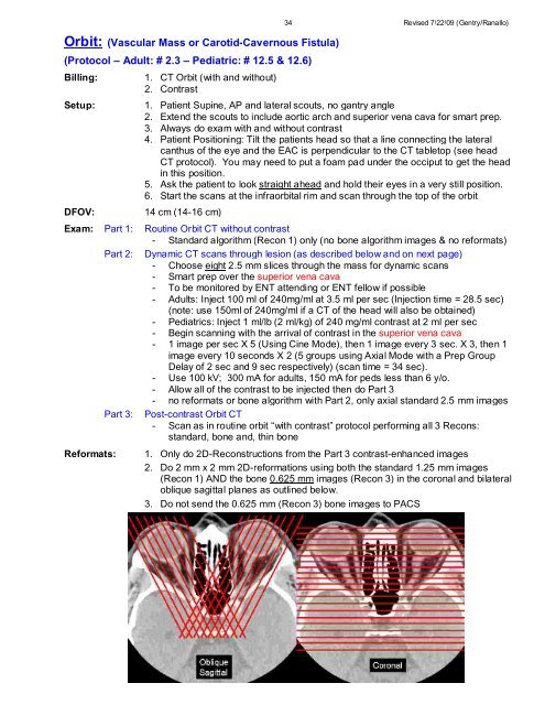
1. Only do 2D-Reconstructions from the Part 3 contrast-enhanced images<br />
2. Do 2 mm x 2 mm 2D-reformations using both the standard 1.25 mm images<br />
(Recon 1) AND the bone 0.625 mm images (Recon 3) in the coronal and bilateral<br />
oblique sagittal planes as outlined below.<br />
3. Do not send the 0.625 mm (Recon 3) bone images to PACS


