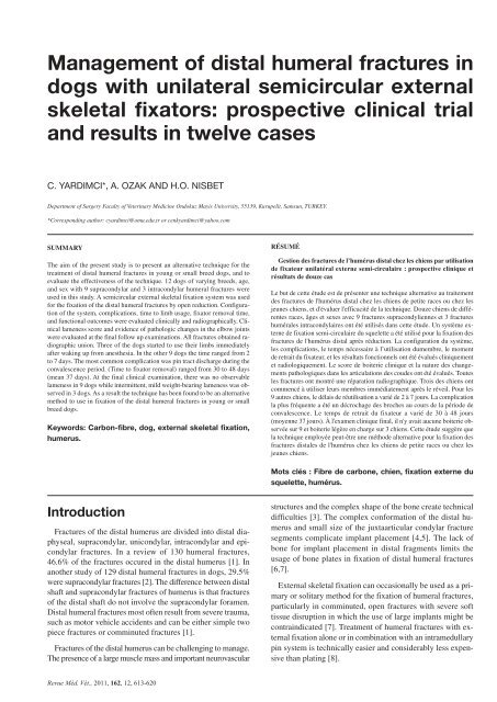
Management Of Distal Humeral Fractures In dogs With
Management Of Distal Humeral Fractures In dogs With
Management Of Distal Humeral Fractures In dogs With

You also want an ePaper? Increase the reach of your titles
YUMPU automatically turns print PDFs into web optimized ePapers that Google loves.
<strong>Management</strong> of distal humeral fractures in<br />
<strong>dogs</strong> with unilateral semicircular external<br />
skeletal fixators: prospective clinical trial<br />
and results in twelve cases<br />
C. YARDIMCI*, A. OZAK AND H.O. NISBET<br />
Department of Surgery Faculty of Veterinary Medicine Ondokuz Mayis University, 55139, Kurupelit, Samsun, TURKEY.<br />
*Corresponding author: cyardimci@omu.edu.tr or cenkyardimci@yahoo.com<br />
SUMMARY<br />
The aim of the present study is to present an alternative technique for the<br />
treatment of distal humeral fractures in young or small breed <strong>dogs</strong>, and to<br />
evaluate the effectiveness of the technique. 12 <strong>dogs</strong> of varying breeds, age,<br />
and sex with 9 supracondylar and 3 intracondylar humeral fractures were<br />
used in this study. A semicircular external skeletal fixation system was used<br />
for the fixation of the distal humeral fractures by open reduction. Configuration<br />
of the system, complications, time to limb usage, fixator removal time,<br />
and functional outcomes were evaluated clinically and radiographically. Clinical<br />
lameness score and evidence of pathologic changes in the elbow joints<br />
were evaluated at the final follow up examinations. All fractures obtained radiographic<br />
union. Three of the <strong>dogs</strong> started to use their limbs immediately<br />
after waking up from anesthesia. <strong>In</strong> the other 9 <strong>dogs</strong> the time ranged from 2<br />
to 7 days. The most common complication was pin tract discharge during the<br />
convalescence period. (Time to fixator removal) ranged from 30 to 48 days<br />
(mean 37 days). At the final clinical examination, there was no observable<br />
lameness in 9 <strong>dogs</strong> while intermittent, mild weight-bearing lameness was observed<br />
in 3 <strong>dogs</strong>. As a result the technique has been found to be an alternative<br />
method to use in fixation of the distal humeral fractures in young or small<br />
breed <strong>dogs</strong>.<br />
Keywords: Carbon-fibre, dog, external skeletal fixation,<br />
humerus.<br />
RÉSUMÉ<br />
Gestion des fractures de l’humérus distal chez les chiens par utilisation<br />
de fixateur unilatéral externe semi-circulaire : prospective clinique et<br />
résultats de douze cas<br />
Le but de cette étude est de présenter une technique alternative au traitement<br />
des fractures de l'humérus distal chez les chiens de petite races ou chez les<br />
jeunes chiens, et d'évaluer l'efficacité de la technique. Douze chiens de différentes<br />
races, âges et sexes avec 9 fractures supracondyliennes et 3 fractures<br />
humérales intracondylaires ont été utilisés dans cette étude. Un système externe<br />
de fixation semi-circulaire du squelette a été utilisé pour la fixation des<br />
fractures de l'humérus distal après réduction. La configuration du système,<br />
les complications, le temps nécessaire à l’utilisation dumembre, le moment<br />
de retrait du fixateur, et les résultats fonctionnels ont été évalués cliniquement<br />
et radiologiquement. Le score de boiterie clinique et la nature des changements<br />
pathologiques dans les articulations des coudes ont été évalués. Toutes<br />
les fractures ont montré une réparation radiographique. Trois des chiens ont<br />
commencé à utiliser leurs membres immédiatement après le réveil. Pour les<br />
9 autres chiens, le délais de réutilisation a varié de 2 à 7 jours. La complication<br />
la plus fréquente a été un décrochage des broches au cours de la période de<br />
convalescence. Le temps de retrait du fixateur a varié de 30 à 48 jours<br />
(moyenne 37 jours). À l'examen clinique final, il n'y avait aucune boiterie observée<br />
sur 9 et boiterie légère en charge sur 3 chiens. Cette étude suggère que<br />
la technique employée peut-être une méthode alternative pour la fixation des<br />
fractures distales de l'humérus chez les chiens de petite races ou chez les<br />
jeunes chiens.<br />
Mots clés : Fibre de carbone, chien, fixation externe du<br />
squelette, humérus.<br />
<strong>In</strong>troduction<br />
<strong>Fractures</strong> of the distal humerus are divided into distal diaphyseal,<br />
supracondylar, unicondylar, intracondylar and epicondylar<br />
fractures. <strong>In</strong> a review of 130 humeral fractures,<br />
46.6% of the fractures occured in the distal humerus [1]. <strong>In</strong><br />
another study of 129 distal humeral fractures in <strong>dogs</strong>, 29.5%<br />
were supracondylar fractures [2]. The difference between distal<br />
shaft and supracondylar fractures of humerus is that fractures<br />
of the distal shaft do not involve the supracondylar foramen.<br />
<strong>Distal</strong> humeral fractures most often result from severe trauma,<br />
such as motor vehicle accidents and can be either simple two<br />
piece fractures or comminuted fractures [1].<br />
<strong>Fractures</strong> of the distal humerus can be challenging to manage.<br />
The presence of a large muscle mass and important neurovascular<br />
structures and the complex shape of the bone create technical<br />
difficulties [3]. The complex conformation of the distal humerus<br />
and small size of the juxtaarticular condylar fracture<br />
segments complicate implant placement [4,5]. The lack of<br />
bone for implant placement in distal fragments limits the<br />
usage of bone plates in fixation of distal humeral fractures<br />
[6,7].<br />
External skeletal fixation can occasionally be used as a primary<br />
or solitary method for the fixation of humeral fractures,<br />
particularly in comminuted, open fractures with severe soft<br />
tissue disruption in which the use of large implants might be<br />
contraindicated [7]. Treatment of humeral fractures with external<br />
fixation alone or in combination with an intramedullary<br />
pin system is technically easier and considerably less expensive<br />
than plating [8].<br />
Revue Méd. Vét., 2011, 162, 12, 613-620
614 YARDIMCI (C.) AND COLLABORATORS<br />
External skeletal fixation has the advantage of adjustibility<br />
after surgery a unique advantage over plate- or pin fixation,<br />
frames may be adjusted or reinforced in the postoperative period.<br />
The technique is also less invasive than plate fixation,<br />
and allows better access to wounds than external coaptation<br />
does [9]. Application of traditional circular external fixation<br />
systems is limited proximal to the elbow and stifle, as the<br />
proximity of the trunk interferes with placement of complete<br />
rings [10].<br />
As there is an absence of clear, longitudinal safe corridors<br />
in the canine humerus and as the regional anatomy of the forelimb<br />
is complex, it is not possible to maintain a direct axial<br />
alignment of the pins inserted in the safe areas and lines [11].<br />
This hampers the use of conventional linear external fixation<br />
systems in humeral fractures. Because of this limitation a semicircular<br />
external skeletal fixation system (SESFS * ) has been<br />
developed which enables multi-plane half pin insertion [12].<br />
Three or 4 half pins may be connected to each arch of the<br />
SESFS. <strong>In</strong> this system, because half pins can only perpendicularly<br />
be inserted to the bone, in order to avoid of encountering<br />
of the pins at the same arch, only two pin application (one<br />
above, one below) is possible if bicortical application is desired.<br />
But the system allows more than two half pin insertions in<br />
unicortical applications.<br />
Our purpose is to report the clinical features and outcome<br />
of 12 <strong>dogs</strong> with supracondylar or intracondylar humeral fractures<br />
that were stabilized with SESFS to provide an alternative<br />
method for management of the distal humeral fractures in<br />
young or small breed <strong>dogs</strong>.<br />
the owners of the animals were advised to use local antibiotic<br />
spray ‡‡ for possible pin tract infections during the convalescence<br />
period. Postoperative external coaptation and exercise<br />
restriction was not carried out in any of the <strong>dogs</strong>.<br />
The principal connecting elements of SESFS are the 6-<br />
holed 45° (180 mm Ø, 1/8 ring arch, 7x18x85 mm) or 5-holed<br />
40° (180 mm Ø, 1/9 ring arch, 7x18x80mm) carbon-fibre<br />
arches. The other components of the system composed of 6<br />
mm Ø threaded rods (80, 100, 120, 150 mm length), half pin<br />
fixation bolts for 3 and 4 mm Ø half pins, 6 mm Ø hex nuts,<br />
and 3 and 4 mm Ø negative profile end-threaded half pins. <strong>In</strong><br />
some <strong>dogs</strong> 3 mm Ø biopsy punch was used for circular stab<br />
incision of skin for closed application of the half pins (figure 1).<br />
All the components except arches were made of 316L stainless<br />
steel. Depending on fracture type, and body weight of the patient,<br />
3 or 4 arched configurations were used. The 40° and 45°<br />
carbon-fibre arches were used in <strong>dogs</strong> weighting
TREATMENT OF DISTAL HUMERAL FRACTURES 615<br />
epicondyle. The deep fascia of the brachium was incised along<br />
the cranial border of the triceps. The deep fascia was undermined<br />
to allow cranial retraction of the brachiocephalicus<br />
muscle and the cephalic vein and exposure of the radial nerve<br />
(figure 2.a). To obtain a better exposure of the distal portion<br />
of the bone, brachiocephalicus muscle retracted cranially and<br />
brachialis muscle and radial nerve retracted caudally [13]. <strong>In</strong><br />
intracondylar fractures, fracture area was exposed following<br />
osteotomy of the tuber olecrani. Then fracture was identified,<br />
debrided, and anatomically reduced and maintained with<br />
bone-holding forceps. Then Kirshner wires or an intercondylar<br />
lag screw was applied from slight proximal of the lateral epicondyle<br />
in order not to interfere with the condylar half pin.<br />
Following reduction of the fracture, the first half pin was<br />
perpendicularly and bicortically applied from slightly distal<br />
of bony protuberance of lateral epicondyle with low speed<br />
power-drill after predrilling with with 2,7mm Ø drill for 3mm<br />
Ø half pin and 3,5 mm Ø drill for 4 mm Ø half pin. The diameter<br />
of the half pin chosen was, in general, the largest that<br />
was thought could be placed across the condyle. Then the second<br />
half pin was applied to the lateral surface of the distal<br />
part (1-2 cm to the fracture line) of the proximal fragment and<br />
secured to the frame. Once acceptable reduction of the fracture<br />
was achieved, the construct was completed by placing additional<br />
one or two half pins to the craniolateral surface of proximal<br />
humerus in closed fashion (figure 2.b). Before closure of the<br />
surgery site, half pin fixation bolts were secured to the carbonfibre<br />
arches firmly and all nuts were controlled for being<br />
firmly attached to the frame. <strong>In</strong> all <strong>dogs</strong>, stability of the immobilized<br />
fracture fragments was checked under flexion, extension,<br />
and rotation of the elbow joint. Surgery site was<br />
closed routinely. <strong>In</strong> the anatomical reconstruction of the fractures<br />
no adjunctive implants were used, except Kirschner<br />
wires and intercondylar lag screws used in intracondylar fractures.<br />
Postoperative radiographs were obtained and then pinskin<br />
tension adjustments were made, if necessary, while<br />
animals were under general anesthesia. At the time of the discharge,<br />
owners were instructed to clean the pin tract-skin interface<br />
every other day, use daily antibiotic spray. Weekly<br />
recheck examinations and radiographic assessments were performed<br />
and the fixator was removed following interfragmental<br />
bony bridgings were seen. Also goniometric examinations of<br />
both operated and contralateral elbow joints were done at final<br />
follow up examinations and the discrepancy between maximum<br />
flexion ranges were recorded. Radiological and final clinical<br />
assessments were performed by a surgeon aware of<br />
previous surgery.<br />
Evidence of pathologic changes in the joint (osteoarthritis<br />
score) (0-4) were scored as follows: No radiographic evidence<br />
of articular pathology (0), joint effusion/soft tissue changes<br />
without evidence of osteoarthritis (OA) (1), evidence of<br />
early/minimal OA (2), moderate OA changes (3), severe OA<br />
changes (4) [14].<br />
At the final follow up examination, a clinical lameness score<br />
(0-5) was given as follows: No observable lameness (0), intermittent,<br />
mild weight-bearing lameness with little if any<br />
change in gait (1), consistent, mild weight-bearing lameness<br />
with little change in gait (2), moderate weight-bearing lameness<br />
– obvious lameness with noticable “head bob” and<br />
change in gait (3), severe weight-bearing lameness – “toe-touching”<br />
only (4), non-weight-bearing (5) [14].<br />
FIGURE 2: (a) The segment on the lateral aspect of the humerus occupied by the brachialis muscle, from apex of the proximal safe area<br />
to the apex of the distal safe area, represents an unsafe corridor due to the presence of the radial nerve (white arrow) (b) <strong>In</strong> SESFS,<br />
the first two pins (1, 2) inserted to the lateral surface of distal humerus. Once acceptable reduction of the fracture was achieved, the<br />
construct was completed by placing additional one or two half pins (3) to the craniolateral surface of proximal humerus in closed<br />
fashion.<br />
Revue Méd. Vét., 2011, 162, 12, 613-620
616 YARDIMCI (C.) AND COLLABORATORS<br />
Results<br />
<strong>In</strong>formation regarding each animal’s signalment and history,<br />
fracture description, fixator configuration, time elapsed between<br />
fracture occurence and repair, duration of the operation<br />
procedure, complications, concurrent injuries, time to limb<br />
usage and fixator removal, osteoarthritis score, and the lameness<br />
score at the final follow up examination are listed in<br />
Table I.<br />
The animals ranged in age from 3 months to 18 months<br />
(mean 7.5 months). Body weights ranged from 5 to 13 kg<br />
(mean 9.4 kg). Distribution of the fractures according to the<br />
fracture type were 6 supracondylar simple (figure 3.a-b), 3 supracondylar<br />
comminuted, and 3 intracondylar. The history<br />
was motor vehicle accident in 9 <strong>dogs</strong>, and falling from an elevated<br />
height in 3 <strong>dogs</strong>. Affected limb was right in 8 <strong>dogs</strong> and<br />
left in 4 <strong>dogs</strong>. There was no evidence of neurological dysfunction<br />
in any of the dog.<br />
Three <strong>dogs</strong> had concurrent orthopedic injuries. Case no. 3<br />
had contralateral supracondylar femur and femoral neck fracture,<br />
and ipsilateral mid-diaphyseal oblique tibial fracture. Tibial<br />
and supracondylar femoral fractures were both stabilized with<br />
SESFS and excision arthroplasty was performed to the femoral<br />
neck fracture. Case no. 7 was presented with ipsilateral stable<br />
Signalment Description Fixator Duration Duration Postoperative Concurrent Time to OA LS<br />
Case of the fracture configuration from injury of the complications injuries limb<br />
no History to surgery procedure usage/fixator<br />
(days) (minute) removal (days<br />
following surgery)<br />
4-m-old, 5 kg, Supracondylar, 40°, 3 arched,<br />
1 F, Cocker Spaniel FEH simple, R 2P: 3mm Ø 2 ETHP 2 46 None None ♣/40 0 0<br />
1D: 3mm Ø 1 ETHP<br />
4-m-old, 9 kg, M, 40°, 3 arched, ■ Mild pin tract<br />
2 Labrador Retriever FEH <strong>In</strong>tracondylar, L 2P: 3mm Ø 3 ETHP 2 72 discharge None 2/32 1 0<br />
1D: 4mm Ø 1 ETHP<br />
Left supracondylar<br />
5-m-old, 8 kg, M, Supracondylar, 40°, 3 arched, femur and collum<br />
3 English Pointer MVA simple, R 2P: 3mm Ø 2 ETHP 1 45 None femoris fracture, 4/32 0 0<br />
1D: 4mm Ø 1 ETHP right mid-diaphyseal<br />
tibia fracture<br />
6-m-old, 11 kg, M, Supracondylar, 45°, 3 arched, Malunion<br />
4 Mix breed MVA communited, L 2P: 3mm Ø 3 ETHP 14 51 deformity None 2/30 2 1<br />
1D: 4mm Ø 1 ETHP<br />
11-m-old, 12kg, F, Supracondylar, 45°, 3 arched, Mild pin tract<br />
5 Mix breed MVA simple, R 2P: 3mm Ø 3 ETHP 1 50 discharge None 4/35 0 0<br />
1D: 4mm Ø 1 ETHP<br />
3-m-old, 5kg, M, Supracondylar, 40°, 3 arched,<br />
6 Turkish Kangal MVA simple, R 2P: 3mm Ø 2 ETHP 2 41 None None ♣/30 0 0<br />
1D: 3mm Ø 1 ETHP<br />
6,5-m-old, 13kg, F, Supracondylar, 45°, 4 arched, R, Mc-III-IV<br />
7 German Shep. MVA comminuted, R 3P: 4mm Ø 3 ETHP 1 48 None fracture 7/42 1 0<br />
1D: 4mm Ø 1 ETHP<br />
5-m-old, 7 kg, M, Supracondylar, 40°, 3 arched,<br />
8 Mix breed MVA comminuted, L 2P: 3mm Ø 2 ETHP 2 56 None None ♣/36 1 0<br />
1D: 4mm Ø 1 ETHP<br />
18-m-old, 8kg, F, Supracondylar, 40°, 3 arched,<br />
9 Terrier MVA simple, L 2P: 3mm Ø 2 ETHP 3 62 None None 3/42 0 0<br />
1D: 4mm Ø 1 ETHP<br />
9-m-old, 12 kg, M, 45°, 4 arched, ■ R, 6-7 th rib fracture,<br />
10 Mix breed MVA <strong>In</strong>tracondylar, R 3P: 3mm Ø 3 ETHP 4 88 None pneumothorax 4/40 1 1<br />
1D: 4mm Ø 1 ETHP<br />
6-m-old, 10 kg, F, Supracondylar, 45°, 3 arched,<br />
11 Turkish Kangal MVA simple, R 2P: 4mm Ø 2 ETHP 1 53 None None 3/35 0 0<br />
1D: 4mm Ø 1 ETHP<br />
12 14-m-old, 13 kg, F, 45°, 3 arched, ■ Mild pin tract<br />
English Setter FEH <strong>In</strong>tracondylar, R 3P: 4mm Ø 3 ETHP 4 96 discharge None 5/48 2 1<br />
1D: 4mm Ø 1 ETHP<br />
F: female, M: male, m:month, FEH: falling from an elevated height, MVA: motor vehicle accident, R: right, L: left, P: proximal, D: distal, Ø: diameter,<br />
ETHP: end threaded half pin, Mc: metacarpus, ■: adjunctive fixation with Kirschner wires or lag screw application, ♣: immediately after waking up<br />
from anesthesia, OA: osteoarthritis score at final follow up examination, LS: Lameness score at final follow up examination.<br />
TABLE I: Preoperative and postoperative details of 12 <strong>dogs</strong> with distal humeral fracture managed with SESFS.<br />
§§<br />
Synulox palatable 50 mg, Pfizer, Belgium<br />
Revue Méd. Vét., 2011, 162, 12, 613-620
TREATMENT OF DISTAL HUMERAL FRACTURES 617<br />
metacarpus III-IV fracture, which were splinted. Case no. 10<br />
was presented with ipsilateral 6-7 th rib fracture which were<br />
complicated with pneumothorax. A chest tube was placed and<br />
stayed in place until pneumothorax was resolved. External<br />
fixation of humeral fracture was carried out after this process.<br />
Four <strong>dogs</strong> experienced complications after fixator application.<br />
The most common complication was pin tract discharge<br />
during the convalescence period. This local inflamation did<br />
not complicate with lameness in any of the dog. The <strong>dogs</strong> developed<br />
minor pin tract discharge were responded to improved<br />
cleaning of pin-skin interfaces and oral antibiotic §§ administration<br />
based on cultures and sensitivities. Case no. 4 was<br />
diagnosed with failed fracture repair due to collapse of the attempted<br />
coaptation. <strong>In</strong> this dog the elapsed time from fracture<br />
to repair was 14 days. At the initial examination decreased<br />
fracture mobility was observed. <strong>In</strong> the surgical treatment process,<br />
bone fragments were fixed to the frame without any reduction<br />
attempt in order not to harm the regenerated<br />
fibrocartilaginous callus formation. At the final follow up examination,<br />
despite radiographically significant malunion deformity,<br />
the dog revealed intermittent, mild weight-bearing<br />
lameness.<br />
Duration from injury to surgery was ranged 1 to 14 days.<br />
All of the fractures obtained union. Three of the <strong>dogs</strong> started<br />
use the limb immediately after waking up from anesthesia (figure<br />
4.a). <strong>In</strong> the other <strong>dogs</strong> the time ranged from 2 to 7 days<br />
(figure 4.b). Time to fixator removal ranged from 30 to 48<br />
days (mean 37 days).<br />
<strong>In</strong> present study, two of the 40° and three of the 45° three<br />
arched frames were reused after vapour sterilization (15 minutes<br />
at 130°C). There was no visible damage associated with<br />
reusing any of the arches, and no structural failures occured.<br />
Final clinical and radiological evaluation was performed<br />
between 34 to 56 weeks after removal of the fixator. <strong>In</strong> all<br />
<strong>dogs</strong>, 5 to 15 degrees of decrease in flexion range in the elbow<br />
FIGURE 3: Preoperative medio-lateral (a) and cranio-caudal (b) x-rays of case no.1. Immediate postoperative cranio-caudal x-rays after<br />
fixation with three, 3 mm Ø ETHP and 40°, 3 arched SESFS (c). At the final follow-up radiographic examination on postoperative<br />
44 th week, there was no evidence of articular pathology observed in both cranio-caudal (d) and medio-lateral (e, f) x-rays.<br />
Revue Méd. Vét., 2011, 162, 12, 613-620
618 YARDIMCI (C.) AND COLLABORATORS<br />
joint of the operated limb was observed according to the goniometric<br />
examination of the operated limb at final follow up<br />
examinations. At the final follow up examination, there was<br />
no evidence of articular pathology in 6 <strong>dogs</strong> (figure 3) but soft<br />
tissue changes or early minimal osteoarthritis was observed<br />
in the other 6 <strong>dogs</strong>. According to the final clinical examination,<br />
there was no observable lameness in 9 <strong>dogs</strong> while intermittent,<br />
mild weight-bearing lameness was observed in 3 <strong>dogs</strong>.<br />
Discussion<br />
The canine humerus is a concentric bone and concentric<br />
bones were defined as those completely, or almost completely,<br />
surrounded by muscle masses and not offering safe corridors<br />
for pin insertion. The muscle bulk around the humerus and<br />
the proximity of the chest wall preclude the use of the stronger<br />
type II or III fixators [15]. A safe area exists in the craniolateral<br />
aspect of the proximal humerus which is palpable subcutaneously<br />
and not covered by muscle masses. The next<br />
distal palpable point is represented by the lateral epicondylar<br />
crest on the lateral aspect of the distal fourth of the humerus,<br />
which ends in the lateral epicondyle [11]. The division of the<br />
humeral diaphysis into lateral and medial condyles and the<br />
presence of the supracondylar foramen limit the bone available<br />
implant placement in the distal fragment during internal<br />
fixation [16]. Special care has to be exercised in the distal humerus<br />
to avoid intra-articular pin placement and impaired<br />
elbow movement as pin insertion in the distal half of the olecranon<br />
fossa would interfere with the anconeal process during<br />
full extension of the elbow. The segment on the lateral aspect<br />
of the humerus occupied by the brachialis muscle, from apex<br />
of the proximal safe area to the apex of the distal safe area,<br />
represents an unsafe corridor due to the presence of the radial<br />
nerve. <strong>In</strong> order to reduce the incidence of premature pin loosening,<br />
maximal pin to bone contact and bone purchase can<br />
be achieved by inserting the pins at a proper angulation; pins<br />
used in the proximal safe area are ideally inserted with a slight<br />
craniolateral to caudomedial while the condylar pins inserted<br />
lateral to medial direction [11]. Semicircular external skeletal<br />
fixation system enables multi-plane half pin application from<br />
the same frame and this explains why SESFS is preferred and<br />
used in present study.<br />
FIGURE 4: Fixator tolerance was good to excellent in all <strong>dogs</strong>. Clinical view of case no.1 immediately after the operation (a) and case<br />
no.11 on postoperative 3 rd day (b).<br />
FIGURE 5: Cranio-caudal x-rays of case no. 2 preoperatively (a) and immediately after the operation (b). Medio-lateral x-ray immediately<br />
after removal of the fixator on postoperative 32 th day (c). Medio-lateral x-ray (d) and clinical view of the dog on postoperative 25 th<br />
week (e).<br />
Revue Méd. Vét., 2011, 162, 12, 613-620
TREATMENT OF DISTAL HUMERAL FRACTURES 619<br />
There are advantages and disadvantages to use of low-stiffness<br />
ESF constructs. Low-stiffness ESF configurations allow for<br />
axial micromotion and bone loading at the fracture site, which<br />
are beneficial to healing; however, they can contribute to high<br />
shear stress at the bone-pin interface during weight-bearing<br />
resulting in high strain and premature pin loosening [17]. It<br />
may be prudent to avoid low-stiffness frames in heavier <strong>dogs</strong><br />
because forces acting at the bone-pin interface are increased<br />
and as frame stiffness increases, time to bone union increases<br />
[17]. <strong>In</strong>creasing the number of fixation pins from the minimum<br />
of two pins per major fragment increases the area of pin-bone<br />
interface, thus decreasing the incidence of bone resorption and<br />
subsequent pin loosening. The larger pins are stiffer than<br />
smaller pins by a direct relationship to the fourth power of the<br />
radius; thus a small increase in diameter produces a large increase<br />
in stiffness [18]. Single pin fixation of a fragment<br />
contravenes conventional recommendations for external fixation<br />
[8,9]. But fixation of a juxta-articular fragment as in distal humeral<br />
and femoral fractures complicates with restricted pin<br />
insertion area. Because of such limitations, hybrid external<br />
fixation systems were designed and started to use [19]. <strong>In</strong> the<br />
study performed by KIRBY et al. [10], fixation of femoral<br />
and humeral fractures were made with linear-circular hybrid<br />
external skeletal fixators in 21 <strong>dogs</strong> and 8 cats. <strong>In</strong> 5 of the<br />
<strong>dogs</strong>, the fracture type was supracondylar and/or intracondylar.<br />
The fixation of the condyles were made with single threaded<br />
pin in 4 <strong>dogs</strong> and with multiple half pins (2.4 mm Ø) in one<br />
dog. <strong>In</strong> the present study, a single negative profile ETHP -in<br />
the largest diameter that was thought could be- was placed bicortically<br />
across the condyle. This biomechanical factor resulted<br />
in additional load sharing by the external skeletal<br />
fixator during weightbearing, specifically placing substantial<br />
stress on the distal condylar ETHP. <strong>In</strong> order to avoid pin loosening<br />
due to excessive strain on the condylar ETHP, the<br />
weights of the <strong>dogs</strong> were limited to 13 kg. Although any exercise<br />
or training restriction, except jumping from an elevated<br />
height was instructed to any of the owner the use of a single<br />
pin in distal fragment did not complicated with reduction failure<br />
in any of the <strong>dogs</strong>.<br />
An adequate blood supply is necessary for bone to carry out<br />
its normal physiological function. It has been suggested that<br />
the porosity observed under plates could be attributed to a response<br />
to cortical flow alteration induced following placement<br />
of the plate [20]. The altered flow may alter the microenvironment<br />
in cortical bone beneath plates inducing vascular ingrowth<br />
and bone resorption [8]. <strong>In</strong> locking plate systems<br />
(LCP) it is not necessary to press a locking plate against the<br />
bone as conventional plating systems, a feature which protects<br />
periosteal vasculature and removes the need for accurate<br />
contouring of the implant. <strong>In</strong> a study repair of humeral Y-T<br />
fractures were done with a new “String of Pearls” locking<br />
plate system [21]. Two plate fixation was performed in all 13<br />
<strong>dogs</strong> with a mean weight of 22.8 kg and results were good to<br />
excellent except one poor dog. Requirement of a more extensive<br />
exposure and necessity of a new surgical procedure for<br />
removal of the plates are two disadvantages of bone plates<br />
when compared with ESF.<br />
Optimal fixation of articular fractures involves restoration<br />
of joint congruity and through precise anatomical reduction<br />
and stable fixation of articular component of the fracture while<br />
minimising soft tissue trauma and allowing for early mobilisation<br />
of the joint [22]. The complex conformation of the distal<br />
humerus and small size of the juxtaarticular condylar fracture<br />
segments complicate implant placement [4, 5, 23]. Fracture<br />
stability is dependent on interfragmentary friction and on the<br />
integrity of the supracondylar components of the fracture repair<br />
[24]. Fixation by screws inserted in lag fashion is generally<br />
used to stabilize humeral condylar fractures to provide interfragmentary<br />
compression and promote primary bone healing<br />
[4, 6, 25]. A condylar ETHP does not achieve the adequate<br />
compression between the fractured condyles. So, in present<br />
study, the treatment procedure of 3 intracondylar fractures<br />
was achieved in two steps: fixation of intercondylar fracture<br />
with Kirschner wires or a lag screw and stabilization of the<br />
supracondylar fracture with SESFS. Because of the restricted<br />
area in lateral condylar surface, the intercondylar lag screw<br />
was applied from slightly proximal of the lateral epicondyle<br />
in order not to interfere with the condylar half pin. <strong>In</strong> a recent<br />
study, humeral condylar fractures were repaired with selfcompressing<br />
Orthofix pins in 23 <strong>dogs</strong> [26]. As a future project,<br />
by using the self-compressing Orthofix pin in distal arch, intracondylar<br />
fractures can be treated by SESFS with no adjunctive<br />
fixation.<br />
Condylar humeral fractures carry guarded prognosis and<br />
have a high incidence of complications including fixation failure,<br />
refracture and development of post-traumatic osteoarthritis<br />
however, prognosis for leg function after repair of distal diaphyseal<br />
and or supracondylar fractures is good to excellent<br />
[4]. Reduced elbow flexion and post-traumatic osteoarthritis<br />
are consistent findings in <strong>dogs</strong> that have had surgical stabilisation<br />
of articular fractures of the distal humerus, but do not<br />
appear to correlate with a poor clinical outcome [2, 27]. Nine<br />
<strong>dogs</strong> in present study had an excellent functional outcome without<br />
lameness. Although there was no pain in range of motion,<br />
clinical examination revealed 5 to 15 degrees reduced<br />
flexion in comparison to the contralateral elbow in all <strong>dogs</strong>.<br />
<strong>In</strong> a study evaluating the long-term results of humeral condylar<br />
fracture repair 13 <strong>dogs</strong>, it was found that all of the elbows developed<br />
post-traumatic osteoarthtritis, however, there was no<br />
correlation between the presence of radiographic osteoarthritis<br />
and limb function [27]. <strong>In</strong> present study, although 6 of the<br />
<strong>dogs</strong> showed evidence of osteoarthritis, only 3 of them revealed<br />
intermittent mild lameness. According to present authors, it<br />
may be possible that young <strong>dogs</strong> are more capable of recovering<br />
from articular fractures without developing degenerative joint<br />
disease.<br />
Ideally, patients with distal humeral fracture should be operated<br />
on as soon as possible after they have been thoroughly<br />
evaluated and appropriately stabilized. Surgical repair with<br />
either open or closed reduction is recommended within 24 to<br />
72 hours [14]. Decreased range of joint motion is a major<br />
complication associated with distal humeral fractures, so early<br />
return of limb use is important. Early ambulation not only induce<br />
fracture healing by promoting axial micromotion also prevent<br />
disuse atrophy and muscle contractures in fracture patients<br />
[28, 29]. The primary goal of using SESFS in these case series<br />
was achieving early ambulation and use of the fractured limb.<br />
Carbon-fibre materials are lightweight, x-ray transparent<br />
and sufficiently stable under vapour sterilization conditions.<br />
Revue Méd. Vét., 2011, 162, 12, 613-620
620 YARDIMCI (C.) AND COLLABORATORS<br />
The x-ray transparency enables traditional view radiographs<br />
without interference from overlapping arches which is more<br />
important in medio-lateral projections [30]. <strong>In</strong> present study,<br />
presence of carbon arches enhanced our ability to assess fracture<br />
healing on the lateral radiographic projections. A 6 holed,<br />
45°carbon-fiber arch weighes 12.5 g. An aluminum and a<br />
316L stainless steel arch the same size as carbon-fiber arch<br />
weighes approximately 20 g and 50 g respectively. So a standard<br />
4 arched frame weighes 120 g, 150 g, and 270 g if the<br />
arches are made of carbon-fiber, aluminum and 316L stainless<br />
steel respectively. Although carbon-fiber is an expensive orthopedic<br />
material, durability and reusability of the system reduce<br />
the cost. These superiorities augment the worth of<br />
carbon-fiber in veterinary orthopedics.<br />
As a result it was concluded that unilateral SESFS found to<br />
be practical and efficient in the treatment of the distal humeral<br />
fractures of young or small breed <strong>dogs</strong> weighing less than or<br />
equal to 13 kg. This is the preliminary report about the treatment<br />
of distal humeral fractures in <strong>dogs</strong> with a single distal<br />
pin frame designed SESFS in which early postoperative and<br />
long term results seem to be favorable.<br />
Acknowledgment<br />
We thank Atilim Teknik, Samsun, Turkey for providing the<br />
ESF systems used in this study.<br />
References<br />
1. - BARDET J.F., HOHN R.B., RUDY L.R., et al.: <strong>Fractures</strong> of the humerus<br />
in <strong>dogs</strong> and cats: A retrospective study of 130 cases. Vet. Surg.,<br />
1983, 12,73-77.<br />
2. - STURGEON C., WILSON A.M., MCGUIGAN P., et al.: Triceps tenetomy<br />
and double plate stabilization of “Y-T” fracture of the distal<br />
humeral condyle in three <strong>dogs</strong>. Vet. Comp. Orthop. Traumatol., 2000,<br />
13, 34-40.<br />
3. - WALLACE M.K., BERG J.: Craniolateral approach to the humerus<br />
with transection of the brachialis muscle. Vet. Surg., 1991, 20, 97-99.<br />
4. - VANINI R., SMEAK D.D., OLMSTEAD M.L.: Evaluation of surgical<br />
repair of 135 distal humeral fractures in <strong>dogs</strong> and cats. J. Am. Anim.<br />
Hosp. Assoc., 1988, 24, 537-545.<br />
5. - TURNER T.M.: <strong>Fractures</strong> of distal humerus. <strong>In</strong>: JOHNSON A.L.,<br />
HOULTON J.E.F., VANINI R. (eds): AO Principles of Fracture <strong>Management</strong><br />
in the Dog and Cat. AO Publishing, Switzerland, 2005, pp.:<br />
216-228.<br />
6. - MATTHIESEN D.T., WALTER M.: Surgical management of distal humeral<br />
fractures. Comp. Contin. Educ. Pract. Vet., 1984, 6, 1027-1036.<br />
7. - MATTHIESEN D.T.: <strong>Fractures</strong> of the humerus. Vet. Clin. North. Am.<br />
Small. Anim. Pract., 1992, 22, 121-133.<br />
8. - PIERMATTEI D.L., FLO G.L., DECAMP C.E.: <strong>Fractures</strong>: Classification,<br />
diagnosis, and treatment. <strong>In</strong>: PIERMATTEI D.L., FLO G.L.<br />
(eds): Brinker, Piermattei and Flo’s Handbook of Small Animal Orthopedics<br />
and Fracture Repair, 4 th ed MO Saunders, St. Louis, 2006,<br />
pp.:25-159.<br />
9. - MARCELLIN-LITTLE D.J.: External skeletal fixation. <strong>In</strong>: SLATTER<br />
D. (ed): Textbook of Small Animal Surgery, 3 rd ed, Saunders, Philadelphia,<br />
2003, pp.: 1818-1834.<br />
10. - KIRKBY K.A., LEWIS D.D., LAFUENTE M.P., et al.: <strong>Management</strong><br />
of humeral and femoral fractures in <strong>dogs</strong> and cats with linear-circular<br />
hybrid external skeletal fixators. J. Am. Anim. Hosp. Assoc., 2008,<br />
44, 180-197.<br />
11. - MARTI J.M., MILLER A.: Delimitation of safe corridors for the insertion<br />
of external fixator pins in the dog 2: Forelimb. J. Small. Anim.<br />
Pract., 1994, 35, 78-85.<br />
12. - YARDIMCI C, OZAK A, NISBET H.O.: <strong>Management</strong> of femoral<br />
fractures in <strong>dogs</strong> with semicircular external skeletal fixators. Vet.<br />
Surg., 2011, 40, 379-387.<br />
13. - PIERMATTEI D.L.: Approach to the distal shaft of the humerus<br />
through a craniolateral incision. <strong>In</strong>: PIERMATTEI D.L. (ed): An<br />
Atlas of Surgical Approaches to the Bones and Joints of the Dog and<br />
Cat, 3 rd ed, PA Saunders, Philadelphia, 1993, pp.: 139-142.<br />
14. - COOK J.L., TOMLINSON J.L., REED A.N.: Fluoroscopically guided<br />
closed reduction and internal fixation of fractures of the lateral<br />
portion of the humeral condyle: Prospective clinical study of the technique<br />
and results in ten <strong>dogs</strong>. Vet. Surg., 1999, 28, 315-321.<br />
15. - KLAUSE S.E., SCHWARZ P.D., EGGER E.L., et al.: A modification<br />
of the unilateral type I external skeletal fixator configuration for primary<br />
or secondary support of supracondylar humeral and femoral femoral<br />
fractures. Vet. Comp. Orthop. Traumatol., 1990, 3, 130-134.<br />
16. - ARON D.N., PALMER R.H., JOHNSON A.L.: Biological strategies<br />
and a balanced concept for repair of highly comminuted long bone<br />
fractures. Comp. Contin. Educ. Pract. Vet., 1995, 17, 35-49.<br />
17. - GEMMIL T.J., CAVE T.A., CLEMENTS D.N., et al.: Treatment of<br />
canine and feline diaphyseal radial and tibial fractures with low stiffness<br />
external skeletal fixation. J. Small. Anim. Pract., 2004, 45, 85-91.<br />
18. - PIERMATTEI D.L., FLO G.L., DECAMP C.E.: <strong>Fractures</strong> of the humerus.<br />
<strong>In</strong>: PIERMATTEI D.L., FLO G.L., (eds): Brinker, Piermattei<br />
and Flo’s Handbook of Small Animal Orthopedics and Fracture Repair,<br />
4 th ed, MO Saunders, St. Louis, 2006, pp.: 297-324.<br />
19. - LEWIS D.D., CROSS A.R., CARMICHAEL S., et al.: Recent advances<br />
in external fixation. J. Small. Anim. Pract., 2001, 42, 103-112.<br />
20. - PERREN S.M., CORDEY J., RAHN B.A., et al.: Early temporary<br />
porosis of bone induced by internal fixation implants. A reaction to necrosis,<br />
not to stress protection? Clin. Orthop. Relat. Res., 1988, 232, 139-151.<br />
21. - NESS M.G.: Repair of Y-T humeral fractures in <strong>dogs</strong> using paired<br />
‘String of Pearls’ locking plates. Vet. Comp. Orthop. Traumatol.,<br />
2009, 6, 492-497.<br />
22. - HAHN D.M.: Current principles of treatment in the clinical practice<br />
of articular fractures. Clin. Orthop. Relat. Res., 2004, 423, 27-32.<br />
23. - MACIAS C., GIBBONS S.E., MCKEE W.M.: Y-T humeral fractures<br />
with supracondylar comminution in five cats. J. Small. Anim. Pract.,<br />
2006, 47, 89-93.<br />
24. - AU K., MATTERN K.L., LEWIS D.D.: <strong>In</strong>tracondylar humeral fracture<br />
stabilisation in a dog using a transilial rod and external fixation.<br />
J. Small. Anim. Pract., 2008, 49, 148-151.<br />
25. - JOHNSON A.L., HULSE D.A.: <strong>Management</strong> of spesific fractures.<br />
<strong>In</strong>: FOSSUM T.W. (ed), Small Animal Surgery, 2 nd ed , MO Mosby,<br />
St Louis, 2002, pp.: 932-937.<br />
26. - GUILLE A.E., LEWIS D.D., ANDERSON T.P., et al.: Evaluation of<br />
surgical repair of humeral condylar fractures using self-compressing<br />
orthofix pins in 23 <strong>dogs</strong>. Vet. Surg., 2004, 33, 314-322.<br />
27. - GORDON W.J., BESACON M.F., CONZEMIUS M.G., et al.: Frequency<br />
of post-traumatic osteoarthritis in <strong>dogs</strong> after repair of a humeral<br />
condylar fracture. Vet. Comp. Orthop. Traumatol., 2003, 16, 1-5.<br />
28. - LINCOLN J.D.: Treatment of open, delayed union, and nonunion<br />
fractures with external skeletal fixation. Vet. Clin. North. Am. Small.<br />
Anim. Pract., 1992, 22, 195-207.<br />
29. - HARARI J.: Complications of external skeletal fixation. Vet. Clin.<br />
North. Am. Small. Anim. Pract., 1992, 22, 99-107.<br />
30. - MIGLIARESI C., NICOLI F., ROSSI S., et al.: Novel uses of carbon<br />
composites for the fabrication of external fixators. Comp. Sci. Technol.,<br />
2004, 64, 873-883.<br />
Revue Méd. Vét., 2011, 162, 12, 613-620