Gray-scale and color Doppler US findings of amniotic sheets
Gray-scale and color Doppler US findings of amniotic sheets
Gray-scale and color Doppler US findings of amniotic sheets

Create successful ePaper yourself
Turn your PDF publications into a flip-book with our unique Google optimized e-Paper software.
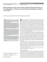
Diagn Interv Radiol DOI 10.4261/1305-3825.DIR.4696-11.1<br />
© Turkish Society <strong>of</strong> Radiology 2011<br />
FETAL IMAGING<br />
ORIGINAL ARTICLE<br />
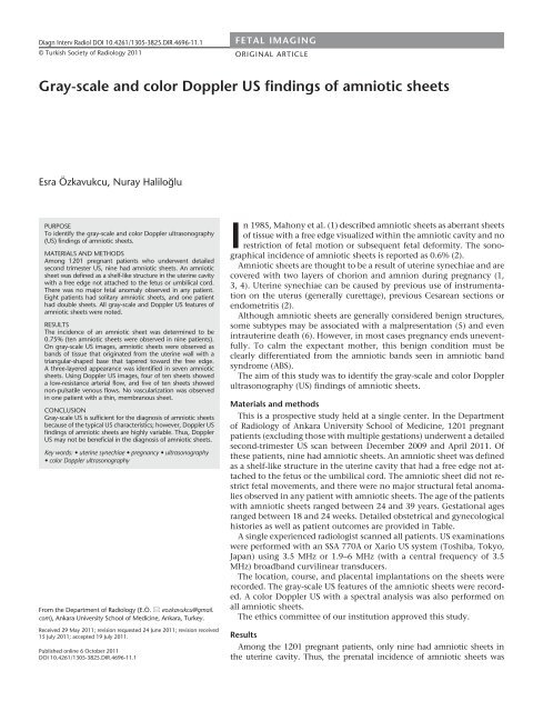
<strong>Gray</strong>-<strong>scale</strong> <strong>and</strong> <strong>color</strong> <strong>Doppler</strong> <strong>US</strong> <strong>findings</strong> <strong>of</strong> <strong>amniotic</strong> <strong>sheets</strong><br />
Esra Özkavukcu, Nuray Haliloğlu<br />
PURPOSE<br />
To identify the gray-<strong>scale</strong> <strong>and</strong> <strong>color</strong> <strong>Doppler</strong> ultrasonography<br />
(<strong>US</strong>) <strong>findings</strong> <strong>of</strong> <strong>amniotic</strong> <strong>sheets</strong>.<br />
MATERIALS AND METHODS<br />
Among 1201 pregnant patients who underwent detailed<br />
second trimester <strong>US</strong>, nine had <strong>amniotic</strong> <strong>sheets</strong>. An <strong>amniotic</strong><br />
sheet was defined as a shelf-like structure in the uterine cavity<br />
with a free edge not attached to the fetus or umbilical cord.<br />
There was no major fetal anomaly observed in any patient.<br />
Eight patients had solitary <strong>amniotic</strong> <strong>sheets</strong>, <strong>and</strong> one patient<br />
had double <strong>sheets</strong>. All gray-<strong>scale</strong> <strong>and</strong> <strong>Doppler</strong> <strong>US</strong> features <strong>of</strong><br />
<strong>amniotic</strong> <strong>sheets</strong> were noted.<br />
RESULTS<br />
The incidence <strong>of</strong> an <strong>amniotic</strong> sheet was determined to be<br />
0.75% (ten <strong>amniotic</strong> <strong>sheets</strong> were observed in nine patients).<br />
On gray-<strong>scale</strong> <strong>US</strong> images, <strong>amniotic</strong> <strong>sheets</strong> were observed as<br />
b<strong>and</strong>s <strong>of</strong> tissue that originated from the uterine wall with a<br />
triangular-shaped base that tapered toward the free edge.<br />
A three-layered appearance was identified in seven <strong>amniotic</strong><br />
<strong>sheets</strong>. Using <strong>Doppler</strong> <strong>US</strong> images, four <strong>of</strong> ten <strong>sheets</strong> showed<br />
a low-resistance arterial flow, <strong>and</strong> five <strong>of</strong> ten <strong>sheets</strong> showed<br />
non-pulsatile venous flows. No vascularization was observed<br />
in one patient with a thin, membranous sheet.<br />
CONCL<strong>US</strong>ION<br />
<strong>Gray</strong>-<strong>scale</strong> <strong>US</strong> is sufficient for the diagnosis <strong>of</strong> <strong>amniotic</strong> <strong>sheets</strong><br />
because <strong>of</strong> the typical <strong>US</strong> characteristics; however, <strong>Doppler</strong> <strong>US</strong><br />
<strong>findings</strong> <strong>of</strong> <strong>amniotic</strong> <strong>sheets</strong> are highly variable. Thus, <strong>Doppler</strong><br />
<strong>US</strong> may not be beneficial in the diagnosis <strong>of</strong> <strong>amniotic</strong> <strong>sheets</strong>.<br />
Key words: • uterine synechiae • pregnancy • ultrasonography<br />
• <strong>color</strong> <strong>Doppler</strong> ultrasonography<br />
From the Department <strong>of</strong> Radiology (E.Ö. eozkavukcu@gmail.<br />
com), Ankara University School <strong>of</strong> Medicine, Ankara, Turkey.<br />
Received 29 May 2011; revision requested 24 June 2011; revision received<br />
15 July 2011; accepted 19 July 2011.<br />
Published online 6 October 2011<br />
DOI 10.4261/1305-3825.DIR.4696-11.1<br />
In 1985, Mahony et al. (1) described <strong>amniotic</strong> <strong>sheets</strong> as aberrant <strong>sheets</strong><br />
<strong>of</strong> tissue with a free edge visualized within the <strong>amniotic</strong> cavity <strong>and</strong> no<br />
restriction <strong>of</strong> fetal motion or subsequent fetal deformity. The sonographical<br />
incidence <strong>of</strong> <strong>amniotic</strong> <strong>sheets</strong> is reported as 0.6% (2).<br />
Amniotic <strong>sheets</strong> are thought to be a result <strong>of</strong> uterine synechiae <strong>and</strong> are<br />
covered with two layers <strong>of</strong> chorion <strong>and</strong> amnion during pregnancy (1,<br />
3, 4). Uterine synechiae can be caused by previous use <strong>of</strong> instrumentation<br />
on the uterus (generally curettage), previous Cesarean sections or<br />
endometritis (2).<br />
Although <strong>amniotic</strong> <strong>sheets</strong> are generally considered benign structures,<br />
some subtypes may be associated with a malpresentation (5) <strong>and</strong> even<br />
intrauterine death (6). However, in most cases pregnancy ends uneventfully.<br />
To calm the expectant mother, this benign condition must be<br />
clearly differentiated from the <strong>amniotic</strong> b<strong>and</strong>s seen in <strong>amniotic</strong> b<strong>and</strong><br />
syndrome (ABS).<br />
The aim <strong>of</strong> this study was to identify the gray-<strong>scale</strong> <strong>and</strong> <strong>color</strong> <strong>Doppler</strong><br />
ultrasonography (<strong>US</strong>) <strong>findings</strong> <strong>of</strong> <strong>amniotic</strong> <strong>sheets</strong>.<br />
Materials <strong>and</strong> methods<br />
This is a prospective study held at a single center. In the Department<br />
<strong>of</strong> Radiology <strong>of</strong> Ankara University School <strong>of</strong> Medicine, 1201 pregnant<br />
patients (excluding those with multiple gestations) underwent a detailed<br />
second-trimester <strong>US</strong> scan between December 2009 <strong>and</strong> April 2011. Of<br />
these patients, nine had <strong>amniotic</strong> <strong>sheets</strong>. An <strong>amniotic</strong> sheet was defined<br />
as a shelf-like structure in the uterine cavity that had a free edge not attached<br />
to the fetus or the umbilical cord. The <strong>amniotic</strong> sheet did not restrict<br />
fetal movements, <strong>and</strong> there were no major structural fetal anomalies<br />
observed in any patient with <strong>amniotic</strong> <strong>sheets</strong>. The age <strong>of</strong> the patients<br />
with <strong>amniotic</strong> <strong>sheets</strong> ranged between 24 <strong>and</strong> 39 years. Gestational ages<br />
ranged between 18 <strong>and</strong> 24 weeks. Detailed obstetrical <strong>and</strong> gynecological<br />
histories as well as patient outcomes are provided in Table.<br />
A single experienced radiologist scanned all patients. <strong>US</strong> examinations<br />
were performed with an SSA 770A or Xario <strong>US</strong> system (Toshiba, Tokyo,<br />
Japan) using 3.5 MHz or 1.9–6 MHz (with a central frequency <strong>of</strong> 3.5<br />
MHz) broadb<strong>and</strong> curvilinear transducers.<br />
The location, course, <strong>and</strong> placental implantations on the <strong>sheets</strong> were<br />
recorded. The gray-<strong>scale</strong> <strong>US</strong> features <strong>of</strong> the <strong>amniotic</strong> <strong>sheets</strong> were recorded.<br />
A <strong>color</strong> <strong>Doppler</strong> <strong>US</strong> with a spectral analysis was also performed on<br />
all <strong>amniotic</strong> <strong>sheets</strong>.<br />
The ethics committee <strong>of</strong> our institution approved this study.<br />
Results<br />
Among the 1201 pregnant patients, only nine had <strong>amniotic</strong> <strong>sheets</strong> in<br />
the uterine cavity. Thus, the prenatal incidence <strong>of</strong> <strong>amniotic</strong> <strong>sheets</strong> was
a<br />
b<br />
Figure 1. a, b. Sagittal <strong>US</strong> views <strong>of</strong> the patient no. 2 showing an <strong>amniotic</strong> sheet originating from the uterine wall with a triangular-shaped base<br />
(arrows, a) <strong>and</strong> a tapered, membrane-like <strong>amniotic</strong> sheet (curved arrow, b).<br />
determined to be 0.75%. All <strong>amniotic</strong><br />
<strong>sheets</strong> were classified as incomplete<br />
<strong>sheets</strong>, according to the classification<br />
outlined by Tan et al. (6). Eight patients<br />
had solitary <strong>amniotic</strong> <strong>sheets</strong>, whereas<br />
one patient had two <strong>sheets</strong>. Five <strong>amniotic</strong><br />
<strong>sheets</strong> were located in the transverse<br />
plane, whereas five <strong>sheets</strong> were longitudinally<br />
positioned in the uterine cavity.<br />
On the gray-<strong>scale</strong> <strong>US</strong>, <strong>amniotic</strong> <strong>sheets</strong><br />
were observed as b<strong>and</strong>s <strong>of</strong> tissue originating<br />
from the uterine wall with a triangular-shaped<br />
base (Fig. 1a). Amniotic<br />
<strong>sheets</strong> tapered toward the free edge,<br />
with some resembling the thinness <strong>of</strong><br />
a membrane (Fig. 1b). Sonographically,<br />
three layers were identified in seven<br />
<strong>of</strong> the ten <strong>sheets</strong> with two echogenic<br />
lines <strong>and</strong> a hypoechoic line located in<br />
between (Fig. 2). Placental extension <strong>of</strong><br />
Figure 2. A sagittal <strong>US</strong> view <strong>of</strong> the patient no. 3 showing the three-layered <strong>amniotic</strong> sheet<br />
(arrow) with two echogenic lines <strong>and</strong> a hypoechoic line located in between.<br />
Table. The obstetrical <strong>and</strong> gynecological history <strong>of</strong> study patients<br />
Previous Previous Previous<br />
Spontaneous Fetal Mode <strong>of</strong> delivery<br />
No. Gravidity Parity D/C C/S uterine surgery<br />
abortion outcome (indication)<br />
1 4 1 1 0 0 1 N C/S (cephalopelvic disproportion)<br />
2 3 2 0 0 0 0 N C/S (breech presentation)<br />
3 2 0 1 0 0 0 N Vaginal delivery<br />
4 1 0 0 0 0 0 N Vaginal delivery<br />
5 3 1 1 0 0 0 N/A N/A<br />
6 1 0 0 0 0 0 N/A N/A<br />
7 3 1 1 0 0 0 N Vaginal delivery<br />
8 3 1 0 0 0 1 N C/S (previous C/S)<br />
9 2 0 1 0 0 0 N/A N/A<br />
D/C, dilatation <strong>and</strong> curettage; C/S, Cesarean section; N, normal; N/A, not available (two patients were lost during follow-up, <strong>and</strong> one has not yet delivered)<br />
ii • Diagnostic <strong>and</strong> Interventional Radiology<br />
Özkavukcu <strong>and</strong> Haliloğlu
Figure 3. A sagittal <strong>US</strong> view <strong>of</strong> the patient no. 1 showing the placenta<br />
(arrow) extending into the <strong>amniotic</strong> sheet.<br />
Figure 4. A sagittal <strong>US</strong> view <strong>of</strong> the patient no. 5 showing the “thick”<br />
free edge <strong>of</strong> the incomplete <strong>amniotic</strong> sheet (arrows).<br />
a<br />
b<br />
Figure 5. a, b. A sagittal <strong>US</strong> view (a) <strong>of</strong> the patient no. 2 showing a relatively thick <strong>amniotic</strong> sheet (arrows) with placental extension. A <strong>color</strong><br />
<strong>Doppler</strong> <strong>US</strong> view (b) showing substantial arterial vascularization. Spectral analysis is not shown.<br />
the <strong>amniotic</strong> <strong>sheets</strong> was noted in three<br />
<strong>of</strong> the nine patients with <strong>amniotic</strong><br />
<strong>sheets</strong> (Fig. 3), <strong>and</strong> we did not observe<br />
any “bulbous tips” (Fig. 4). Fetuses always<br />
moved freely, independent <strong>of</strong> the<br />
<strong>amniotic</strong> <strong>sheets</strong>, <strong>and</strong> no major malformations<br />
were noted in any fetus.<br />
On the <strong>color</strong> <strong>Doppler</strong> <strong>US</strong>, the patients<br />
with relatively thick <strong>sheets</strong> also<br />
had placental implantations on the<br />
<strong>amniotic</strong> <strong>sheets</strong>, <strong>and</strong> substantial arterial<br />
vascularization (similar to that observed<br />
at the uteroplacental junction)<br />
was also noted (Fig. 5). However, thin<br />
parts <strong>of</strong> the <strong>sheets</strong> showed poor vascularization<br />
on the <strong>color</strong> <strong>and</strong> power<br />
<strong>Doppler</strong> <strong>US</strong> images. A spectral analysis<br />
<strong>of</strong> these thin <strong>sheets</strong> revealed a lowresistance<br />
arterial flow (in four <strong>of</strong> ten<br />
<strong>sheets</strong>) (Fig. 6) or non-pulsatile venous<br />
flow (in five <strong>of</strong> ten <strong>sheets</strong>) (Fig. 7). The<br />
heart rates obtained from the arterial<br />
flows were always <strong>of</strong> maternal origin,<br />
suggesting that the <strong>amniotic</strong> <strong>sheets</strong> are<br />
<strong>of</strong> a maternal rather than fetal origin<br />
(Fig. 6b). No vascularization was noted<br />
on the <strong>Doppler</strong> <strong>US</strong> in one patient with<br />
a thin, membranous sheet.<br />
Discussion<br />
In our study, the incidence <strong>of</strong> <strong>amniotic</strong><br />
<strong>sheets</strong> was 0.75%. This percentage<br />
is comparable to those reported in<br />
previous retrospective studies (2, 5–7).<br />
There are a few reasons for the negligible<br />
difference. First, a single radiologist<br />
with 10 years <strong>of</strong> experience in obstetrical<br />
<strong>US</strong> imaging performed all <strong>US</strong><br />
scans in our study; thus, there were no<br />
interobserver variations. Furthermore,<br />
<strong>amniotic</strong> <strong>sheets</strong> were specifically examined<br />
for during all <strong>US</strong> scans.<br />
Tan et al. (6) classified <strong>amniotic</strong><br />
<strong>sheets</strong> into complete <strong>and</strong> incomplete<br />
<strong>sheets</strong>. A complete sheet only has a<br />
small perforation that is not visible<br />
on <strong>US</strong> scans, whereas an incomplete<br />
sheet has a free-floating edge. They<br />
suggested that although the incomplete<br />
<strong>sheets</strong> are benign, complete <strong>amniotic</strong><br />
<strong>sheets</strong> may be associated with<br />
intrauterine death. The authors hypothesized<br />
that the small defects on<br />
the complete <strong>sheets</strong> may predispose<br />
the mother <strong>and</strong> fetus to cord prolapse<br />
<strong>and</strong> intrauterine death (6). None <strong>of</strong><br />
our patients had a complete sheet<br />
or poor pregnancy outcome, which<br />
suggests that complete <strong>sheets</strong> are extremely<br />
rare.<br />
<strong>Gray</strong>-<strong>scale</strong> <strong>and</strong> <strong>color</strong> <strong>Doppler</strong> <strong>US</strong> <strong>findings</strong> <strong>of</strong> <strong>amniotic</strong> <strong>sheets</strong> • iii
a<br />
b<br />
The most important differential diagnosis<br />
<strong>of</strong> <strong>amniotic</strong> <strong>sheets</strong> is ABS. ABS<br />
is thought to result from a defect in<br />
the <strong>amniotic</strong> membrane that exposes<br />
the fetus to the chorionic cavity.<br />
iv • Diagnostic <strong>and</strong> Interventional Radiology<br />
Figure 6. a, b. A sagittal<br />
gray-<strong>scale</strong> <strong>US</strong> view (a)<br />
<strong>of</strong> the patient no. 5<br />
showing the double<br />
<strong>amniotic</strong> <strong>sheets</strong> (arrows),<br />
located close to one<br />
another (C, cervix). A<br />
<strong>color</strong> <strong>Doppler</strong> <strong>US</strong> <strong>and</strong><br />
spectral analysis (b) <strong>of</strong><br />
the thicker <strong>amniotic</strong><br />
sheet revealed a lowresistance<br />
arterial flow<br />
<strong>and</strong> a heart rate <strong>of</strong><br />
maternal origin.<br />
Figure 7. A <strong>color</strong> <strong>Doppler</strong> <strong>US</strong> <strong>of</strong> the patient no. 6 <strong>and</strong> spectral analysis <strong>of</strong> the <strong>amniotic</strong> sheet<br />
showing a non-pulsatile venous flow.<br />
Serious fetal malformations can occur<br />
in ABS, such as limb, trunk, vertebral,<br />
cranial, facial, <strong>and</strong> abdominal abnormalities<br />
(1). The <strong>amniotic</strong> b<strong>and</strong>s in<br />
ABS are observed as membranes that<br />
flap with fetal movements or attach to<br />
the fetus <strong>and</strong> restrict fetal movement<br />
(1). Conversely, <strong>amniotic</strong> <strong>sheets</strong> do not<br />
attach to the fetus or umbilical cord,<br />
<strong>and</strong> the fetus can move independently.<br />
The number <strong>of</strong> <strong>amniotic</strong> b<strong>and</strong>s in ABS<br />
can range from one to many. Amniotic<br />
<strong>sheets</strong> are usually solitary, but multiple<br />
<strong>sheets</strong> have also been reported<br />
(8). There are no major structural fetal<br />
anomalies that accompany <strong>amniotic</strong><br />
<strong>sheets</strong>. Because <strong>amniotic</strong> <strong>sheets</strong> consist<br />
<strong>of</strong> two layers <strong>of</strong> chorion <strong>and</strong> amnion,<br />
whereas <strong>amniotic</strong> b<strong>and</strong>s consist<br />
<strong>of</strong> only a single layer <strong>of</strong> amnion (9),<br />
<strong>amniotic</strong> <strong>sheets</strong> appear thicker than<br />
<strong>amniotic</strong> b<strong>and</strong>s (3). Amniotic <strong>sheets</strong><br />
are similar to the inter-twin membrane<br />
seen in dichorionic-di<strong>amniotic</strong> twins<br />
(3). This layered <strong>and</strong> relatively thick<br />
shelf-like appearance was noted in all<br />
<strong>of</strong> our patients, with the exception<br />
<strong>of</strong> one patient who had a thin, taut<br />
sheet. Additionally, the sheet base was<br />
similar to the “twin peak sign” seen in<br />
dichorionic-di<strong>amniotic</strong> twins. Other<br />
useful sonographic features <strong>of</strong> <strong>amniotic</strong><br />
<strong>sheets</strong> are the broad base at the<br />
origin on the uterine wall <strong>and</strong> the bulbous<br />
free edge or tip (1, 3, 9, 10). In our<br />
study, the triangular broad base was always<br />
present, except in the one patient<br />
with a thin, membranous <strong>amniotic</strong><br />
sheet; no patients exhibited a bulbous<br />
free edge. The free edge was difficult to<br />
see <strong>and</strong> required scanning in different<br />
planes. The free edges always appeared<br />
thick <strong>and</strong> taut rather than flapping.<br />
Occasionally, the placenta appeared<br />
to be implanted on the <strong>amniotic</strong> sheet<br />
(9, 10). Korbin et al. (10) found that<br />
the placenta appeared to be implanted<br />
on the <strong>amniotic</strong> sheet in 26.1% <strong>of</strong> the<br />
cases. They also concluded that pregnancy<br />
outcomes are similar in patients<br />
with <strong>and</strong> without placental implantation<br />
on the <strong>amniotic</strong> sheet (10). In our<br />
study, three <strong>of</strong> the nine patients had<br />
placental implantation on the <strong>amniotic</strong><br />
sheet.<br />
Previous publications regarding<br />
<strong>Doppler</strong> analysis <strong>of</strong> the <strong>amniotic</strong><br />
<strong>sheets</strong> were case reports (11, 12), thus<br />
making this the first original article<br />
on <strong>Doppler</strong> analysis <strong>of</strong> the <strong>amniotic</strong><br />
<strong>sheets</strong> in a series <strong>of</strong> patients. Some<br />
authors claimed that <strong>color</strong> <strong>Doppler</strong><br />
<strong>US</strong> imaging with spectral analysis has<br />
an important role in the differential<br />
diagnosis <strong>of</strong> intrauterine membranes<br />
<strong>of</strong> undetermined origin during pregnancy<br />
(11, 12). We also showed that<br />
Özkavukcu <strong>and</strong> Haliloğlu
all <strong>amniotic</strong> <strong>sheets</strong> that had an arterial<br />
flow revealed a maternal heart rate, indicating<br />
the maternal origin <strong>of</strong> these<br />
benign structures; however, five <strong>of</strong><br />
ten <strong>sheets</strong> showed only venous flows.<br />
Because the <strong>Doppler</strong> <strong>findings</strong> varied,<br />
the use <strong>of</strong> <strong>Doppler</strong> <strong>US</strong> to diagnose<br />
<strong>amniotic</strong> <strong>sheets</strong> is debatable. Other<br />
conditions that can mimic <strong>amniotic</strong><br />
<strong>sheets</strong> include chorio<strong>amniotic</strong> separation,<br />
vanished twin, uterine septum,<br />
<strong>and</strong> circumvallate placenta (2, 3). The<br />
course <strong>of</strong> the sheet in the uterus may<br />
help to differentiate an <strong>amniotic</strong> sheet<br />
from a septate uterus. The septum <strong>of</strong><br />
a septate uterus is <strong>of</strong>ten in the fundus<br />
<strong>and</strong> is oriented in the sagittal plane (8,<br />
10). Amniotic <strong>sheets</strong> in our study were<br />
oriented either in the transverse or sagittal<br />
planes. Korbin et al. (10) suggested<br />
that the myometrial tissues can <strong>of</strong>ten<br />
be seen extending into the base <strong>of</strong> the<br />
septum in a septate uterus <strong>and</strong> that<br />
previous radiological examinations <strong>of</strong><br />
the uterus may also be helpful in differential<br />
diagnosis. Nonetheless, it may<br />
not always be possible to differentiate a<br />
septate uterus from an <strong>amniotic</strong> sheet<br />
using only <strong>US</strong> scans.<br />
There were two major limitations<br />
in our study. First, the structure <strong>of</strong> an<br />
<strong>amniotic</strong> sheet has not been proven<br />
histopathologically. Second, there was<br />
a small number <strong>of</strong> patients enrolled in<br />
our study.<br />
In conclusion, gray-<strong>scale</strong> <strong>US</strong> <strong>findings</strong><br />
alone are sufficient for the diagnosis <strong>of</strong><br />
<strong>amniotic</strong> <strong>sheets</strong> because they have typical<br />
<strong>US</strong> appearances. <strong>Doppler</strong> <strong>US</strong> images<br />
<strong>of</strong> <strong>amniotic</strong> <strong>sheets</strong> show the variable<br />
flow patterns <strong>of</strong> arterial or venous<br />
origins; thus, <strong>Doppler</strong> <strong>US</strong> may not be<br />
beneficial in the diagnosis <strong>of</strong> <strong>amniotic</strong><br />
<strong>sheets</strong>.<br />
Conflict <strong>of</strong> interest disclosure<br />
The authors declared no conflicts <strong>of</strong> interest.<br />
References<br />
1. Mahony BS, Filly RA, Callen PW, et al. The<br />
<strong>amniotic</strong> b<strong>and</strong> syndrome: antenatal sonographic<br />
diagnosis <strong>and</strong> potential pitfalls.<br />
Am J Obstet Gynecol 1985; 152:63–68.<br />
2. Sistrom CL, Ferguson JE. Abnormal membranes<br />
in obstetrical ultrasound: incidence<br />
<strong>and</strong> significance <strong>of</strong> <strong>amniotic</strong> <strong>sheets</strong> <strong>and</strong><br />
circumvallate placenta. Ultrasound Obstet<br />
Gynecol 1993; 3:249–255.<br />
3. R<strong>and</strong>el SB, Filly RA, Callen PW, et al.<br />
Amniotic <strong>sheets</strong>. Radiology 1988; 166:633–<br />
636.<br />
4. Stamm E, Waldstein G, Thickman D, et<br />
al. Amniotic <strong>sheets</strong>: natural history <strong>and</strong><br />
histology. J Ultrasound Med 1991; 10:501–<br />
504.<br />
5. Lazebnik N, Hill LM, Many A, et al. The<br />
effect <strong>of</strong> <strong>amniotic</strong> sheet orientation on<br />
subsequent maternal <strong>and</strong> fetal complications.<br />
Ultrasound Obstet Gynecol 1996;<br />
8:267–271.<br />
6. Tan KB, Tan TY, Tan JV, et al. The <strong>amniotic</strong><br />
sheet: a truly benign condition?<br />
Ultrasound Obstet Gynecol 2005; 26:639–<br />
643.<br />
7. Ball RH, Buchmeier SE, Longnecker M.<br />
Clinical significance <strong>of</strong> sonographically<br />
detected uterine synechiae in pregnant<br />
patients. J Ultrasound Med 1997; 16:465–<br />
469.<br />
8. Brown DL, Felker RE, Emerson DS.<br />
Intrauterine shelves in pregnancy: sonographic<br />
observations. AJR Am J Roentgenol<br />
1989; 153:821–824.<br />
9. Finberg HJ. Uterine synechiae in pregnancy:<br />
exp<strong>and</strong>ed criteria for recognition<br />
<strong>and</strong> clinical significance in 28 cases. J<br />
Ultrasound Med 1991; 10:547–555.<br />
10. Korbin CD, Benson CB, Doubilet PM.<br />
Placental implantation on the <strong>amniotic</strong><br />
sheet: effect on pregnancy outcome.<br />
Radiology 1998; 206:773–775.<br />
11. Abuhamad AZ, Romero R, Shaffer WK, et<br />
al. The value <strong>of</strong> <strong>Doppler</strong> flow analysis in<br />
the prenatal diagnosis <strong>of</strong> <strong>amniotic</strong> <strong>sheets</strong>. J<br />
Ultrasound Med 1992; 11:623–624.<br />
12. Sherer DM, Lysikiewicz AJ. <strong>Doppler</strong> flow<br />
velocimetry assisted diagnosis <strong>of</strong> an intrauterine<br />
synechia during pregnancy. Am J<br />
Perinatol 2002; 19:421–426.<br />
<strong>Gray</strong>-<strong>scale</strong> <strong>and</strong> <strong>color</strong> <strong>Doppler</strong> <strong>US</strong> <strong>findings</strong> <strong>of</strong> <strong>amniotic</strong> <strong>sheets</strong> • v