Schweizer Archiv für Neurologie und Psychiatrie ... - Sanp.ch
Schweizer Archiv für Neurologie und Psychiatrie ... - Sanp.ch
Schweizer Archiv für Neurologie und Psychiatrie ... - Sanp.ch

Create successful ePaper yourself
Turn your PDF publications into a flip-book with our unique Google optimized e-Paper software.
Posters SSNR<br />
3]. The aim of this study is to assess the reversibility of these<br />
diffusion <strong>ch</strong>anges following temporal lobe surgery, by applying<br />
a novel voxel-based tract-based spatial statistics (TBSS) [4]<br />
te<strong>ch</strong>nique for whole-brain analysis of fractional anisotropy (FA).<br />
Material and methods: The study included 23 patients with<br />
unilateral HS. Twelve patients <strong>und</strong>erwent temporal lobe surgery.<br />
Follow-up MRI was done in a mean interval of 4 months. Preprocessing<br />
and statistics analysis of FA maps with TBSS was<br />
done using FSL tools [5]. Pre-operative FA asymmetry in all 23<br />
patients was assessed within subjects between lesional and<br />
contralateral hemispheres with TBSS with threshold-free cluster<br />
enhancement (TFCE)[6] correction. The post-operative dataset<br />
of 12 patients was compared with the corresponding preoperative<br />
dataset using voxel-wise and volume of interest (VOI)<br />
statistical analysis with a paired t-test.<br />
Results: Within a mean interval time of 6.3 months after surgery,<br />
the majority of the included patients was seizure free (n = 10,<br />
83.3%). The pre-operative comparison between lesional and<br />
contralateral hemispheres showed a significant FA reduction in<br />
the ipsilateral hippocampus, fornix and corpus callosum. Voxelwise<br />
comparison between post and pre-operative dataset did<br />
not show supra-threshold voxels. VOI statistical analysis showed<br />
significant FA decrease in ipsilateral fornix and significant<br />
increase in contralateral fornix and bilateral white matter after<br />
surgery.<br />
Conclusion: The ipsi-lesional fornix showed decreased<br />
FA after surgery, consistent with Wallerian degeneration. In<br />
contrast, contra-lesional fornix and bilateral large white matter<br />
fiber tracts demonstrated increasing FA indicating recovery of<br />
axonal integrity. This might be due to the removal of spreading<br />
epileptogenic activity after temporal lobe surgery. References:<br />
1. Assaf B, Mohamed F, Abou-Khaled K, et al. Diffusion Tensor<br />
Imaging of the Hippocampal Formation in Temporal Lobe<br />
Epilepsy. AJNR. 2003;24:1857–62. 2. Con<strong>ch</strong>a L, Beaulieu C,<br />
Gross D. Bilateral limbic diffusion abnormalities in unilateral<br />
temporal lobe epilepsy. Ann Neurology. 2004;57:188–96.<br />
3. Thivard L, Lehéricy S, Krainik A, et al. Diffusion tensor<br />
imaging in medial temporal lobe epilepsy with hippocampal<br />
sclerosis. NeuroImage. 2005;28:682–90. 4. Smith S, Jenkinson<br />
M, Johansen-Berg H, et al. Tract-based spatial statistics:<br />
Voxelwise analysis of multi-subject diffusion data. NeuroImage.<br />
2006;31:1487–505. 5. Smith S, Jenkinson M, Woolri<strong>ch</strong> M, et al.<br />
Advances in functional and structural MR image analysis and<br />
implementation as FSL. NeuroImage. 2004;23:208–19. 6. Smith<br />
S, Ni<strong>ch</strong>ols T. Threshold-free cluster enhancement: Addressing<br />
problems of smoothing, threshold dependance and localisation in<br />
cluster inference. NeuroImage. 2009;1:83–98.<br />
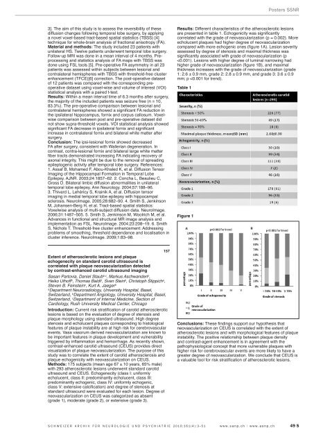
Results: Different <strong>ch</strong>aracteristics of the atherosclerotic lesions<br />
are presented in table 1. E<strong>ch</strong>ogenicity was significantly<br />
correlated with the grade of neovascularization (p = 0.002). More<br />
e<strong>ch</strong>olucent plaques had higher degree of neovascularization<br />
compared with more e<strong>ch</strong>ogenic ones (figure 1A). Lesion severity<br />
assessed by degree of stenosis and maximal thickness was<br />
significantly associated with grade of neovascularization (p<br />