apd_mgz_p1-1-4
apd_mgz_p1-1-4
apd_mgz_p1-1-4

Create successful ePaper yourself
Turn your PDF publications into a flip-book with our unique Google optimized e-Paper software.
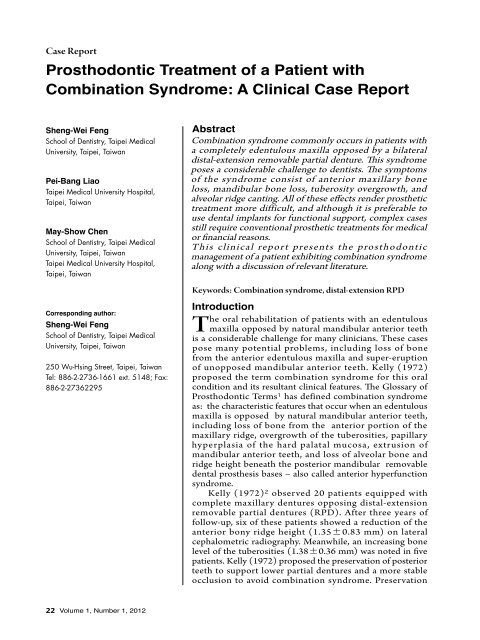
Case Report<br />
Prosthodontic Treatment of a Patient with<br />
Combination Syndrome: A Clinical Case Report<br />
Sheng-Wei Feng<br />
School of Dentistry, Taipei Medical<br />
University, Taipei, Taiwan<br />
Pei-Bang Liao<br />
Taipei Medical University Hospital,<br />
Taipei, Taiwan<br />
May-Show Chen<br />
School of Dentistry, Taipei Medical<br />
University, Taipei, Taiwan<br />
Taipei Medical University Hospital,<br />
Taipei, Taiwan<br />
Corresponding author:<br />
Sheng-Wei Feng<br />
School of Dentistry, Taipei Medical<br />
University, Taipei, Taiwan<br />
250 Wu-Hsing Street, Taipei, Taiwan<br />
Tel: 886-2-2736-1661 ext. 5148; Fax:<br />
886-2-27362295<br />
Abstract<br />
Combination syndrome commonly occurs in patients with<br />
a completely edentulous maxilla opposed by a bilateral<br />
distal-extension removable partial denture. This syndrome<br />
poses a considerable challenge to dentists. The symptoms<br />
of the syndrome consist of anterior maxillary bone<br />
loss, mandibular bone loss, tuberosity overgrowth, and<br />
alveolar ridge canting. All of these effects render prosthetic<br />
treatment more difficult, and although it is preferable to<br />
use dental implants for functional support, complex cases<br />
still require conventional prosthetic treatments for medical<br />
or financial reasons.<br />
This clinical repor t presents the prosthodont ic<br />
management of a patient exhibiting combination syndrome<br />
along with a discussion of relevant literature.<br />
Keywords: Combination syndrome, distal-extension RPD<br />
Introduction<br />
The oral rehabilitation of patients with an edentulous<br />
maxilla opposed by natural mandibular anterior teeth<br />
is a considerable challenge for many clinicians. These cases<br />
pose many potential problems, including loss of bone<br />
from the anterior edentulous maxilla and super-eruption<br />
of unopposed mandibular anterior teeth. Kelly (1972)<br />
proposed the term combination syndrome for this oral<br />
condition and its resultant clinical features. The Glossary of<br />
Prosthodontic Terms 1 has defined combination syndrome<br />
as: the characteristic features that occur when an edentulous<br />
maxilla is opposed by natural mandibular anterior teeth,<br />
including loss of bone from the anterior portion of the<br />
maxillary ridge, overgrowth of the tuberosities, papillary<br />
hyperplasia of the hard palatal mucosa, extrusion of<br />
mandibular anterior teeth, and loss of alveolar bone and<br />
ridge height beneath the posterior mandibular removable<br />
dental prosthesis bases – also called anterior hyperfunction<br />
syndrome.<br />
Kelly (1972) 2 observed 20 patients equipped with<br />
complete maxillary dentures opposing distal-extension<br />
removable partial dentures (RPD). After three years of<br />
follow-up, six of these patients showed a reduction of the<br />
anterior bony ridge height (1.350.83 mm) on lateral<br />
cephalometric radiography. Meanwhile, an increasing bone<br />
level of the tuberosities (1.380.36 mm) was noted in five<br />
patients. Kelly (1972) proposed the preservation of posterior<br />
teeth to support lower partial dentures and a more stable<br />
occlusion to avoid combination syndrome. Preservation<br />
22 Volume 1, Number 1, 2012
Case Report<br />
of posterior occlusion and avoidance of<br />
anterior hyperfunction are considered the<br />
primar y treatment suggestions for this<br />
complex condition. Saunder et al (1979) 3<br />
and Jameson (2001) 4 suggested the use of an<br />
alternative tooth form and occlusal concept<br />
(linear occlusion) and minimum anterior<br />
contact for reducing further bone loss caused<br />
by hyperfunction of anterior teeth. Previous<br />
studies advocated osseointegrated implantretained<br />
or implant-supported prostheses to<br />
change the occlusal force distribution and<br />
decrease the traumatic stress to the alveolar<br />
bone resulting from combination syndrome. 5<br />
T h e p r e s e n t r e p o r t d e t a i l s t h e<br />
prosthodontic management of a specific<br />
patient exhibiting symptoms of combination<br />
syndrome.<br />
Case Report<br />
A 73-year-old male patient was referred to<br />
the Dentistry Department of Taipei Medical<br />
University Hospital in Taipei, Taiwan, for<br />
restorative treatment. The patient's chief<br />
complaints were inadequate retention of<br />
maxillary complete denture and inability<br />
to chew comfortably. No major systemic<br />
diseases or drug allergies were reported. On<br />
examination, the patient had an edentulous<br />
maxilla and nine natural mandibular anterior<br />
teeth (Figure 1). Clinically, the patient<br />
displayed anterior bone loss and flabby<br />
tissue of the maxillary ridge, overgrowth of<br />
the maxillary tuberosities, and over-erupted<br />
mandibular anterior teeth (Figure 2). The<br />
patient rejected any surgery and implant<br />
therapy due to financial considerations. The<br />
patient agreed to have a new complete denture<br />
and a mandibular removable partial denture<br />
Figure 1. Panoramic radiograph showing<br />
a typical case of combination syndrome<br />
with severe resorption of the anterior<br />
maxillary and super-eruption of unopposed<br />
mandibular anterior teeth.<br />
(a)<br />
(b)<br />
(c)<br />
(d)<br />
(e)<br />
Figure 2.: (a) Occlusal view of maxillary<br />
arch. (b) Occlusal view of mandibular<br />
arch showing tooth crowding. (c)<br />
Right-side view revealing the retained<br />
canine. (d) Preoperative frontal view<br />
showing greater bone resorption of the<br />
premaxillary area. (e) Left-side view<br />
indicating sufficient restoration space.<br />
Journal of Prosthodontics and Implantology 23
Case Report<br />
after some discussion.<br />
Initial therapy included oral hygiene<br />
instructions, caries control, and nonsurgical<br />
periodontal therapy. At the first clinical<br />
appointment for prosthodontic treatment,<br />
a preliminary impression of the maxillary<br />
and mandibular arches was made w ith<br />
irreversible hydrocolloid materials (Hydrogum,<br />
Zhermack®, Badia Polesine, Italy) and poured<br />
with dental stone. A custom tray was fabricated<br />
for the maxillary complete denture impression<br />
and a wax relief was applied to the anterior<br />
flabby tissue area. A green modeling compound<br />
(GC Corp, Tokyo, Japan) was then used to<br />
obtain accurate denture border position and<br />
seal. The definitive impression of the maxillary<br />
arch was made w ith v inyl polysilox ane<br />
impression material (Virtual®, Ivoclar Vivadent,<br />
Schaan, Italy) ( Figure 3a). A definitive cast was<br />
created with type III dental stone.<br />
A Kennedy Class I RPD of the mandibular<br />
arch was designed after surveying the cast.<br />
Following tooth preparation, the definitive<br />
impression of the RPD framework was made<br />
with vinyl polysiloxane material (Aquasil,<br />
Dentsply Caulk, Milford, Delaware, USA).<br />
The altered-cast technique was promulgated<br />
to improve the stability of the mandibular<br />
RPD and correct any errors incurred at the<br />
first impression stage. After the face-bow<br />
transfer, the maxillary and mandibular master<br />
casts were mounted in centric relation on a<br />
semi-adjustable articulator (Whip Mix 3040,<br />
Louisville, Kentucky, USA). In addition,<br />
an intraoral Gothic Arch Tracer (Simplex®,<br />
Dentsply, New York, USA) was applied to<br />
verif y accurate and reproducible occlusal<br />
vertical dimension (OVD) and centric relation<br />
(CR)(Figure 3c).<br />
The selection of maxillary anterior artificial<br />
teeth was determined by patient gender and<br />
personality. Balanced occlusion was indicated<br />
for this case to assure an even distribution<br />
of occlusal force and prevent occlusal<br />
interferences on the residual ridge. The tooth<br />
arrangement was checked for esthetics and CR<br />
position and then submitted for processing.<br />
After prescrip, both casts were remounted,<br />
adjusted, and polished. At a subsequent<br />
appointment, the finished prostheses were<br />
delivered and minimal occlusal adjust ment<br />
was needed. The patient was pleased with their<br />
appearance and chewing ability. A maintenance<br />
program including oral hygiene instruction and<br />
prosthesis home care was established. At the<br />
18-month maintenance visit, no complications<br />
were observed.<br />
Discussion<br />
Treatment of patients with an edentulous<br />
ma x illa opposed to natural mandibular<br />
anterior teeth and a distal-extension RPD is<br />
considered a challenge for dental practitioners.<br />
Combination syndrome has a prevalence rate<br />
(a)<br />
(b)<br />
Figure 3.: (a) Maxillary impression prepared by mucostatic impression technique. (b) Altered-cast impression for the distalextension<br />
RPD. (c) Gothic arch tracer was applied to record the CR position.<br />
(c)<br />
(a)<br />
(b)<br />
Figure 4.: Frontal and lateral views of the finished prostheses at the time of delivery. Even occlusal contacts and minimum<br />
anterior contacts were provided for this case.<br />
(c)<br />
24 Volume 1, Number 1, 2012
Case Report<br />
of approximately 24% for denture patients 6 .<br />
Therefore, it is necessar y for dentists to<br />
understand the particular problems of patients<br />
and provide a comprehensive treatment plan.<br />
Increasing pressure on the premaxillary<br />
alveolar ridge and loss of adequate posterior<br />
occlusal contacts are impor tant factors<br />
in relation to combination syndrome 6, 7 .<br />
The bone loss in the midline of the maxilla<br />
observed by Kelly (1972) was 0.43 mm/<br />
year. López-Roldán etal (2009) 8 and Barber<br />
etal (1990) 9 reported similar results (0.32<br />
mm/year and 0.36 mm/year, respectively)<br />
among patients wearing a maxillary complete<br />
denture and mandibular overdentures on two<br />
implants, a situation in which the prosthetics<br />
are biomechanically similar to Kelly's cases.<br />
Maximum support of the denture-bearing<br />
area, preservation of the mandibular posterior<br />
abutment, and balanced occlusion were all<br />
proposed to prevent bone loss and excess<br />
pressure on the anterior maxillary alveolar<br />
ridge. Similarly, Van Waas etal (1993) 10<br />
suggested the avoidance of total tooth<br />
extraction, the preservation of a few teeth, and<br />
the use of overdentures.<br />
In the present case, the mucostatic<br />
impression technique w ith relief at the<br />
anterior maxillary flabby tissue was applied<br />
to accurately record the entire functional<br />
denture-bearing area (Figure 3a). In addition,<br />
the maxillary right canine root was retained<br />
f o r p reser vat i o n o f a l veolar b o n e and<br />
proprioception. Meanwhile, a proper occlusal<br />
plane, the balancing of tooth contacts during<br />
excursive movements, the elimination of<br />
anterior contacts, and remounting techniques<br />
were used to gain better distribution of<br />
occlusal force and reduce stress on the anterior<br />
maxillary alveolar ridge.<br />
The ef fect of mandibular status on<br />
maxillary ridge resorption has been widely<br />
discussed and investigated. Carlsson etal<br />
(1967) 11 compared bone resorption of the<br />
anterior maxillar y alveolar ridge among<br />
patients with maxillary complete dentures<br />
and three different mandibular statuses: (1) a<br />
mandibular complete denture; (2) mandibular<br />
anterior teeth with bilateral extension RPD;<br />
and (3) mandibular teeth only. Greater bone<br />
resorption was found in the groups that had<br />
anterior mandibular teeth with or without<br />
an RPD when compared to the group with<br />
mandibular teeth only. However, small and<br />
insignificant changes of the bone height were<br />
described over five-years of follow-up in<br />
patients with a maxillary complete denture<br />
opposed by a bar-retained mandibular RPD 12 .<br />
Other studies 13, 14 showed no significant<br />
differences and proposed that the individual<br />
variations were larger, but the experimental<br />
data revealed that greater bone resorption<br />
occurred among patients with unilateral or<br />
bilateral RPD. To prevent the occlusal and<br />
enhance the treatment of cs, we propose that<br />
(1) the distal-extension mandibular RPD<br />
may serve a negative role for the deterioration<br />
of combination syndrome 15 ; and (2) the<br />
application of dental implants in edentulous<br />
areas, especially at premolar or molar regions,<br />
could provide better posterior support 16 .<br />
References<br />
1. The glossary of prosthodontic terms. J Prosthet Dent 2005; 94: 10-92.<br />
2. Kelly E. Changes caused by a mandibular removable partial<br />
denture opposing a maxillary complete denture. J Prosthet Dent<br />
1972; 27: 140-50.<br />
3. Saunders TR, Gillis RE Jr, Desjardins RP. The maxillary complete<br />
denture opposing the mandibular bilateral distal-extension partial<br />
denture: treatment considerations. J Prosthet Dent 1979; 41: 124-8.<br />
4. Jameson WS. The use of linear occlusion to treat a patient with<br />
combination syndrome: a clinical report. J Prosthet Dent 2001;<br />
85: 15-9.<br />
5. Tolstunov L. Management of biomechanical complication of<br />
implant-supported restoration of a patient with combination<br />
syndrome: a case report. J Oral Maxillofac Surg. 2009; 67: 178-88.<br />
6. Shen K, Gongloff RK. Prevalence of the 'combination syndrome'<br />
among denture patients. J Prosthet Dent 1989; 62: 642-4.<br />
7. Palmqvist S, Carlsson GE, Owall B. The combination syndrome:<br />
a literature review. J Prosthet Dent 2003; 90: 270-5.<br />
8. López-Roldán A , Abad DS, Ber tomeu IG, Castillo EG,<br />
Otaolaurruch ES. Bone resorption processes in patients wearing<br />
overdentures. A 6-years retrospective study. Med Oral Patol Oral<br />
Cir Bucal 2009; 14: 203-9.<br />
9. Barber HD, Scott RF, Maxson BB, Fonseca RJ. Evaluation of<br />
anterior maxillary alveolar ridge resorption when opposed by the<br />
transmandibular implant. J Oral Maxillofac Surg 1990; 48: 1283-7.<br />
10. Van Waas MA, Jonkman RE, Kalk W, Van 't Hof MA, Plooij J, Van<br />
Os JH. Differences two years after tooth extraction in mandibular<br />
bone reduction in patients treated with immediate overdentures or<br />
with immediate complete dentures. J Dent Res 1993; 72: 1001-4.<br />
11. Carlsson GE, Bergman B, Hedegård B. Changes in contour of<br />
the maxillary alveolar process under immediate dentures. A<br />
longitudinal clinical and x-ray cephalometric study covering 5<br />
years. Acta Odontol Scand 1967; 25: 45-75.<br />
12. Uçtali S, Hasanreisolu U, Ieri H. Cephalometric evaluation of<br />
maxillary complete, mandibular fixed-removable partial prosthesis:<br />
a 5-year longitudinal study. J Oral Rehabil 1997; 24:164-9.<br />
13. Crum RJ, Rooney GE Jr. Alveolar bone loss in overdentures: a<br />
5-year study. J Prosthet Dent 1978; 40: 610-3.<br />
14. Tallgren A. The continuing reduction of the residual alveolar<br />
ridges in complete denture wearers: a mixed-longitudinal study<br />
covering 25 years. J Prosthet Dent 1972; 27: 120-32.<br />
15. Palmqvist S, Carlsson GE, Owall B. The combination syndrome:<br />
a literature review. J Prosthet Dent 2003; 90: 270-5.<br />
16. Keltjens HM, Kayser AF, Hertel R , Battistuzzi PG. Distal<br />
extension removable partial dentures supported by implants<br />
and residual teeth: considerations and case reports. Int J Oral<br />
Maxillofac Implants 1993; 8: 208-13.<br />
17. Carlsson GE. Responses of jawbone to pressure. Gerodontology<br />
2004; 21: 65-70.<br />
Journal of Prosthodontics and Implantology 25