notchincisive
Aurum - OCCLUSAL PLANE INDEX Ver 2
Aurum - OCCLUSAL PLANE INDEX Ver 2

Create successful ePaper yourself
Turn your PDF publications into a flip-book with our unique Google optimized e-Paper software.
ARCHITECTING THE OCCLUSAL PLANE<br />
Clayton A. Chan, D.D.S., M.I.C.C.M.O<br />
Director of Neuromuscular Dental Studies, Las Vegas Institute for Advanced Dental Studies<br />
It is known that dental occlusion is influenced by changes in the cant of the occlusal plane.<br />
Studies have defined the geometric and mathematical relationships between dental occlusion and<br />
rotations of the occlusal plane in the sagittal view. As a general clinical guide, each degree of<br />
rotation of the occlusal plane will result in a half millimeter change in the dental occlusal<br />
relationship. This is of importance, because changes in the cant of the occlusal plane are<br />
sometimes unintentional, as well as intentional, during occlusal therapy. Earlier studies have also<br />
documented that the occlusal plane rotates naturally upward and forward approximately 6<br />
degrees during growth and development. This phenomenon tends to develop a Class II dental<br />
relation and therefore has important implications for the developing dentition.<br />
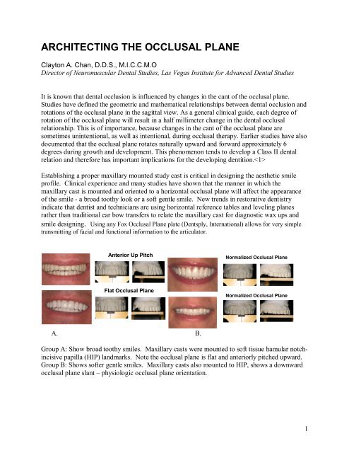
Establishing a proper maxillary mounted study cast is critical in designing the aesthetic smile<br />
profile. Clinical experience and many studies have shown that the manner in which the<br />
maxillary cast is mounted and oriented to a horizontal occlusal plane will affect the appearance<br />
of the smile a broad toothy look or a soft gentle smile. New trends in restorative dentistry<br />
indicate that dentist and technicians are using horizontal reference tables and leveling planes<br />
rather than traditional ear bow transfers to relate the maxillary cast for diagnostic wax ups and<br />
smile designing. Using any Fox Occlusal Plane plate (Dentsply, International) allows for very simple<br />
transmitting of facial and functional information to the articulator.<br />
Anterior Up Pitch<br />
Normalized Occlusal Plane<br />
Flat Occlusal Plane<br />
Normalized Occlusal Plane<br />
A. B.<br />
Group A: Show broad toothy smiles. Maxillary casts were mounted to soft tissue hamular <strong>notchincisive</strong><br />
papilla (HIP) landmarks. Note the occlusal plane is flat and anteriorly pitched upward.<br />
Group B: Shows softer gentle smiles. Maxillary casts also mounted to HIP, shows a downward<br />
occlusal plane slant – physiologic occlusal plane orientation.<br />
1
Relating the Maxillary Arch to the Cranium<br />
There are many reference planes the restorative and orthodontic clinician uses in the assessment<br />
of the maxillary arch to the cranial base. The following is a partial list:<br />
• SN Plane – A line from sella to nasion – considered to represent the cranial base.<br />
• Frankfurt Horizontal Plane – Porion to Orbitale (Bony)<br />
• Camper’s Plane – Acanthionexternal auditory meatus plane (Bony)<br />
• OPPMI Plane Odontoid Process – Pterygomaxillary Fissure – Incisive Foramen<br />
(Bony)<br />
• HIP Line – Hamular Notch – Incisive Papilla (Soft Tissue) – “Transit Line Plane”<br />
• Ala Tragus Line – Ala of nose to tragus of ear (Soft Tissue)<br />
• Many others<br />
All these references change over time based on research.<br />
Nasion<br />
Porion<br />
Sella<br />
Nasion<br />
Porion<br />
Sella<br />
Orbitale<br />
Orbitale<br />
OPPMI<br />
OPPMI<br />
OCCLUSAL PLANE<br />
Physiologic Occlusal Plane Frontal View Pathologic Occlusal Plane<br />
Figure 1<br />
Inclination of the Occlusal Plane (IOP)<br />
The most common plane used is Frankfurt plane (porionorbitale). It was first conceived for the<br />
orientation of skulls in anthropology in the late nineteenth century. Ferrario (1994), in previous<br />
studies have shown that in natural head posture (NHP), the Frankfurt plane is extended, with the<br />
orbitale higher than the tragus or transverse horizontal axis. Men showed an upward tendency<br />
and females showed a downward tendency. This study implied an overly steep angulation of the<br />
occlusal plane with the incisal edges of the maxillary anteriors placed inferiorly when compared<br />
to NHP. It was concluded that the two Frankfurt planes were never coincident in all<br />
subjects; the tragus was always lower and more anterior than the porion.<br />
On average, the angle tragusorbitaleporion was about 6°.<br />
Ciancaglini (2003) when comparing 14 healthy versus 14 TMD young adults with normal<br />
occlusion reported:<br />
2
• No significant deviation from the horizontal was observed for the interpupillary axis and<br />
occlusal plane.<br />
• In lateral view, the Frankfurt plane was upwardorientated relative to the true horizontal<br />
in TMD group (mean angular deviation 2.8 degrees, 95% CI, 1.04.6 degrees ).<br />
• The occlusal and Camper planes were downwardorientated in both groups (P < 0.0001)<br />
• Inclination of occlusal plane tended to be smaller in TMD subjects (mean difference<br />
between groups, 3.8 degrees, 95% CI, 7.60.1 degrees ).<br />
• Furthermore, data suggests, within this population, TMD might be mainly associated with<br />
head posture rather than with craniofacial morphology. See Figure 1.<br />
The Journal of Prosthodontic Dentistry has reported Camper’s Plane (Acanthionexternal<br />
auditory meatus plane, boney) is frequently used for the purpose of establishing the ala tragus<br />
plane. Ideally, the alatragus plane is considered to be parallel to the occlusal plane. “The<br />
occlusal plane is at an angle of approximately 10 degrees relative to the Frankfort horizontal<br />
plane”. <br />
The Journal of Prosthodontics also reported:<br />
1. The inclination of the occlusal plane (IOP) is one of the key factors governing occlusal<br />
balance.<br />
2. Determination of IOP is an important step before equilibrating complete dentures,<br />
comprehensive restorative dentistry and orthodontic procedures. <br />
Chan (2002, 2005) demonstrated by computerized mandibular scanning (CMS), EMG signaling<br />
before and after TENS and with ICAT radiographic imaging that as the mandible moves anterior<br />
along an optimized isotonic path of closure the head tilt’s downward, thus changing the<br />
orientation of the occlusal plane from a flatter occlusal plane (pathologic) as referenced from a<br />
horizontal level baseline to a more angled (6 degrees) occlusal plane (physiologic).<br />
Eye Posture, Head Posture & MaxillaryMandibular Positioning<br />
Dental literature has often used the horizontal level as a reference for analysis of the occlusal<br />
plane both in the frontal and sagittal/lateral views, bipupilar plane, otic plane, as well as head<br />
posture. The orientation of the maxillary cast should be accurately reproduced clinically and<br />
transferred to the laboratory technician’s occlusal analyzing table at the bench both referenced to<br />
horizontal level.<br />
Vision plays a significant role in balance. Approximately twenty percent of the nerve fibers from<br />
the eyes interact with the vestibular system. The interpupillary orientation of the eyes should be<br />
centered within the orbits of the cranium when the cervical neck and head posture is normalized.<br />
The eyes are key sense organs to assist in coordinated balance control and spatial relationships<br />
maintenance of the human body.<br />
In an effort to adjust to the vertical misalignment of the eyes, the person will frequently tip their<br />
head to mechanically help align the eyes. This may often be a result of a posterior malalignment<br />
of the mandible to the cranium (see figure 2). This in turn can cause a tilting up of the head and<br />
3
posteriorizing of the mandible. Ear congestion feelings, resultant dizziness and balance disorders<br />
can result.<br />
Figure 2<br />
Otic plane relates to the sense of balance and equilibrium because it relates to the semicircular<br />
canals. This sense of equilibrium allows us to know the position of the head in space and to the<br />
rest of the body. Mechanoreceptors in the cervical spine and mandible will react to changes in<br />
the cranial, cervical and mandibular posture in an attempt to keep these horizontal relationships<br />
intact.<br />
Occlusal Plane Determination<br />
Traditionally most restorative aesthetic clinicians have paid more attention to the frontal<br />
horizontal plane axis (interpupillary, otic and frontal occlusal) as they related to the long axis of<br />
the face. The use of the classic stick bites and symmetry bites have been used to capture these<br />
two dimensional relationships to register the frontal horizontal levelness of their patients<br />
maxillary arches. This visual subjective assessment by the dentist has been used as a standard<br />
reference check to determine the maxillary arch levelness frontally for years when<br />
communicating with the laboratory technician.<br />
Although this may help aid the technician to mount the maxillary cast in the frontal horizontal<br />
planes, it fails to give an accurate relationship in the sagittal or lateral axis, especially when<br />
realizing that it is the posterior occlusal plane slant (pitch axis) that is critical when designing the<br />
curvature and angle of the smile line (bicuspids to molars) as referenced to the surrounding lip<br />
borders of the oral cavity.<br />
Figure 3: Note the left and right occlusal plane slant of the above patient when seen from the<br />
lateral view (referenced to horizontal level).<br />
4
Most laboratory technicians have found that when using these devices that the maxillary<br />
mountings often did not match the accompanying frontal smile photographs. With years of<br />
laboratory mounting experience, the technician customarily set the stick bite aside and mounted<br />
the maxillary cast to match the photograph in the frontal plane by their trained eyes. Further, it<br />
left in question the angle or slant of the posterior occlusal plane (pitch axis) as it related to the<br />
sagittal horizontal plane relative to a level table.<br />
One of the most important objectives in maxillary mounting is to replicate the maxillary teeth<br />
orientation as it is seen sagittally/laterally from the side view of the patient. This side view of the<br />
occlusal plane can be easily observed when asking the patient to smile with their head at<br />
horizontal level with the pupils of the eyes centered of the orbits looking at the horizon (straight<br />
ahead) and pronouncing the letter “E”.<br />
This occlusal plane angle is critical for optimal smile designing and must be accurately captured<br />
to correctly mount the maxillary cast, referencing it to the horizontal occlusal analyzing table for<br />
proper occlusal plane analysis.<br />
Diagnosing the Maxillary Cast Mountings<br />
The maxillary cast mountings can be very diagnostic as to indicate whether there exists<br />
unresolved cranial to mandibular muscular imbalances. When the head position and eye<br />
orientation within the orbits are in a pathologic position an accommodative response will result<br />
in a forward head posture (effecting the cervical spine relationship – kyphosis) with an<br />
accompany “abnormal mandibular jaw closure pattern” (G. Wolford). The head tilt will be<br />
upward contributing the cervical neck aches and pain with an anatomically flatter to an upward<br />
anterior slanting occlusal plane as referenced from horizontal level (Figure 1). ICAT<br />
radiographic scans will confirm that the boney reference from the odontoid process through the<br />
pterygomaxillary fissure and anterior to the incisive foramen will be abnormally level. Thus,<br />
when mounting the maxillary cast via the comparable hamularincisive papilla (HIP) soft tissue<br />
references it will present as a very flat to anteriorly upward pitched occlusal plane (57.6%). <br />
Patient’s who are neuromuscularly stabilized and craniomandibularcervically balanced will<br />
present with a more normalize head posture (head tilt downward), effecting the cervical spine<br />
relationship – lordosis, with an accompanying isotonic jaw closure pattern. ICAT radiographic<br />
imaging clearly demonstrates (a line through the odontoid process, pterygomaxillary fissure and<br />
anterior to the incisive foramen) a downward slant (87.5%). The occlusal plane will also be<br />
more parallel to these boney references confirming the HIP reference also slants downward in<br />
relationship to horizontal level. This physiologic occlusal relationship must be accurately<br />
recorded and represented in the laboratory maxillary mounting if an optimal smile line is to be<br />
designed to match nature’s occlusal plane. Occlusalcervical, craniomandibular relationships<br />
and tooth widthlength proportions can be achieved to natures design via visual analysis of the<br />
various leveling planes. The trained and experienced laboratory technicians realize these facts.<br />
5
HIP Plane Before Diagnostic Wax UP<br />
Figure 4: Maxillary cast was mounted to classic HIP soft tissue landmarks. The flat occlusal<br />
plane referenced to a horizontal level table would be indicative of a pathologic forward head<br />
orientation with underlying unresolved musculoskeletal cranium to mandibular occlusal posture.<br />
OPI (Fox Plane) Before Diagnostic Wax Up<br />
Figure 5: Maxillary cast mounted using the OPI (Occlusal Plane Index/Fox Plane). Notice the<br />
occlusal plane slants downward (6 degrees) as referenced to the horizontal table, indicative of a<br />
more normalized head posture supported by an optimized mandibular position.<br />
6
Figure 6:<br />
Before Treatment Occlusal Plane at CO<br />
(Flat) – Level HIP<br />
After Treatment Occlusal Plane at Myocentric<br />
(Angled) – Downward slant HIP<br />
A. B.<br />
A: ICAT scan shows a level/flat occlusal plane parallel to the boney odontoidincisive foramen<br />
(HIP) before neuromuscular stabilization (pathologic occlusal plane orientation). Patient is<br />
unposed, at the habitual centric occlusal mandibular posture.<br />
B: ICAT scan shows a more normalized downward slant boney odontoidincisive foramen (HIP)<br />
occlusal plane after neuromuscular orthotic treatment (physiologic occlusal plane orientation), at<br />
an optimized myocentric occlusal mandibular posture.<br />
7
Figure 7<br />
A: HIP Plane Mount and Diagnostic Wax Up – Note the shorter<br />
upper posterior teeth with built in curve of Spee. Central incisors are<br />
waxed off the level table to compensate to create a proper tooth length<br />
and width. Emergence profile of maxillary central incisor will be more<br />
pronounced.<br />
More Tooth<br />
Reduction<br />
Less Tooth<br />
Reduction<br />
B: OPI (Fox Plane) Mount and Diagnostic Wax Up – Note a more<br />
normalized posterior crown length also with a built in curve of Spee. Maxillary<br />
central incisor root angulation is more idealized. The central incisor length and<br />
width is referenced from the occlusal analyzing table and waxed as indicated<br />
without compensating the anterior incisor wax up from the level table.<br />
Recording the Occlusal Plane Angle with the OPI Using A Fox Plane<br />
A simple and reasonable clinical technique using the well known Fox Plane (Dentsply, Trubyte)<br />
can be used to record the maxillary arch with the patients head at horizontal level (Figure 8).<br />
A<br />
Figure 8<br />
B<br />
8
A<br />
B<br />
Figure 9<br />
Clinical Technique – This Is How I Do It<br />
1. First – with the patient standing straight and the head positioned with eyes looking straight<br />
ahead looking at the horizon, make sure the sagittal head tilt is with the eyes in the center of<br />
the orbits. (Natures leveling bubbles). This will assist in getting the head correctly oriented<br />
to level. Subjectively assess the long axis of the face. The interpupillary eyes should not be<br />
used alone to reference to frontal horizontal level, since some patient’s eyes may be different<br />
from one side to the other. Ear levelness, eye brow heights, nose orientations and corner of<br />
the lips may not always be reliable references for facial symmetry.<br />
2. Syringe any fast set polyvinyl (30 second bite registration material) on the Fox Plane bite<br />
fork and insert it into the mouth upward against the maxillary anterior teeth. Do not press the<br />
posterior region of the bite fork up on the upper posterior occlusal surfaces! It is important<br />
to have the patient keep their head level when opening the lower jaw and the eyes looking<br />
straight ahead. Check to confirm the pupils are centered of the eye sockets/orbits). See<br />
Figure 9A.<br />
3. Orient the Fox Plane to level and perpendicular to the long axis of the face as well as level<br />
sagittally/lateral level to the ground (Figure 9B).<br />
4. Allow the polyvinyl material to set firm while holding Fox Plane with light finger pressure<br />
anteriorly. Take a moment to confirm frontal and sagittal levelness to the ground. If the<br />
recording does not look right repeat the above steps until correctly leveled and recorded.<br />
After the PV material hardens, remove the Fox Plane and occlusal plane index (OPI) from<br />
the mouth.<br />
5. Peal away the PV occlusal plane index (OPI) from the Fox Plane bite fork and place the OPI<br />
on any level mounting table and oriented it to the center/midline. (Orienting the Fox Plane<br />
with the OPI directly on the analyzing table for mounting can also be done). Place the upper<br />
dental cast into the index registration and mount the upper cast.<br />
9
6. After the upper cast is set and mounted remove the OPI from the mounting table and evaluate<br />
the occlusal table slant or angulation (pitch) as it relates to the horizontal table.<br />
7. Mount the lower cast to the upper via the Myocentric bite registration.<br />
Now you have the upper and lower casts mounted physiologically and accurately, relating the<br />
patient’s maxilla and mandible on any articulating “model holder”.<br />
Occlusal Plane Index (OPI)/ Level Fox Plane<br />
Figure 10 A<br />
Figure 10 B: Note pre treatment diagnostic casts indicate a 56 degree occlusal plane angle.<br />
10
OPI gives the laboratory an easy starting reference to build the crowns with 6 10 degree<br />
occlusal plane with little pre operative stone model occlusal reduction. Altering the level<br />
occlusal table at the bench is no longer necessary to create the proper posterior crown length.<br />
Figure 11<br />
A. B.<br />
A. A six degree slant of the occlusal plane referenced from horizontal level is easily waxed with<br />
curve of Spee and curve of Wilson to artistically create a soft smile line. B. Note a more even<br />
distribution of the upper and lower crown length in the posterior region due to proper occlusal<br />
plane determination and recording with the OPI (Fox Plane).<br />
Figure 12<br />
11
Figure 13: A soft gentle smile line is created based on an optimized mandibular position<br />
and a properly mounted maxillary cast mount via the OPI (Fox Plane) technique (not based<br />
on a soft tissue HIP mount/ flat, see Figure 6 – before and after ICAT).<br />
Figure 14: Left: Before treatment smile: Note the maxillary frontal plane downward left<br />
cant. Right: Maxillary finished restorations. Note the corrected occlusal smile line.<br />
Before Treatment<br />
After Treatment<br />
12
Finished maxillary restorations<br />
The patient selected shade 110, 040, 030 from Ivoclar’s Chromoscope shade guide. Heavy<br />
translucency at the incisal third along with a gradual transition to the gingival and proximal. The<br />
natural surface texture is created to give realism alone with internal sculpted dentinal lobes.<br />
Conclusion<br />
The Occlusal Plane Index (OPI)/ Fox Plane is a simple technique to effectively record the<br />
maxillary occlusal plane angle (slant) for a more accurate diagnostic mount and evaluation when<br />
referenced to any horizontal occlusal table analyzer. Kois, Leary, Jankelson, and others have<br />
used versions of the Fox Plane to align the maxillary arch successfully for years. This technique<br />
is designed to better assess the maxillary occlusal cants, asymmetries and occlusal discrepancies<br />
when referenced to the horizontal ground when the patient’s head is correctly oriented looking at<br />
a level horizontal position. It assists both the dentist and laboratory technician to better<br />
communicate a more representative occlusal plane orientation for occlusal waxing and smile<br />
design. It is a simple and inexpensive technique to use, allowing an easy accurate transfer to any<br />
occlusal analyzing table via the OPI. It minimizes guessing and a need to alter the occlual pitch<br />
or angle of the occlusal plane in the laboratory. It allows for a more proportional distribution and<br />
crown length ratio between the upper and lower posterior crowns and prevents the need to<br />
excessively reduce the maxillary posterior occlusion during crown preparation.<br />
Dr. Clayton A. Chan is dedicated to share his passion and teaches the neuromuscular<br />
principles that have worked for him. He is an educator to thousands of dentist all around the<br />
world, inspiring them to take their practices to another level. He is considered by many an<br />
authority on Neuromuscular Dentistry and Occlusion. He practice’s in Las Vegas, Nevada<br />
where he focuses on Aesthetic Dental Orthopedics, orthodontics and TMJ, implementing both the<br />
gnathological and neuromuscular principles. He is Director of Neuromuscular Dentistry at the<br />
Las Vegas Institute for Advanced Dental Studies.<br />
13
References:<br />
1. Braun S, Legan, HL: Changes in occlusion related to the cant of the occlusal plane. Am J<br />
Orthod Dentofacial Orthop. 1997 Nov; 112 (5):17A20A.<br />
2. Virgilio F. Ferrario, MD, Chiarella Sforza, MD, Domenica Germanò, MD, Luca L.<br />
Dalloca, DMD, Alessandro Miani Jr., MD: Head posture and cephalometric analyses: An<br />
integrated photographic/radiographic technique, The Journal of Prosthetic Dentistry,<br />
Volume 106, Number 3, September 1994.<br />
3. Ciancaglini R, ColomboBolla G, Gherlone EF, Radaelli G.: Orientation of craniofacial<br />
planes and temporomandibular disorder in young adults with normal occlusion. J Oral<br />
Rehabil. 2003 Sep;30(9):87886.<br />
4. The Glossary of Prosthodontic Terms, Seventh Edition (GPT7), The Journal of<br />
Prosthodontic Dentistry, Volume 81, Number 1, January 1999.<br />
5. The Inclination of the Occlusal Plane, J. Prosth. Dent., Volume 87, Number 2, February<br />
2002.<br />
6. Investigative Clinical Research for Neuromuscular Dental Technology: HIP Research<br />
Investigative Study, Four studies March 2004, October 2004, May 2005 and June 2005.<br />
82 participating dental laboratory technicians, 154 maxillary model casts, Las Vegas<br />
Institute for Advanced Dental Studies, Las Vegas, Nevada.<br />
14