notchincisive
Aurum - OCCLUSAL PLANE INDEX Ver 2
Aurum - OCCLUSAL PLANE INDEX Ver 2

Create successful ePaper yourself
Turn your PDF publications into a flip-book with our unique Google optimized e-Paper software.
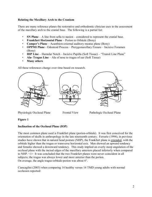
Relating the Maxillary Arch to the Cranium<br />
There are many reference planes the restorative and orthodontic clinician uses in the assessment<br />
of the maxillary arch to the cranial base. The following is a partial list:<br />
• SN Plane – A line from sella to nasion – considered to represent the cranial base.<br />
• Frankfurt Horizontal Plane – Porion to Orbitale (Bony)<br />
• Camper’s Plane – Acanthionexternal auditory meatus plane (Bony)<br />
• OPPMI Plane Odontoid Process – Pterygomaxillary Fissure – Incisive Foramen<br />
(Bony)<br />
• HIP Line – Hamular Notch – Incisive Papilla (Soft Tissue) – “Transit Line Plane”<br />
• Ala Tragus Line – Ala of nose to tragus of ear (Soft Tissue)<br />
• Many others<br />
All these references change over time based on research.<br />
Nasion<br />
Porion<br />
Sella<br />
Nasion<br />
Porion<br />
Sella<br />
Orbitale<br />
Orbitale<br />
OPPMI<br />
OPPMI<br />
OCCLUSAL PLANE<br />
Physiologic Occlusal Plane Frontal View Pathologic Occlusal Plane<br />
Figure 1<br />
Inclination of the Occlusal Plane (IOP)<br />
The most common plane used is Frankfurt plane (porionorbitale). It was first conceived for the<br />
orientation of skulls in anthropology in the late nineteenth century. Ferrario (1994), in previous<br />
studies have shown that in natural head posture (NHP), the Frankfurt plane is extended, with the<br />
orbitale higher than the tragus or transverse horizontal axis. Men showed an upward tendency<br />
and females showed a downward tendency. This study implied an overly steep angulation of the<br />
occlusal plane with the incisal edges of the maxillary anteriors placed inferiorly when compared<br />
to NHP. It was concluded that the two Frankfurt planes were never coincident in all<br />
subjects; the tragus was always lower and more anterior than the porion.<br />
On average, the angle tragusorbitaleporion was about 6°.<br />
Ciancaglini (2003) when comparing 14 healthy versus 14 TMD young adults with normal<br />
occlusion reported:<br />
2