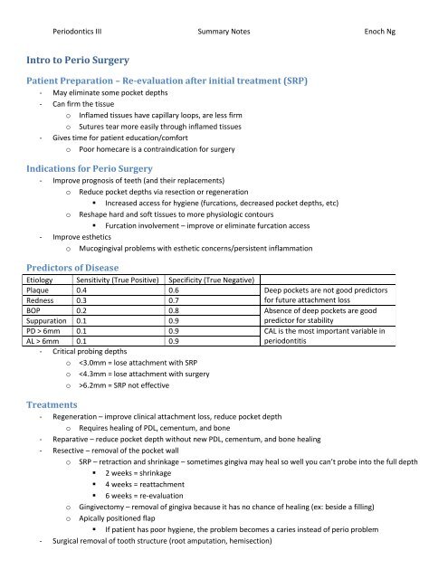
Perio_III
Perio_III
Perio_III

You also want an ePaper? Increase the reach of your titles
YUMPU automatically turns print PDFs into web optimized ePapers that Google loves.
<strong>Perio</strong>dontics <strong>III</strong> Summary Notes Enoch Ng<br />
Intro to <strong>Perio</strong> Surgery<br />
Patient Preparation – Re-evaluation after initial treatment (SRP)<br />
- May eliminate some pocket depths<br />
- Can firm the tissue<br />
o Inflamed tissues have capillary loops, are less firm<br />
o Sutures tear more easily through inflamed tissues<br />
- Gives time for patient education/comfort<br />
o Poor homecare is a contraindication for surgery<br />
Indications for <strong>Perio</strong> Surgery<br />
- Improve prognosis of teeth (and their replacements)<br />
o Reduce pocket depths via resection or regeneration<br />
• Increased access for hygiene (furcations, decreased pocket depths, etc)<br />
o Reshape hard and soft tissues to more physiologic contours<br />
• Furcation involvement – improve or eliminate furcation access<br />
- Improve esthetics<br />
o Mucogingival problems with esthetic concerns/persistent inflammation<br />
Predictors of Disease<br />
Etiology Sensitivity (True Positive) Specificity (True Negative)<br />
Plaque 0.4 0.6 Deep pockets are not good predictors<br />
Redness 0.3 0.7<br />
for future attachment loss<br />
BOP 0.2 0.8 Absence of deep pockets are good<br />
Suppuration 0.1 0.9<br />
predictor for stability<br />
PD > 6mm 0.1 0.9 CAL is the most important variable in<br />
AL > 6mm 0.1 0.9<br />
periodontitis<br />
- Critical probing depths<br />
o
<strong>Perio</strong>dontics <strong>III</strong> Summary Notes Enoch Ng<br />
Treatment Selection<br />
- Overall diagnosis, goals of surgery<br />
- Access<br />
- History of surgery<br />
- Pocket form<br />
- Esthetics<br />
o Anterior teeth – single rooted, patient compliance is huge<br />
o Interproximal bone loss = lose papillae<br />
- Blood supply<br />
Surgical Procedure<br />
Method Selection/Considerations<br />
- Regenerative methods<br />
o Papillae preservation<br />
o Sulcular flaps<br />
o Modified widman flap (to maintain<br />
papillae) – very little tissue loss if done<br />
properly<br />
- Resective methods<br />
o Gingivectomy<br />
o APF<br />
- Smoking considerations<br />
- Informed consent<br />
- Sedation/anesthesia<br />
o Local anesthesia – keep surgery painless<br />
o Inhalation – antianxiety delivery of N 2 O,<br />
safest method of delivery<br />
o Oral sedation – individually variable<br />
o Conscious sedation<br />
o General anesthesia<br />
- Emergency equipment<br />
Procedure<br />
- Premedication<br />
o Prophylactic antibiotics for surgery<br />
o NSAIDS for pain, reduce inflammation<br />
o Anti-anxiety medications<br />
o Chlorhexidine rinse pre and post-op to<br />
decrease aerosol exposure<br />
o Steroids to reduce inflammation<br />
- Tissue Management<br />
o Be gentle and careful<br />
o Observe patient at all times<br />
o Use sharp instruments to avoid<br />
masticating tissue<br />
- Surgical Dressings<br />
• ZO-Eugenol packs<br />
• Non-eugenol packs<br />
• Retention of packs<br />
o Should remain in place for 1 week<br />
o Allow Coe-Pak to harden for 3h before<br />
eating<br />
o Do not disturb pack (ex: brushing, flossing)<br />
- Post-Op<br />
o Printed instructions<br />
o Return appointment<br />
o Repacking<br />
o Tooth mobility<br />
o Mouth care between procedures<br />
o<br />
o<br />
Probing<br />
Root sensitivity<br />
• Desensitizing agents include<br />
homecare and in office products<br />
Anterior Mental Nerve Loop<br />
- Generally 0.5-3.1mm anterior to mental foramen<br />
- 28% of cases 0.4-2.2mm anterior to mental foramen<br />
- 86-90% Caucasians have anterior loop (mental nerve exiting in posterior direction)<br />
- 45% Blacks have mental nerve exiting at right angle to foramen
<strong>Perio</strong>dontics <strong>III</strong> Summary Notes Enoch Ng<br />
Intro to <strong>Perio</strong> Surgery II<br />
Anatomy<br />
- Nerves and arteries usually run superior to mylohyoid muscle, but may run inferiorly<br />
- Lingual concavity – estimate how superior it is for implant placement<br />
- An incision following the central grooves to reach the Mn 3 rd molars will cut into muscles, nerves, and arteries.<br />
The incision should turn and follow the ramus posterior superiorly<br />
- Lingual nerve position is variable – so long as work is done in the keratinized tissue, should be okay. Outside the<br />
keratinized tissue = greater risk<br />
- It can be difficult to close a flap without creating a lingual flap, but care should be taken because nerve/artery<br />
bundles run on the lingual side of the ramus<br />
o Never do a sharp sectioning on the lingual side, because you don’t know where the danger zone is<br />
o Mylohyoid release –allows for the lingual side to the brought superiorly<br />
Blood Supply and Tissue Survival<br />
- Blood Supply<br />
o Full or partial thickness flaps are both useable<br />
o Partial thickness flaps in thin tissue may cause flap death<br />
o Recipient site vascularity affects survival of thin flaps<br />
o Velvet incision = off angled incision by the papilla to allow a large enough piece of tissue for suturing<br />
- Full periosteal horizontal incision<br />
o 24h – disturbance to gingiva coronal to incision<br />
o 48h – local superficial gingival necrosis, overall tissue perfused by perio and intraosseous vessels<br />
o Significance = most of the blood supply comes apical to coronal<br />
• Blood supply from the roots/bones = tissue heals like a scrape, top tissue sloughs<br />
• In a free gingival graft, lots of keratinized tissue is gained but no height<br />
• Height is gained by position tissue coronally (envelop flap with releasing incisions)<br />
- Internal bevel incision between gingiva and periosteum<br />
o 24h – no change<br />
o Significance = healing is fast because of good blood perfusion<br />
- Full thickness flap made, not reflected vs reflected and replaced<br />
o 24h – similar disturbance, reflected flap had 50% greater reduction<br />
o 96h – reflected flap had poorer appearance<br />
o Significance = reflecting flaps decreases blood perfusion to elevated tissue<br />
- FTF reflected, vertical incisions beyond the mucogingival margins, with test group incision length 2x control<br />
o 24h – test group had poorer healing, marginal tissue necrosis<br />
o Significance = critical length:width ratio = 2:1<br />
- Flaps placed over recession areas, test flap 50% narrower than control<br />
o 24h – test flap had 50% reduced circulation<br />
o 7days – cleft-like tissue loss around gingival margin of test flap<br />
• Overly long flaps had some ischemic marking at sutures<br />
o Significance = excess tension or excess flexion decreases flap healing
<strong>Perio</strong>dontics <strong>III</strong> Summary Notes Enoch Ng<br />
Gingival Surgical Procedures<br />
Limited to gingiva, does not involve underlying osseous structures<br />
- Gingival curettage – removal of gingival wall of perio pocket to separate out diseased soft tissue<br />
• Aka = excisional new attachment procedure, ultrasonic curettage, caustic drugs<br />
o Inadvertent curettage – happens with SRP<br />
o No clinical value (2002) – healing is by long junctional epithelium, no new attachment is gained<br />
- Gingivectomy<br />
o Indications<br />
• Elimination of suprabony pockets in firm, fibrous tissue (SRP usually clears edematous pockets)<br />
• Elimination of gingival enlargements<br />
• Elimination of suprabony abscesses<br />
o<br />
o<br />
o<br />
o<br />
Before they become infrabony ones<br />
• Access for restorative dentistry<br />
<br />
<br />
Biologic width important in esthetic areas<br />
Body wants around 2mm between bone and gingiva (beware of gingival rebound)<br />
Biologic width = 2.04mm, supra-alveolar tissues (dentogingival junction) = 2.73mm<br />
• Esthetics<br />
<br />
<br />
Ideal width = CI 25% wider than laterals, 10% wider than canines<br />
Ideal height = CI and canine 20% longer than laterals<br />
o Ratio = 1.2/1.0<br />
Surgical stents – want to know if the stent is for what tooth structure is showing, or<br />
where the crown margin is going to be placed (margin can be slightly in pocket)<br />
Want to preserve some keratinized tissue, do not want to remove all of it<br />
• Keratinized gingiva = pocket depth + attached gingiva (mucogingival junction)<br />
Measure pocket depth, mark it to cause it to bleed. Incision is apical to bleeding point, 45 o bevel to root<br />
Healing<br />
• Initially – acute PMN infiltrate and some necrosis, formation of initial protective clot<br />
<br />
<br />
<br />
<br />
12-24h – epithelial cells at margin migrate into granulation tissue and beneath the<br />
necrotic tissue<br />
24h – increased CT and angioblasts below surface layer<br />
4-16 days – vasodilation and vascularity start to decrease until normal<br />
o Epithelium grows at 0.5mm/day<br />
5-14days – surface epthelialization complete, keratinization incomplete<br />
7 weeks – complete repair of CT<br />
Limiting Factors<br />
• Amount of keratinized gingiva<br />
• Esthetics and esthetic maintenance<br />
• Access to osseous defects for correction<br />
• Less post-op pain if procedure allows for primary closure<br />
• Gingiva may rebound without racial pigmentation – risk for patients with racial pigmentation
<strong>Perio</strong>dontics <strong>III</strong> Summary Notes Enoch Ng<br />
o Electrosurgery<br />
• Good hemorrhage control<br />
• Bad for patients with poorly shielded cardiac pacemakers<br />
• Must be limited to superficial procedures – can cause necrosis if tip touches bone or cementum<br />
Different if only trying to coagulate – can be done at lower temperature<br />
o Chemosurgery<br />
• Difficult to control depth<br />
• Slower healing<br />
• NOT recommended<br />
- Gingivoplasty<br />
o Possible to do APF, gingivectomy, or combined techniques<br />
o Edematous tissue – treat with SRP<br />
o If extremely fibrous and interferes with access – consider treating with gingivectomy<br />
• Gingivectomy can recontour at the margin if adequate keratinized gingiva is present<br />
o Apically Positioned Flap – needs firm tissue, conserve keratinized gingiva<br />
• Common on palatal side – may end up sitting up in a point during crown lengthening. If it<br />
doesn’t stay down, a blood clot can form there and cause tissue rebound. Tissue must lay down<br />
and be positioned apically to prevent rebound<br />
o Combined technique<br />
• Cut border to a regular border first, then place a flap and position apically<br />
• If gingival rebound is from drug use for systemic problems, insurance will pay for multiple<br />
gingivectomies<br />
• Use packs to prevent hematoma formation to prevent gingival rebound, gives esthetic outcome<br />
- Gingival flap<br />
o The exception from all the others. Full thickness flaps may touch osseous structure but should not<br />
contour it or affect it<br />
Treatment considerations<br />
- Functional/esthetic compromise of adjacent teeth<br />
o Opening interdental spaces<br />
o Creating excessively “long” teeth<br />
- Gingival diseases<br />
o Modified by medications<br />
o<br />
<br />
<br />
Difedipine<br />
Cyclosporine<br />
Phenytoin (Dilantin)<br />
• Drug influenced gingival diseases<br />
<br />
Drug influenced gingival enlargements (inflammatory drug induced hyperplasia)<br />
Drug influenced gingivitis (Oral contraceptive associated, other)<br />
• Drugs that cause<br />
Dental plaque induced gingival diseases<br />
• Usually seen in young patients rather than older patients<br />
• In patient has poor hygiene, tissue will have a greater rebound effect<br />
• Beware handicapped patients – their hygiene provider must be informed of importance of care
<strong>Perio</strong>dontics <strong>III</strong> Summary Notes Enoch Ng<br />
<strong>Perio</strong>dontal Flap Surgery<br />
Surgical Procedures<br />
- Gingivectomy<br />
- <strong>Perio</strong>dontal flaps<br />
- Osseous contouring<br />
- Bone grafts<br />
- Laterally sliding flaps<br />
- Free gingival grafts<br />
<strong>Perio</strong>dontal Flaps<br />
- Increase access to root<br />
- Reduce Pocket Depth<br />
- Expose areas for regeneration<br />
- Crown lengthening<br />
Incision Design<br />
- Internal bevel scalloped (modified Widman) – most basic horizontal incision<br />
• Thins the gingiva, conserves attached gingiva, removes pocket lining epithelium<br />
o Incision to periosteum to detach flap<br />
o Scalloping to loosen pocket epithelium from tooth<br />
o Horizontal incision to remove pocket lining epithlium<br />
- Vertical Incisions<br />
o Placed at line angle<br />
o Should not be at apex of gingival sulcus or interproximally<br />
• Loss of papilla or may cause gingival defect<br />
o Usually not placed on palatal side, Mn lingual, or nasopalatine areas (vessels, esthetic zones)<br />
- Blades<br />
o #15 – good for newbs, sharp curved blade<br />
o #11 – sharp straight end with pointed tip<br />
o #12/12B – curved (like a sickle) with tip, good for distal wedge<br />
Flaps<br />
- Full thickness – goes through periosteum, might get up to 0.5mm bone loss because bone is thin<br />
- Partial thickness – goes through CT, some CT and all periosteum remains attached to osseous structure<br />
- Conventional flap – removes pocket epithelium<br />
- Sulcular incision – used when you don’t want to lose attached gingiva or in esthetic zones (anterior region)<br />
- Repositioned flap – replacing the flap back to where it was before (modified Widman surgeries)<br />
- Apically positioned flap – used after 4-6week post-op probing after SRP assuming pocket depths don’t improve<br />
Crown Lengthening<br />
- Restorative margin cannot be closer than 2mm to crestal bone, or will disrupt osseous structure<br />
- Sounding bone – probing through the biologic width to the bone, gives an idea of what bone contour is like<br />
- Biologic width – expose 3-4mm of tooth coronal to bone during surgery to accommodate 2mm biologic width<br />
- Modified Widman – internal bevel primary incision, then scalloping and removal of desired gingiva<br />
o Buck or orban knives helpful for removing interproximal tissue<br />
- Suturing – slight exposure interproximally is okay. Cortical bone is backed up by cancellous bone<br />
- Coe pack placed to help with healing, prevent rebound<br />
o Left for 3-4 days, up to 7-10 days<br />
o Post-surgical hemorrhage controlled by pressure, sutures, clotting, packs<br />
o Remove pack to ID bleeding source/stop bleeding if necessary<br />
- Chlorhexidine rinses (both pre and post-op)<br />
- Takes 4 weeks to heal
<strong>Perio</strong>dontics <strong>III</strong> Summary Notes Enoch Ng<br />
Free Gingival Grafting<br />
- Coe packs can be used to cover donor site<br />
- Take 10 days to get granulated, 6 weeks to heal<br />
- Tissue dries and contracts quickly once it is removed from donor site<br />
- Pain management<br />
Mild 200-400mg ibuprofen 650mg aspirin 650-1000mg acetaminophen<br />
Moderate 600-800mg ibuprofen 400mg ibuprofen with 60mg<br />
codeine or equivalent<br />
600-1000mg acetaminophen<br />
with 60mg codeine or equivalent<br />
Severe<br />
600-800mg ibuprofen with 10mg<br />
oxycodone or equivalent<br />
1000mg acetaminophen with<br />
10mg oxycodone or equivalent<br />
Distal Wedge Technique<br />
- Best done with #12 blade<br />
- Can be triangular, rectangular, or scalloped incisions<br />
- Incisions diverge from each other, allows full thickness flaps to “collapse” so there isn’t extra bundling when<br />
sutured back together<br />
- Used to eliminate distal pockets
<strong>Perio</strong>dontics <strong>III</strong> Summary Notes Enoch Ng<br />
Treatment of Osseous Defects<br />
- Resection<br />
- Debridement<br />
- Grafting<br />
Types of Defects<br />
- Dehiscence – root exposure connected to the rest of the tooth<br />
- Fenestration – window of root exposued<br />
- Positive architecture – interproximal bone more coronal than radicular bone<br />
- Negative architecture – interproximal bone more apical than radicular bone<br />
- Infrabony defect – base of pocket apical to crest of alveolar bone<br />
- Infrabony Pockets (negative architecture)<br />
o 1 osseous wall<br />
o 2 osseous walls – aka crater, most common osseous defect, usually because patient doesn’t floss well<br />
o 3 osseous walls<br />
Furcations<br />
CEJ to opening of furcation<br />
Mn molars<br />
Mx molars<br />
- Buccal side = 3mm - ML side = 3mm<br />
- Lingual side = 4mm - Buccal side = 4mm<br />
- DL side = 5mm<br />
- As much as possible, best NOT to open furcation (hard area to clean)<br />
Other Surgical Considerations<br />
- Widow’s Peak<br />
o If you leave bone adjacent to tooth surface by line angle (tendency to leave bone by line angle)<br />
o More likely to have pocketing, reduction of bone by line angles to give smooth reduction reduces<br />
pocketing during healing<br />
- Radiographic Limitations – 2D image, can’t diagnose periodontitis or determine number of walls in defect<br />
- Sounding bone (transgingival probing) – probing through the attached gingiva to find out where the bone is<br />
o Used to discover osseous defects and what they are like, can help determine number of walls in defect<br />
Osseous Resection<br />
- Often combined with APF – gives a very predictable outcome for reducing/eliminating pocket depths<br />
- Indications<br />
o Wide 3 wall defects<br />
o Interproximal craters (2 wall defects)<br />
o Hemiseptums (1 wall defects)<br />
o Furcations – no blood supply except apically<br />
o Thick alveolar bone<br />
- Ostectomy – shaping to ideal form which may sacrifice some supportive bone (bone adjacent to tooth)<br />
o Crown lengthening – removes some bone from proximal teeth for contouring)<br />
- Osteoplasty – reshaping without sacrificing supporting bone<br />
o Tori removal
<strong>Perio</strong>dontics <strong>III</strong> Summary Notes Enoch Ng<br />
Regeneration/Repair<br />
- Repair – healing of a wound by tissue that does not fully restore architecture or function<br />
o Long junctional epithelium<br />
- Regeneration – reproduction of a lost part resulting in new bone, cementum, and PDL<br />
Bone Grafts<br />
- NEED good blood supply<br />
- Fix periodontal defects<br />
o For 1 wall defects, may try osseous resectioning instead of grafting because of low blood supply<br />
- Alveolar ridge augmentation<br />
- Fill extraction sites<br />
- Sinus augmentation<br />
Types of Grafts<br />
o Best in narrow 3 wall defects – best for regeneration probability (blood supply)<br />
• Can condition root surface with tetracycline by soaking cotton pellet or mixing tetracycline into<br />
allograft reconstitute<br />
o Levels of Fill<br />
• Overfill<br />
• Standard fill (at level of alveolar ridge)<br />
• Underfill<br />
o Flaps should be placed back so that the graft is complete covered<br />
- Autograft – self (may give regeneration)<br />
o Graft Sources<br />
• Mx tuberosity<br />
• Mn ramus<br />
• Chin<br />
• Tori<br />
• Edentulous ridges<br />
• Extraction sockets – wait 8-12 weeks after surgery for harvesting bone<br />
o Osseous Caogulum<br />
• Bone dust and blood – autogenous<br />
• From exostoses and edentulous ridges<br />
• Use a round carbide bur (#6 or 8), pack coagulum into defect<br />
Low speed – 5,000-30,000rpm<br />
• Can also use rongeurs to clip/cut bone<br />
<br />
<br />
<br />
250-750um = predictable resorbed and good at inducing periodontium regeneration<br />
>1000um = probably be rejected by body, cause bony spicules forced out of gingiva<br />
<strong>Perio</strong>dontics <strong>III</strong> Summary Notes Enoch Ng<br />
- Allograft – same species, usually cadaver (may give regeneration)<br />
o Graft Sources (Must be reconstituted with saline before use)<br />
• Iliac cancellous bone<br />
<br />
Freeze dried (50% fill)<br />
Decalcifiled freeze dried (cortical bone better – more morphogenic protein)<br />
o Advantages<br />
• No second site morbidity<br />
• Preservation of patient’s tissue<br />
• Reduced surgical time<br />
• Availability, quantity, predictability, utility<br />
• Lack of adverse reactions<br />
o Disadvantage – risk of disease transfer is 1/1-8million<br />
- Xenograft – different species, usually bovine (usually repair over regeneration)<br />
- Alloplast – synthetic material (usually repair over regeneration)<br />
o Sclera<br />
o Cartilage<br />
o Plaster of Paris (CaSO 4 )<br />
o Plastic materials (HTR polymers)<br />
o Calcium phosphate<br />
• Hydroxyapatite<br />
• Tricalcium phosphate<br />
o Coral derived materials<br />
o Glass granules<br />
o Other<br />
Conclusions<br />
- 3mm (60%) bone fill – may be possible to get 4mm (75%) bone fill<br />
- Total regeneration is not possible<br />
- Allografts are safe<br />
- Crestal bone fill is not consistently possible – difficulty because of blood supply<br />
- Regeneration best with DFDBA/autogenous bone<br />
- Growth factors show future promise in promoting regeneration/repair
<strong>Perio</strong>dontics <strong>III</strong> Summary Notes Enoch Ng<br />
Guided Tissue Regeneration<br />
- Allows repopulation of a periodontal defect by cells capable of forming new connective tissue attachment and<br />
alveolar bone<br />
o Epithelium – extremely fast, 0.5mm/day – races down and prevents PDL from forming after SRP<br />
o Gingival connective tissue – slow growth<br />
o Cementum/PDL – slow growth<br />
o Alveolar bone – slow growth<br />
- GTR barrier membrane allows gingival CT, bone, and PDL to form by blocking epithelium from reaching all of the<br />
defect area<br />
- Ideal Membranes<br />
o Absorbable<br />
o Biocompatible<br />
o Cell occlusive – blocks epithelium from getting through<br />
o Space maintenance<br />
o Tissue integration<br />
o Clinically manageable<br />
Indications<br />
- Class II Furcations<br />
- Bone recession<br />
- Alveolar ridge preservation<br />
- Sinus augmentation<br />
- Grafting for implants<br />
- Sinus repair after perforation from extraction<br />
- Bone augmentation after infection<br />
- Some mucogingival defects<br />
Contraindications<br />
- Insufficient bone height/width<br />
o Very difficult to bulk build bone vertically<br />
because of lack of blood supply<br />
Furcations<br />
- For grade II furcations, better if grafting and GTR is combined<br />
- Membrane should be 2mm away from CEJ, but should completely cover the furcation<br />
- If membrane is exposed, procedure may be compromised. Better to completely cover membrane<br />
- Enamel projections must be removed (usually via bur) to allow for regeneration<br />
Barriers<br />
- 1 o generation – non-resorbable<br />
o Gore-Tex<br />
• Polytetrafluoroethylene<br />
• 2 nd stage surgery needed – 4-6 weeks later to remove membrane<br />
<br />
Use small incision, do not disturb tissue<br />
Cover new tissue with flap<br />
• Extremely biocompatible<br />
• Multiple transgingival and submerged configurations to fit defect orientation<br />
Go at least 2-3mm laterally out from defect<br />
<br />
<br />
Membrane should be completed covered by gingiva<br />
Can be titanium reinforced (TR) to maintain shape – better space making in non-space<br />
making defects
<strong>Perio</strong>dontics <strong>III</strong> Summary Notes Enoch Ng<br />
- 2 o generation – resorbable<br />
Guidor – membrane<br />
- Polylactic acid + citric acid ester<br />
- Degrades in 4-6 weeks<br />
Biomend – membrane<br />
- Collagen<br />
- Hemostatic, chemotactic for fibroblast<br />
- Easily manipulated<br />
- Degradation in 4 weeks<br />
Capset – suspension, reconstitute in sterile saline<br />
- Calcium sulfate<br />
- Placed over bone grafts to cover site and<br />
allow for soft tissue healing<br />
- Degradation in 3-4 weeks<br />
Resolute – membrane<br />
- Polyglycolic acid and polylactic acid-co-glycolic acid<br />
- Degradation in 8 weeks<br />
Emdogain – reconstituted into a gel, syringe dispensed<br />
- Enamel matrix protein<br />
- Amelogenins (procine – from pigs) – regeneration of<br />
perio defects, associated with tooth growth<br />
- Surface cementum forming cells<br />
- User friendly<br />
o Debride root surface<br />
Atrisorb<br />
o<br />
o<br />
PrefGel (with EDTA) to condition tooth surface<br />
Apply Emdogain – gives clinical attachment and<br />
alveolar bone over 12 months<br />
- 3 o generation – resorbable and antimicrobial (4% doxycycline)<br />
o Atrisorb-D – free flowing gel, syringe dispensed<br />
• Excellent seal against tooth – best outcome as it blocks epithelial migration through space<br />
Autologous Platelet Concentrate<br />
- Spray over membrane to increase healing<br />
Draw 30-50mL blood<br />
Processed in centrifuge to concentrate platelets and<br />
growth factors to accelerate healing<br />
- Take platelet rich plasma (lots of growth factors)<br />
- Concentrate those growth factors for use<br />
Spray membrane with this suspension<br />
- Concentrated platelets (10x)<br />
- Concentrated growth factors<br />
- Increased cell recruitment<br />
- Increased cell division<br />
- Accelerated wound healing<br />
Surgical Guidelines for GTR<br />
- Use full thickness flaps<br />
- Need primary coverage of membranes<br />
- Chlorhexidine rinses for 4-6 weeks<br />
- Antibiotic coverage for 14 days<br />
- Gentle brushing for 3 weeks<br />
Affected By<br />
- Systemic factors<br />
- Hygiene, smoking<br />
- Technical factors<br />
- Membrane exposure<br />
- Post-operative infection<br />
Conclusions<br />
- Bioabsorbable<br />
- Good for class II furcations, infrabony defects<br />
- Is technique sensitive<br />
- Oral hygiene is important, smoking has adverse effects<br />
- Stable for >8 years (>10-15 years)
<strong>Perio</strong>dontics <strong>III</strong> Summary Notes Enoch Ng<br />
Multirooted Teeth<br />
- Furcation involvement in general = greater chance of tooth loss<br />
- Patients with good hygiene means furcation involvement has no effect on tooth loss<br />
o Hygiene is vital for furcation involved teeth<br />
- Most commonly lost teeth due to periodontitis<br />
o Mx molars – average CEJ to furcation = 4mm<br />
o Mn molars<br />
• Enamel projections most common in Mx and Mn 2 nd molars<br />
o Mx first premolars<br />
- There are often osseous cavities by furcations<br />
Treatment Options<br />
- Odontoplasty – recontour the tooth and cover with a crown. Tooth should already be RCTed or patient will have<br />
lots of sensitivity<br />
- SRP – use a cavitron in furcation; much better than hand instruments or cavitron alone<br />
o Furcations with PD>4mm less favourable than flat surfaces for SRP treatment<br />
o SRP can cause attachment loss (therefore disease progression) if not done properly<br />
- Open debridement<br />
o More calculus is left behind in furcations than non-furcated areas<br />
o True for both open and closed curettage<br />
- APFs<br />
o Increases easier access to furcation areas for hygiene<br />
o Tunneling – creating class <strong>III</strong> furcation to allow for cleaning<br />
• May be problematic – big increased risk of caries<br />
<br />
<br />
6.7% extracted because of caries<br />
4.7% hemisected because of caries<br />
23.5% developed caries<br />
- Root amputation/hemisection<br />
o Usually failure is from endodontic reasons, not periodontic ones<br />
• <strong>Perio</strong>dontic failures usually in Mx molars<br />
• Most Mn molars failed due to root fracture<br />
o Most often, hemisected teeth (Mn molars) served as distal bridge abutments<br />
o Topical fluorides and hygiene vital in root hemisections – increased risk of caries<br />
- Regenerative therapy (GTR)<br />
o Class II furcations treated with GTR if there is some infrabony part to lesion<br />
o Clinical/radiographic response generally similar between allogenic bone grafts, GTR, and growth factors<br />
(emdogain)<br />
o Class <strong>III</strong> furcations do NOT respond well to surgical treatment<br />
• Best to maintain non-surgically or have tooth extracted
<strong>Perio</strong>dontics <strong>III</strong> Summary Notes Enoch Ng<br />
<strong>Perio</strong> Plastic Surgery<br />
- Clinical attachment level = most important parameter for determining perio status, developing treatment plan<br />
- Probing depths help determine what type of treatment to go with<br />
Definitions<br />
- Mucogingival defect – departure from normal dimension and morphology of the relationship between gingiva<br />
and alveolar bone<br />
- Mucogingival surgery – perio surgical procedure to correct defects in morphology, position, and/or amount of<br />
gingiva<br />
- <strong>Perio</strong> plastic surgery – surgical procedure to correct or eliminate anatomic, developmental, or traumatic<br />
deformities of gingiva and alveolar mucosa<br />
Criteria for perio plastic surgery<br />
- Surgical site free of plaque, calculus, inflammation<br />
o Good hygiene important to keep perio problem from become a caries problem<br />
- Maintain adequate blood supply<br />
- Be familiar with anatomy of recipient and donor site<br />
- Ensure stable grafting tissue<br />
- Minimize trauma to surgical site<br />
Keratinized Gingiva<br />
- 1mm required<br />
- 2mm suggested<br />
- 5mm keratinized gingiva with 3mm attach gingiva for subgingival restorations<br />
Indications<br />
- Root Coverage<br />
o Coronally positioned flap<br />
o Semilunar flap<br />
o Laterally positioned flap<br />
o Double papilla flap<br />
o Free Gingival Graft<br />
o GTR<br />
o Connective tissue grafts<br />
- Gingival Augmentation<br />
o Free gingival graft<br />
<br />
<br />
<br />
Increase gingival width<br />
Deepen vestibule<br />
For ridge augmentation<br />
Advantages<br />
- Donor material readily available, even with thin<br />
palates<br />
- Predictable healing<br />
o<br />
o<br />
Disadvantages<br />
- 2 surgical sites<br />
- Color may not match recipient area<br />
- Graft may be overly thick when mature<br />
- Donor site sensitivity<br />
Allow for 25-50% shrinkage of graft<br />
Can be done to increase the amount of keratinized gingiva around a tooth, also make tissue thicker
<strong>Perio</strong>dontics <strong>III</strong> Summary Notes Enoch Ng<br />
Millers Classification for Gingival Recession<br />
- Class I<br />
o Defect coronal to MGJ<br />
o Interproximal bone and papillae intact, no malpositioning<br />
o 100% coverage<br />
- Class II<br />
o Recession or attachment loss apical to MGJ<br />
o Interproximal bone and papillae intact, no malpositioning<br />
o 100% coverage<br />
- Class <strong>III</strong><br />
o Recession or attachment loss at or apical to MGJ<br />
o Some bone loss, papillae loss, or malposition present<br />
o Partial root coverage only expected<br />
- Class IV<br />
o Recession of attachment loss apical to MGJ<br />
o Extreme interproximal bone loss (horizontal bone loss), papillae loss, or extreme malposition present<br />
o No root coverage can be expected<br />
Root Coverage<br />
o Considerations<br />
• Thin tissue – can be augmented to increase thickness for more stability<br />
• Crater defect – 2 osseous walls (one buccal, one lingual)<br />
Cortical bone only, difficult to get bone height because of poor blood supply<br />
- Pedicle Graft<br />
o Laterally positioned flap is the graft<br />
- Free gingival graft<br />
o SRP done, modifiers applied<br />
o Donor tissue (1.5-2.5mm for root coverage) placed between layers of a partial thickness flap<br />
o Sutured into place<br />
- Connective tissue grafts<br />
o Partial thickness flap<br />
o SRP done, tetracycline applied<br />
o Grafted, flap repositioned over graft, sutured<br />
- Connect tissue grafts modified technique<br />
• No vertical incisions when preparing recipient site<br />
• Excellent blood supply to flap<br />
• Avoids scarring in esthetic zones<br />
o Partial thickness flap<br />
o Graft approximated, flap coronally positioned, sutured<br />
o After grafting, will always look bulky but will fade over several months of healing<br />
- Envelop Flap for CT Grafting (Raetzke technique)<br />
o SRP, tetracycline used for surface conditioning<br />
o Papilla undermined to create envelope flap<br />
o Graft placed into envelope, sutured
<strong>Perio</strong>dontics <strong>III</strong> Summary Notes Enoch Ng<br />
Implants<br />
- Impact of Diameter Change – increasing diameter exponentially increases surface area<br />
o 3.7mm = baseline 5.50mm 2 seating area<br />
o 4.0mm = 5% more SA 5.50mm 2 seating area<br />
o 5.0mm = 35% more SA 12.25mm 2 seating area<br />
o 6.0mm = 61% more SA 21.00mm 2 seating area<br />
Biologic Width<br />
- Bone level<br />
o Vertical bone loss is determined by position of the microgap at implant/abutment interface<br />
o Absent an interface, rough smooth border determines position of alveolar crest<br />
- Probing Depths<br />
o Implants do not have gingival fibers that attach – pocket goes right to bone<br />
• Gingvodental and transseptal fibers do not exist in gingiva surround implant abutment<br />
o Some implants (ex: microlock) have tiny microthreads that sometimes may allow CT attachment<br />
Emergence Profile<br />
- Coronal aspect of implant should be 2-3mm apical to CEJ of adjacent teeth<br />
o 3mm deep from CEJ of proximal teeth in esthetic zones<br />
- Internal connection gives retention<br />
- Scalloped design sometimes shows 2 rabbit ears over the gingiva – bad esthetics<br />
- Papillae height for contact point to alveolar crest<br />
o 3mm apart to preserve the alveolar crest peak interdentally<br />
- Platform switching gives moves the implant/abutment interface away from horizontally, allowing bone to<br />
remain at height instead of receding<br />
Bony Sockets and Implants<br />
- Minimum 1mm bone both buccal and lingual to implant<br />
o Prefer 2mm buccal bone for esthetics<br />
o Implant may be placed slightly to the palatal to give buccal bone width<br />
- If the socket is >2mm, it’s worth grafting<br />
- If no grafting is done, lose around 5mm of bone in the anterior<br />
o 2/3 of this bone is lost in the first 3 months<br />
- Immediately post-extraction, place an ovate pontic to help maintain tissue scalloping and papillae form<br />
- Ridge expansion can be done if needed after socket healing
<strong>Perio</strong>dontics <strong>III</strong> Summary Notes Enoch Ng<br />
Implant <strong>Perio</strong>/Recall<br />
- Use plastic scalers so not to scratch the implant<br />
- Before cementing a crown, feel for what the implant threads feel like<br />
o Sometimes cement can feel like the threads<br />
o In some instances, may want to place a flap to remove the cement to ensure the area is clean<br />
- If using a scaler, cover it with a plastic sleeve or something to prevent scratching/damaging implant<br />
- Prophy paste and a rubber cup on a prophy head/handpiece = good for polishing implant bars when removal of<br />
said bars are not indicated<br />
- Woven nylon floss cords are abrasive enough to remove calculus, good for cleaning supracrevicular<br />
abutments/restorations<br />
- Proxi-floss can be adapted to the abutment surface/ridge bars<br />
o Fins are designed to remove plaque<br />
- G-floss – designed for hygienic restorations<br />
- Sponge tip applicators for Chlorhexidine<br />
- <strong>Perio</strong>-aids and end-tufted brushes can remove plaque at subgingival margins<br />
- Specific brushes based on<br />
o Final restoration design<br />
o Access to sulcus<br />
o Patient dexterity
<strong>Perio</strong>dontics <strong>III</strong> Summary Notes Enoch Ng<br />
<strong>Perio</strong>dontal Medicine<br />
- Consider systemic conditions that increase susceptibility to<br />
periodontal disease<br />
- Consider periodontal infections as a risk factor for systemic disease<br />
Systemic Diseases Associated with <strong>Perio</strong>dontitis<br />
Cardiac disease<br />
- Risk – 25% increase of coronary heart disease for patients with<br />
periodontitis<br />
- Men < 50 years – 70% increased risk<br />
- Moderate/Severe periodontitis – 70% increased risk<br />
Stroke/Atherosclerosis<br />
- End result blocks blood flow = O 2 starved cardiac tissue<br />
1. <strong>Perio</strong>dontal infection systemic circulation coronary or carotid arteries atheroma development<br />
2. <strong>Perio</strong>dontal infection production of inflammatory mediators in the gingiva systemic circulation <br />
coronary or carotid arteries atheroma development<br />
- Atherosclerosis<br />
o Bacteria in Atherosclerotic Plaque<br />
• Dental Involved – Actinobacillus, Porphyromonas, Prevotella<br />
• Other – Chlamydia, Cytomegalovirus, Tanarella<br />
o Risk Factors<br />
• Host (Genetics), <strong>Perio</strong>dontitis, Environment (Cholesterol)<br />
Diabetes<br />
- Epidemiology – obesity (BMI) is rising in the USA<br />
o Type I – IDDM<br />
• 126mg/dL<br />
- HbA1c<br />
- Normal
<strong>Perio</strong>dontics <strong>III</strong> Summary Notes Enoch Ng<br />
- Complications<br />
o Blindness<br />
o Kidney disease<br />
• Polyuria – frequent urination<br />
• Polydipsia – thirsty<br />
• Polyphagia – hungry<br />
o Nerve damage<br />
o Heart diseases/stroke<br />
o <strong>Perio</strong>dontal disease<br />
• PMN function – decreased phagocytosis and chemotaxis<br />
Increased infections<br />
• Collagen metabolism – decreased collagen synthesis by fibroblasts<br />
Increased crosslinking (due to AGE products) decreases cell turnover<br />
o Slower wound healing<br />
- Hyoglycemic Shock – treat with sugar<br />
o Be aware of drug peak activity – greatest risk of hypoglycemia<br />
o Symptoms = Confusion, Shakiness, Agitated, Anxious, Sweating, Dizziness<br />
Low birth weight/premature birth<br />
o Pregnancy increases risks<br />
• Gingivitis by 60-70%<br />
• Heart murmurs by 90%<br />
• Cardiac output increased by 50%<br />
o Inform the patient, good oral hygiene is important, best to treat in 2 nd trimester<br />
o FDA Pregnancy Drug Classifications<br />
A B C D E<br />
Animal No Risk No Risk Toxic Risk Great Risk<br />
Human Unknown Unknown<br />
o Local Anesthesias<br />
• Class B = Lidocaine, prilocaine, etidocaine (_ocaine, except procaine)<br />
• Class C = mepivacaine, bupivacaine, procaine ( _vacaine and procaine)<br />
o Antibiotics<br />
• Class B = penicillin, erythromycin, clindamycin, cephalosporin, metronidazole, azithromycin<br />
o<br />
For erythromycin, avoid Erythromycin Estolate during pregnancy<br />
• Class C = Clarithromycin, Ciproflaxin<br />
• Class D = tetracycline<br />
Analgesics<br />
• Aspirin – Class C/D (3 rd trimester)<br />
• Acetaminophen – class B<br />
Can be combined with codeine, hydrocodone, oxycodone<br />
• Ibuprofen – class B/D (3 rd trimester)<br />
• Propoxyphene (opioid analgesic)
<strong>Perio</strong>dontics <strong>III</strong> Summary Notes Enoch Ng<br />
- Pre-Term Low Birth Weight<br />
• 10% of newborns<br />
•
<strong>Perio</strong>dontics <strong>III</strong> Summary Notes Enoch Ng<br />
Chemotherapeutics<br />
Local vs Systemic Delivery<br />
- Can be used to treat periodontitis<br />
- Should be used as an adjunctive treatment to SRP<br />
- Systemic delivery is as good as local delivery<br />
- Local and systemic can be used at the same time if desired<br />
Local Delivery Advantages<br />
- More concentrated<br />
- Fewer side effects<br />
- Sustained delivery<br />
- Patient compliance<br />
Local Delivery Disadvantages<br />
- More chair side time<br />
- More expensive<br />
- No effect on bacterial reservoirs<br />
- Do NOT use for pregnant patients<br />
Local Delivery Drugs<br />
- Actisite (tetracycline hydrochloride) – still FDA approved, but discontinued. High [tetracycline]<br />
o <strong>Perio</strong> Fiber Therapy – place fiber for 10days (tissue distension after fiber removal)<br />
- <strong>Perio</strong>Chip (2.5mg Chlorhexidine)<br />
o Gelatin carrier – bioabsorbable, pockets >5mm, no refrigeration<br />
o Good for patients allergic to tetracycline, pockets 2mm reduction – 30.3% vs 13.5% SRP alone after 9 months<br />
- Atridox (8.5% Doxycycline)<br />
o Only drug approved to increase CAL (probably through long junctional epithelium)<br />
o Bioabsorbable, controlled release for 7days for pockets >5mm<br />
• Can be packed TOO deeply into pocket, creating discomfort<br />
o 2 syringe mixing – 450mg atrigel to 50mg doxycycline<br />
o Liquid solidifies upon contact with crevicular fluid – sets into wax-like consistency in 1-2min<br />
o Significantly reduces anaerobic bacteria, but doesn’t develop antibiotic resistance<br />
- Arestin (Minocycline)<br />
o Sustained release as a powder, no reconstitution or refrigeration needed, 2 year stability<br />
o 25% more patients went from >6mm PD to 5mm pockets<br />
o NOT shown to be more effective with SRP than SRP alone<br />
Actisite Atridox <strong>Perio</strong>chip Arestin<br />
Ease of Use Moderate Easy Easy Easy<br />
# of Sites Multi (1-2 fibers/tooth) 8-15 (sites/syringe) 8 (1 chip/site) 1 (1 site/carp)<br />
Dressing/Glue Y Y/N N N<br />
GCF Conc ug/mL 1300 1000 125 1000<br />
Release (days) 10 7 10 10<br />
Removal Y N N N
<strong>Perio</strong>dontics <strong>III</strong> Summary Notes Enoch Ng<br />
Systemic Delivery Drugs<br />
- <strong>Perio</strong>stat – only FDA approved oral systemic treatment for chronic periodontitis – suppresses tissue destroying<br />
enzyme activity<br />
o 20mg = no antimicrobial action, no bacterial flora changes and no resistance after 18 months<br />
o Acts as an enzyme suppressor<br />
- Adjunctive to SRP, promote attachment level and decrease pocket depth<br />
- Indications – maintenance patients, refractory/recurrent periodontitis, smokers trying to quit<br />
- Prescription:<br />
o<br />
o<br />
o<br />
o<br />
20mg doxycycline 2x daily, duration up to clinician<br />
1h before meals, take with adequate fluids, do not double up dosage<br />
Efficacy – minimum = 3months, max = 9months<br />
Safety – 12months, follow traditional tetracycline contraindications<br />
Indications for Controlled Delivery<br />
- Pockets >5mm<br />
- BOP<br />
- Not responsive to SRP<br />
- Esthetic concerns (surgery contraindicated)<br />
- Refractory periodontitis<br />
- Medically compromised patients (surgery contraindicated)<br />
- Recent oral cancer<br />
- Uncontrolled diabetes<br />
- Smoking patients<br />
- Dental phobic patients<br />
Oral Manifestations of Uncontrolled Diabetes<br />
- Severe gingival inflammation<br />
- Acute gingival or periodontal abscesses<br />
- Rapidly advancing perio disease<br />
Rx: <strong>Perio</strong>stat<br />
Disp: 180 capsules<br />
Sig : 1 cap BID<br />
Refills: 2<br />
Systemic Antibiotics for <strong>Perio</strong> Therapy<br />
- Not necessary most of the time – offer little/no advantage as adjunct to conventional therapy<br />
- Refractory disease – progressive destruction of perio attachment in spite of good conventional mechanical<br />
therapy<br />
o Doxycycline 100mg (21 tablets)<br />
• q12h for day, then qd until gone<br />
o<br />
Take 1hr before or 2hr after meals<br />
• Decreased absorption by – antacids, NaHCO 3 , Al, Mg, Ca, antidiarrheals, Fe, Zn, food/dairy<br />
• Bacteriostatis, acts on protein synthesis (affects G+ and G-)<br />
• Side effect – tooth discoloration<br />
Amox 500mg and metronidazole 250mg (22 tablets each)<br />
• 2 tabs each, then 1 tab q8h until gone<br />
<br />
<br />
<br />
Metronidazole – may take with or w/o food<br />
Bactericidal (DNA synthesis), obligate anaerobes only<br />
Side effects – GI tract, anticoagulant, disulfiram-like reaction, don’t mix with ^OH
<strong>Perio</strong>dontics <strong>III</strong> Summary Notes Enoch Ng<br />
o Clindamycin 150mg (30 tablets)<br />
• 2 tabs immediately, then 1 tab q6h until gone<br />
<br />
<br />
May take with or w/o food<br />
Bacteriostatic (affects G+ and G-)<br />
Side effects – pseudomembranous colitis (toxin from C. difficile<br />
o Treat with oral vanco if necessary<br />
o Ciprofloxacin 250mg and metronidazole 250mg<br />
• 2 tablets each q12h for 5 days<br />
- Aggressive periodontitis<br />
o Juvenile perio – incisors and first molars affected severely<br />
• Actinobacillus actinomycetemcomitans – resists removal by mechanical debridement, even<br />
when surgically accessed<br />
o Use doxocycline or amox/metronidazole combination for drug therapy<br />
- Systemic Conditions<br />
o Chediak-higashi syndrome<br />
o Down’s syndrome<br />
o Diabetes<br />
o AIDS – AIDS associated necrotizing ulcerative periodontitis<br />
• Gingivitis – chlorhexidine rinse<br />
• <strong>Perio</strong>dontitis (NUG, NUP) – chlorhexidine rinse<br />
o<br />
o<br />
o<br />
o<br />
o<br />
o<br />
Metronidazole therapy (250mg, 1 tab qid for 22tabs)<br />
Cancer<br />
• Routine Care (17-20days after chemotherapy)<br />
WBC > 2000/mm 3<br />
Platelet > 50,000/mm 3<br />
• If catheter present – Amox 2g or clinda 600mg 1h pre-op<br />
Papillon-LeFevre syndrome<br />
• Rx – augmentin (amox 500mg, clavulanic acid 125mg)<br />
• Clavulanic acid – increases amox effectiveness be inactivating beta lactamases<br />
• Don’t want to give tetracyclines to younger patients – may stain permanent dentition<br />
Leukemia<br />
Neutropenia<br />
Hypophosphatasia<br />
Leukocyte adhesion deficiency<br />
Uncontrolled Diabetes<br />
HbA1c<br />
Fasting Blood Glucose<br />
200mg/dL = defer elective treatment, use antibiotic if must treat<br />
- <strong>Perio</strong>stat – no antibacterial activity, inhibits collagenases<br />
o Rx: doxycycline 100mg<br />
o Disp: 21tablets<br />
o Sig: 1 tablet q12h first day, qd after until done
<strong>Perio</strong>dontics <strong>III</strong> Summary Notes Enoch Ng<br />
Pregnancy<br />
- Antibiotics may decrease efficacy of birth control pills<br />
Signs of Infection<br />
- SHaRP<br />
- Pain, redness, edema, pus, fistula<br />
- Fever, increased vitals, lymphadenopathy, malaise, increased WBC count<br />
- Penicillin VK 500mg (28tablets)<br />
o 1 tab q6h<br />
• Pen VK take 1hr before or 2hr after meals<br />
• Amox or augmentin take with or w/o food<br />
• Bactericidal (cell wall synthesis)<br />
• Side effects – allergies, pseudomembranous colitis<br />
o<br />
<br />
<br />
Allergies – 3-10% population<br />
Anaphylactic – 1/7K-25K<br />
Cross-reactivity (cephalosporins) – 3-5%<br />
If pen/amox ineffective within 48-72hr, add in augmentin or metronidazole<br />
• Amox > Pen VK – better absorbed, longer serum half life, may take with food<br />
Peaks at 2h, half life 0.7-1.4hr instead of 30min<br />
- Erythromycin<br />
o Azirthro and erythro – 1hr before or 2hr after meals<br />
o Clarithro – with or w/o food<br />
o Bactericidal – protein synthesis<br />
o Azithro better for perio disease<br />
• Better anaerobic coverage, long serum ½ life, qd only, category B pregnancy<br />
• 250mg BID first day, then qd for 5 days<br />
o Side effects<br />
• Erythro – GI tract and nausea<br />
Antibiotic Principles<br />
- Switch if no response in 48-72h<br />
- Continue 2-3 days after symptoms gone<br />
- Loading dose = double maintenance dose<br />
- If using oral contraceptive, use an additional form of birth control
<strong>Perio</strong>dontics <strong>III</strong> Summary Notes Enoch Ng<br />
Advancements in Surgical Techniques<br />
LASER – light amplification by stimulated emission of radiation<br />
- Wavelength determines character of laser<br />
o Soft Tissue Lasers – Argon, CO 2 , Nd:YAG, diode<br />
• May target pigment, water, bone, etc<br />
• May cause necrosis of hard tissue if used improperly<br />
- Irbium laser can be used for SRP (calculus and endotoxin removal), but has great potential to damage hard tissue<br />
and is NOT shown to be greater benefit for clinical attachment loss than conventional SRP<br />
Advantage<br />
Disadvantage<br />
- Hemostasis<br />
- Precautions for eyes, other tissues<br />
- Bactericidal<br />
- Reflected beam<br />
- Minimal wound contraction - CO 2 can cut tissue, others may cause damage<br />
- Laser oscillation decreases intensity (chance of damaging stuff)<br />
Diode Laser<br />
- Affected by:<br />
o Type of lesion<br />
o Wavelength used<br />
o Vascularity<br />
o Selected mode of operation<br />
o Speed of cutting desired<br />
o Exposure time of tissue to laser<br />
- Operations<br />
o Tissue management/gingivectomy<br />
o Gingival troughing (for impression taking around a crown prep, etc)<br />
o Hemostasis<br />
o Gingival sculpting/gingivectomy<br />
o Crown exposure for orthodontics<br />
o Biopsy (fibromas, etc)<br />
o Frenectomy<br />
Laser Evidence<br />
- Conflicting results, comparisons between relative effectiveness of lasers vs conventional is impossible<br />
- Nd:YAG or Er:YAG lasers may be useful in SRP adjunctive to conventional or stand alone, but no advantage in<br />
gaining clinical attachment loss<br />
iCAT – radiographic stents<br />
- The image on its own is not enough – shows thickness, but doesn’t show if its in the needed position for good<br />
implant placement<br />
Piezoelectic Surgery<br />
- Works like a jackhammer – hits on a hard surface<br />
- Soft tissue has give, so it doesn’t cut soft tissue<br />
- Very useful for sinus lifts, hard tissue surgeries
<strong>Perio</strong>dontics <strong>III</strong> Summary Notes Enoch Ng<br />
Occlusion and Orthodontics<br />
Occlusion<br />
o<br />
o<br />
o<br />
Physiologic – no signs of dysfunction or disease<br />
Traumatic – occlusion associated with dysfunction or disease<br />
• More rapid progression of periodontally involved teeth<br />
• Primary – excessive occlusal force on normal dentition<br />
• Secondary – normal force on a periodontally compromised tooth<br />
Therapeutic – specific interventions designed to treat dysfunction<br />
• Stable endpoint of Mn closure<br />
• Bilateral distribution of occlusal forces<br />
• Axial loading of teeth<br />
- Therapeutic priority – control inflammation<br />
o After inflammation, THEN address residual mobility<br />
- Clinical Features of Occlusal Trauma<br />
o Tooth mobility, increased displacement, stable pattern adaptation<br />
o Tooth migration, pain on percussion, radiographic changes (widened PDL, apical resorption, etc)<br />
o TMJ dysfunction<br />
o Excessive wave facets, fractures<br />
o Fremitus – vibration of palpation<br />
- Treatment<br />
o Evaluate vitality and parafunctional habits<br />
o Occlusal adjustments – prophylactic equilibration is contraindicated<br />
o Splitting<br />
o Orthodontic tooth movement<br />
o Occlusal reconstruction<br />
o Extraction<br />
- Outcome Assessment<br />
o Decreased mobility or stable pattern<br />
o Decreased migration of teeth<br />
o Stable of decreased radiographic changes<br />
o Relief of pain, fremitus, occlusal interferences<br />
o Stable, functional, physiologic and esthetically acceptable occlusion
<strong>Perio</strong>dontics <strong>III</strong> Summary Notes Enoch Ng<br />
<strong>Perio</strong>-Ortho<br />
- Orthodontic Extrusion<br />
• Bracket placement<br />
• Extrusive ortho forces<br />
• Circumcrestal fiberotomy<br />
<br />
<br />
<br />
<br />
Prevents rotational relapse, funneling or lipping of alveolar crest bone<br />
Done every 1-2 weeks while tooth is being extruded, supracrestal gingival fibers are<br />
severed under LA<br />
Speed of extrusion can allow for increased/decreased bone movement<br />
Fast extrusion – 2-3mm/week, soft tissue but not bone movement<br />
Slow extrusion – 2-3mm/month, bone and soft tissue moves with the tooth<br />
• Possible APF<br />
o Advantages<br />
• May eliminate need for osseous resection<br />
• No loss of interproximal papillae<br />
• No post-op sensitivity<br />
o Concerns<br />
• Hard to achieve good emergence profile in final prosthesis<br />
• Complicated hygiene<br />
• Tooth may be difficult to restore/maintain due to root form<br />
- The Fiber Groups and Modeling<br />
• Classical – interradicular, apical, oblique, horizontal, alveolar crest, trans-septal<br />
• Buccal/Lingual – alveolar, alveologingival, dentoperiosteal, cementogingival, circular<br />
• Interproximal – alveologingival, dentoperiosteal, circular, dentogingival<br />
o Fibroblasts – both creates and destroys<br />
• Secretes collagen, elastic, GS<br />
• Destroys collagen, elastic, GC intracellularly and extracellukarky<br />
o Osteoclasts:osteoblasts<br />
• Coupled process<br />
• Systemic factors – hormones, Vit D, etc<br />
• Local factors - GFs, cytokines, etc<br />
- Goals of Ortho Movement<br />
o Force to maximize movement<br />
• Leading side – pressure, resorption, collagen fiber compression<br />
• Trailing side – tension, deposition, collagen fibers stretch<br />
o No pain<br />
o Without root resorption<br />
o Maintains healthy PDL throughout movement
<strong>Perio</strong>dontics <strong>III</strong> Summary Notes Enoch Ng<br />
- Anchorage<br />
o Active element – part that’s moving<br />
o Resistance lement – anchorage<br />
o Molars as anchor teeth, move single tooth and a time, consider large vs small roots<br />
• Usually take 3 teeth to anchor single tooth<br />
• Implants cannot move (no PDL), so can be used to move molars or entire arches<br />
<br />
<br />
Absolute anchorage<br />
Movement simultaneously (not segments, single teeth)<br />
ANY tooth movement (including absolute intrusion<br />
- Surgical Considerations<br />
o Access<br />
o Angle of implant<br />
o Midline suture<br />
o Avoid nasal floor perforation<br />
o Explanation of the implant<br />
o Diagnostic waxup – level the occlusal plane<br />
Wilckodontics<br />
- Corticotomies<br />
o Cut cortical bone, elevate flap<br />
o Lines and dots (marrow space)<br />
o Healing phenomenon<br />
o Less bone mass<br />
o Stimulate osteoblasts and osteoclasts surgically<br />
o Can perform locally (not generally done)<br />
• Just boring into the bone, to stimulate osteoblastic/clastic activity, is fine. Don’t need to remove<br />
cortical bone around the anchorage site<br />
o Makes for much faster tooth movement
<strong>Perio</strong>dontics <strong>III</strong> Summary Notes Enoch Ng<br />
<strong>Perio</strong>-Restorative<br />
- Minimal risk = sealed margins, good proximal contacts, no overhangs<br />
o If any of these are present, this increases risk factors for perio<br />
- Interproximal caries and dental restorations = local risk factors for localized perio attachment loss<br />
o Monitor these sites, take appropriate steps to minimize the risk<br />
o Open contact = food impaction, harder to keep area clean, bacterial trap/retention area<br />
- Widened PDL = should consider occlusal trauma<br />
- Retraction cord = causes REVERSIBLE trauma – heals in about 12 days (2 weeks)<br />
Surgical Instruments<br />
- Blades = good, if you nick the bone it doesn’t cause any problems<br />
- Electrosurg and laser = must be 100% sure you are NOT contacting hard tissue, or it will cause localized necrosis<br />
Contouring<br />
- Over contoured teeth create a bacterial trap, makes it difficult to probe properly to get good measurements<br />
- Under contoured teeth cause food impaction trauma and provide a bacterial trap<br />
- Gingival overgrowth/swelling may be caused from drugs, be aware before offering surgical services<br />
- Infrabony pockets are a big problem = harder to clean, harder to access, etc<br />
Patient Expectations<br />
- Patient esthetic expectations are primarily culturally based<br />
- Metal allergies – use high noble metals to avoid allergic reactions<br />
o Take a good medical history<br />
o Use all ceramics/porcelains to avoid this problem<br />
o Most common metal allergy is nickel<br />
- When splinting teeth, check the occlusion<br />
o When splinted, the teeth may not be mobile. However, a widened PDL may appear for the entire area<br />
• This is a good indication of occlusal trauma/disequilibration
<strong>Perio</strong>dontics <strong>III</strong> Summary Notes Enoch Ng<br />
<strong>Perio</strong> Supportive Therapy<br />
- Purpose of <strong>Perio</strong> Therapy – increase longevity of the person’s natural dentition by preserving supporting<br />
structures of the teeth<br />
- Maintenance and Supportive Therapy – act of continually caring for and preserving the dentition in health and<br />
function<br />
Objectives<br />
- Early recognition of the disease<br />
- Prevention of disease recurrence<br />
- Prevention of further disease advancement<br />
Rational<br />
- Non-surgical therapy (phase I) re-evaluation <br />
perio surgery (phase II) restorative therapy<br />
(Phase <strong>III</strong>) maintenance (phase IV)<br />
o If you code for 4910 (perio recall), be sure<br />
you include WHY so insurance will cover<br />
o <strong>Perio</strong> recall (4910) is NOT a prophy<br />
Major Steps<br />
- Update medical, dental, social history<br />
- Detailed clinical and radiographic examinations<br />
o If you cannot see the bone level in standard bitewings, take vertical bitewings to get bone level<br />
o No pocketing does NOT mean there is no disease – recession is possible<br />
- Determining diagnosis and prognosis<br />
- Review oral hygiene and compliance<br />
- Perform supra and subgingival instrumentation<br />
Maintenance<br />
- Look for mobility, increase probing depths, recession, bleeding, widened PDL, bone loss (Radiographic)<br />
- This is basically a mini exam, to compare findings over time<br />
- Once a perio patient, always a perio patient<br />
o Must use 4910, can NEVER go back to prophy<br />
o <strong>Perio</strong>dontitis is a chronic, lifelong disease<br />
- Without maintenance, therapy is useless<br />
o Limited therapy = instrumentation only, no surgery<br />
- Patient compliance is not reliable<br />
o 17% compliance<br />
o 49% erratic<br />
o 34% none
<strong>Perio</strong>dontics <strong>III</strong> Summary Notes Enoch Ng<br />
<strong>Perio</strong> Therapy Results<br />
- Establish health<br />
- Restore function<br />
- Preserve health<br />
Prevent and Treat Gingivitis<br />
- Usually, no significant calculus builds up in 2 weeks<br />
- Plaque accumulation for 2 weeks is completely reversible<br />
- Health is associated with wealth/country<br />
o Norway = better hygiene and care<br />
o Sri Lanka = worse hygiene/care/condition<br />
• Studies done at age 40, where CAL begins to appear<br />
• Age isn’t an issue, more the accumulation of the disease<br />
<br />
As you get older, the more you accumulate so the greater the progression<br />
Prevent and Treat <strong>Perio</strong>dontitis<br />
- BOP over prolonged period = approx. 70% greater chance for attachment loss/periodontitis<br />
- Gingival recession predominant lesion before age 40<br />
- Highest rate of periodontal disease occurs between ages 50-60<br />
o Pocket periodontology principal mode of destruction between ages 50-60<br />
- ¼ of the population has stable healthy periodontal condition throughout life<br />
- ¾ of the population has slight/moderate perio disease, from 0.02-0.2mm/year<br />
o Cumulative average of 2.44mm as patient approaches 60y/o<br />
o Mean annual risk of initial attachment loss highest between 16-34y/o<br />
• Only 20% of sites continued to lose more attachment<br />
• 4mm)<br />
Teeth Mortality<br />
- Teeth w/o inflamed gingiva maintained average of 51 years<br />
- Teeth w/ inflamed gingiva 46x more likely to be lost<br />
- SRP/Prophy is better than no treatment<br />
o People who receive perio therapy lose much less dentition than those who do not receive perio therapy<br />
• Chance is roughly 5x greater for loss<br />
- Most patients don’t lose teeth, but a few patients lose lots of teeth<br />
o This can skew the data<br />
- <strong>Perio</strong> therapy (surgically positioning apically) may change the problem from a perio to caries/sensitivity<br />
Summary<br />
- Best single action to prevent gingivitis = oral hygiene<br />
- Best ways to treat gingivitis = oral hygiene, calculus removal<br />
- Gingival bleeding/attachment loss = more likely to lose more attachment if there is bleeding<br />
- 5x more tooth loss in untreated population compared to treated population<br />
- The ultimate measure to determine efficacy of perio therapy = tooth retention
<strong>Perio</strong>dontics <strong>III</strong> Summary Notes Enoch Ng<br />
<strong>Perio</strong> for the GP<br />
- Curret perio therapy is provided mostly by specialists<br />
- Practices that employ hygiene:<br />
No Hygienists 1 Hygienist 2 Hygienists 3+ Hygienists<br />
33.8% 24.8% 24.0% 17.4%<br />
- Most patients treated for gum disease are
<strong>Perio</strong>dontics <strong>III</strong> Summary Notes Enoch Ng<br />
Referrals<br />
- Having a good relationship with specialists and general practitioners<br />
o Some of the worst experiences for periodontists are receiving referrals from other providers<br />
- Developing the dental hygienist as a referral source<br />
o Hygienists collect a TON of info, so they’re a good source for suggesting when to refer out a patient<br />
- There are 4 variables that influence number of referrals per month from a GP to a specialist<br />
o Female gender<br />
o Practicing with one other dentist<br />
o Employing 2 or more hygienists<br />
o Being more than 5 miles away from the nearest specialist<br />
- When in doubt, seek periodontal consultation and/or a second opinion<br />
- Provide information relevant to:<br />
o Chief concerns expressed by patient and therapist<br />
o Health status and need for special consideration<br />
o Any past experience with perio treatment<br />
o Anticipated dental therapeutic procedures<br />
o Quality FMX<br />
- Expect to receive back from the specialist:<br />
o <strong>Perio</strong>dontal diagnosis and prognosis<br />
o Proposed perio treatment plan and possible alternatives<br />
o Needs for coordinated treatments including but not limited to: endodontics, TMD, ortho, OMS,<br />
restorative, prosthetic reconstruction, implants, and recommended schedule for perio maintenance<br />
o Patient interest and willingness to accept and follow-up on the proposed perio treatment<br />
- Majority of patients have:<br />
o Early (1-2mm CAL)<br />
o Moderate (3-4mm CAL)<br />
• Chronic periodontitis that are diagnosed early can be typically treated/maintained in by GP<br />
- Aggressive<br />
o Localized or generalized periodontitis in kids or adolescents<br />
o Individuals 30y/o or older with significant bone loss (>4mm or 1/3 of root length)<br />
- Advanced (>5mm CAL) chronc periodontitis with sites depict<br />
o Progressive deepening of pockets with CAL and/or bone loss as seen on radiographs<br />
o Infrabony defects that have the potential for regenerative therapy<br />
o Multiple rooted teeth with more than 4mm CAL with furcation involvement that will complicate therapy<br />
o Mucogingival defects that exhibit progressive recession and/or are an esthetic concern<br />
- Other clinical situations<br />
o Comprehensive perio appraisal prior to initiating ortho or extensive restorative therapy on adults<br />
o Atypical perio diseases associated with immune response or systemic health<br />
o Excision of proliferative or excessive gingival tissues<br />
o Crown lengthening surgery as indicated for restorative and/or esthetic purposes<br />
o Mucogingival defects that exhibit progressive recession and/or are of an esthetic concern<br />
o Tooth/teeth extraction in conjunction with ridge preservation/augmentation<br />
o Exposure of impacted teeth in conjunction with ortho treatment<br />
o Surgical placement of root formed dental implants
<strong>Perio</strong>dontics <strong>III</strong> Summary Notes Enoch Ng<br />
Conclusion<br />
- Main reason for non-compliance is patients don’t attend their own dentist exclusively for maintenance therapy<br />
- Tooth loss and perio deterioration was more marked in this group that patients who in addition attended<br />
specialist office for maintenance therapy<br />
- More is spent on oral health ($71Billion) than cancer ($62Billion)<br />
o Heart conditions have the most ($90Billion)<br />
- Average total expenditure for insurance covered patients is ~ $400<br />
o Only 6-7% of all insured patients reach their yearly maximum<br />
- Around half of all perio procedures are done by non-specialists<br />
o Some preventative procedures are also perio procedures<br />
Background Information and Dental Codes<br />
- Dental benefits plans vary greatly between providers<br />
- Not all carriers legally required to adhere to ADA standard<br />
o Many states designate the ADA as standard for dental carriers, not-for-profit carries (Blue Cross, Delta),<br />
are not governed by such provisions<br />
- Many carriers created contracts based on old CDT codes – until these expire, they will not change over to new<br />
CDT codes<br />
o Insurance carriers typically have large in-house computer systems that are complex and may take time<br />
to re-program<br />
- Dental vs dental hygiene provided services<br />
- Determine overall medical, oral, dental, and perio status<br />
- Use appropriate CDT code regardless of patient insurance<br />
o Clinical evaluation codes<br />
o Preventative codes<br />
o Non-surgical codes<br />
o Surgical codes<br />
o Implant codes<br />
The Leadership Curve<br />
X axis = leadership skill<br />
Y axis = clinical range of clinical care provided<br />
Clinical, organizational, and relationship skills are also<br />
illustrated