Perio_III
Perio_III
Perio_III

Create successful ePaper yourself
Turn your PDF publications into a flip-book with our unique Google optimized e-Paper software.
<strong>Perio</strong>dontics <strong>III</strong> Summary Notes Enoch Ng<br />
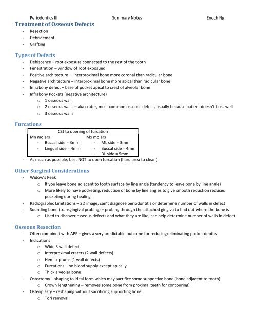
Treatment of Osseous Defects<br />
- Resection<br />
- Debridement<br />
- Grafting<br />
Types of Defects<br />
- Dehiscence – root exposure connected to the rest of the tooth<br />
- Fenestration – window of root exposued<br />
- Positive architecture – interproximal bone more coronal than radicular bone<br />
- Negative architecture – interproximal bone more apical than radicular bone<br />
- Infrabony defect – base of pocket apical to crest of alveolar bone<br />
- Infrabony Pockets (negative architecture)<br />
o 1 osseous wall<br />
o 2 osseous walls – aka crater, most common osseous defect, usually because patient doesn’t floss well<br />
o 3 osseous walls<br />
Furcations<br />
CEJ to opening of furcation<br />
Mn molars<br />
Mx molars<br />
- Buccal side = 3mm - ML side = 3mm<br />
- Lingual side = 4mm - Buccal side = 4mm<br />
- DL side = 5mm<br />
- As much as possible, best NOT to open furcation (hard area to clean)<br />
Other Surgical Considerations<br />
- Widow’s Peak<br />
o If you leave bone adjacent to tooth surface by line angle (tendency to leave bone by line angle)<br />
o More likely to have pocketing, reduction of bone by line angles to give smooth reduction reduces<br />
pocketing during healing<br />
- Radiographic Limitations – 2D image, can’t diagnose periodontitis or determine number of walls in defect<br />
- Sounding bone (transgingival probing) – probing through the attached gingiva to find out where the bone is<br />
o Used to discover osseous defects and what they are like, can help determine number of walls in defect<br />
Osseous Resection<br />
- Often combined with APF – gives a very predictable outcome for reducing/eliminating pocket depths<br />
- Indications<br />
o Wide 3 wall defects<br />
o Interproximal craters (2 wall defects)<br />
o Hemiseptums (1 wall defects)<br />
o Furcations – no blood supply except apically<br />
o Thick alveolar bone<br />
- Ostectomy – shaping to ideal form which may sacrifice some supportive bone (bone adjacent to tooth)<br />
o Crown lengthening – removes some bone from proximal teeth for contouring)<br />
- Osteoplasty – reshaping without sacrificing supporting bone<br />
o Tori removal