Perio_III
Perio_III
Perio_III

You also want an ePaper? Increase the reach of your titles
YUMPU automatically turns print PDFs into web optimized ePapers that Google loves.
<strong>Perio</strong>dontics <strong>III</strong> Summary Notes Enoch Ng<br />
Gingival Surgical Procedures<br />
Limited to gingiva, does not involve underlying osseous structures<br />
- Gingival curettage – removal of gingival wall of perio pocket to separate out diseased soft tissue<br />
• Aka = excisional new attachment procedure, ultrasonic curettage, caustic drugs<br />
o Inadvertent curettage – happens with SRP<br />
o No clinical value (2002) – healing is by long junctional epithelium, no new attachment is gained<br />
- Gingivectomy<br />
o Indications<br />
• Elimination of suprabony pockets in firm, fibrous tissue (SRP usually clears edematous pockets)<br />
• Elimination of gingival enlargements<br />
• Elimination of suprabony abscesses<br />
o<br />
o<br />
o<br />
o<br />
Before they become infrabony ones<br />
• Access for restorative dentistry<br />
<br />
<br />
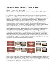
Biologic width important in esthetic areas<br />
Body wants around 2mm between bone and gingiva (beware of gingival rebound)<br />
Biologic width = 2.04mm, supra-alveolar tissues (dentogingival junction) = 2.73mm<br />
• Esthetics<br />
<br />
<br />
Ideal width = CI 25% wider than laterals, 10% wider than canines<br />
Ideal height = CI and canine 20% longer than laterals<br />
o Ratio = 1.2/1.0<br />
Surgical stents – want to know if the stent is for what tooth structure is showing, or<br />
where the crown margin is going to be placed (margin can be slightly in pocket)<br />
Want to preserve some keratinized tissue, do not want to remove all of it<br />
• Keratinized gingiva = pocket depth + attached gingiva (mucogingival junction)<br />
Measure pocket depth, mark it to cause it to bleed. Incision is apical to bleeding point, 45 o bevel to root<br />
Healing<br />
• Initially – acute PMN infiltrate and some necrosis, formation of initial protective clot<br />
<br />
<br />
<br />
<br />
12-24h – epithelial cells at margin migrate into granulation tissue and beneath the<br />
necrotic tissue<br />
24h – increased CT and angioblasts below surface layer<br />
4-16 days – vasodilation and vascularity start to decrease until normal<br />
o Epithelium grows at 0.5mm/day<br />
5-14days – surface epthelialization complete, keratinization incomplete<br />
7 weeks – complete repair of CT<br />
Limiting Factors<br />
• Amount of keratinized gingiva<br />
• Esthetics and esthetic maintenance<br />
• Access to osseous defects for correction<br />
• Less post-op pain if procedure allows for primary closure<br />
• Gingiva may rebound without racial pigmentation – risk for patients with racial pigmentation