Read this Issue. - SASSiT
Read this Issue. - SASSiT
Read this Issue. - SASSiT

Create successful ePaper yourself
Turn your PDF publications into a flip-book with our unique Google optimized e-Paper software.
<strong>SASSiT</strong> <br />
The official<br />
e-journal of<br />
Upper GIT <br />
APRIL 2012<br />
The <br />
NEEDLEHOLDER
CONTENTS <br />
Contributors <br />
“The real gist about GISTs” <br />
Hereditary spherocytosis <br />
Mucinous cystadenoma <br />
Palliative stents for GOO <br />
Dr Bheki Dube <br />
Dr Mark Hampton <br />
Dr Faizel Kimmie <br />
Dr Galya Chinnery <br />
Dr David Thomson <br />
Dr Stefan Hofmeyr <br />
THANK YOU for your time and <br />
outstanding work! <br />
Must read HPB <br />
articles <br />
Crossword <br />
This issue edited and <br />
compiled by: <br />
Dr S Rayamajhi <br />
Next issue:LGIT <br />
Historical facts <br />
The NEEDLEHOLDER <br />
welcomes any <br />
concern or comments about <strong>this</strong> <br />
issue. Every effort has been made <br />
to ensure original work and <br />
accurate referencing. <br />
Special THANK YOU to Dr. <br />
FERHANA GOOL for arranging <br />
a sponsor for the crossword <br />
competition prize!! <br />
Interested in writing a <br />
review or case study? <br />
CONTACT US: <br />
Shreya.r@hotmail.com
SURGICAL CALENDAR<br />
2012 <br />
15 January Closing date for exam entry<br />
19 - 20 March FCS (SA) Intermediate and Final papers<br />
27 - 28 March FCS (SA) Primary papers Country wide<br />
21 - 23 May FCS(SA) intermediate and Final orals<br />
15 June Closing date for exam entry CMSA<br />
25 - 27 June 23rd Biennial Surgical Symposium<br />
10 - 13 July SRS / Registrar Symposium<br />
9 - 11 August 50th SAGES / SASES<br />
21 - 23 August FCS (SA) Intermediate and Final papers<br />
28 - 30 August FCS (SA) Primary papers<br />
5 - 6 October Controversies in Surgery<br />
15 - 17 October FCS(SA) intermediate and Final orals<br />
CMSA<br />
Country wide<br />
Country wide<br />
Durban<br />
CMSA<br />
WITS<br />
Stellenbosch<br />
Durban<br />
Country wide<br />
Country wide<br />
Pretoria<br />
Gauteng<br />
Colorectal Symposium
“The real gist about GISTS” <br />
What a Surgeon Should Know About Gastrointestinal <br />
Stromal Tumors <br />
Dr Bheki Dube, Registrar, General Surgery <br />
UCT/WSU <br />
A gastrointestinal stromal tumor (GIST) is a<br />
rare mesenchymal neoplasia of the<br />
gastrointestinal tract. GISTs were first<br />
described as a separate entity from a collection<br />
of non-epithelial malignancies of the GI tract<br />
in the 1980s and 1990s based on pathologic<br />
and clinical behavior. The discovery of<br />
activating tyrosine-kinase (KIT) mutations as a<br />
near-uniform occurrence in these tumors<br />
greatly influenced the classification and<br />
therapeutic management of these tumors.<br />
Registrar <br />
REVIEW <br />
Epidemiology<br />
Gastrointestinal stromal tumors (GISTs) are<br />
the most frequently encountered mesenchymal<br />
tumor of the gastrointestinal tract. They<br />
represent about 5% of all sarcomas and 80% of<br />
all mesenchymal neoplasms of the<br />
gastrointestinal tract.<br />
According to a Swedish population-based<br />
study, the annual incidence of GISTs is<br />
estimated to be 1.5 per 100 000 and<br />
prevalence 12.9 per 100 000. As many as 5000<br />
to 6000 new cases are diagnosed each year in<br />
the United States. The median age of onset is<br />
about 60 with no clear-cut gender predilection.
The most commonly encountered GIST is<br />
the sporadic form. Familial GISTs occur<br />
as a result of germline mutations in either<br />
the KIT or platelet- derived growth factor<br />
receptor-α (PDGFR-α) proto-oncogenes.<br />
GIST can also occur in patients with<br />
neurofibromatosis type 1 (NF-1) and in<br />
young women as part of a syndrome that<br />
includes paragangliomas, pulmonary<br />
chondromas, and gastric GISTs (Carney<br />
triad).<br />
Pathogenesis and Molecular Basis<br />
of GISTs<br />
GISTs are thought to derive from the<br />
interstitial cells of Cajal; innervated<br />
cells associated with Auerbach’s plexus.<br />
Their behavior ranges from benign to<br />
malignant, and there are several histologic<br />
subtypes, including spindle cell (70%),<br />
epithelioid (20%), and pleomorphic<br />
types.<br />
Ninety- five percent of GISTs have gainof-<br />
function KIT mutations or PDGFR-α<br />
mutations. KIT encodes for a<br />
transmembrane receptor glycoprotein with<br />
tyrosine kinase function and normally<br />
participates in cell growth and survival.<br />
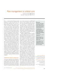
Major morphologic variants of GISTs. (A) Spindle cell and (B) epithelioid.<br />
The insets show diffuse and dot-like KIT immunoreactivity.
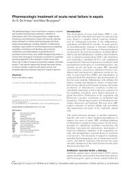
Most KIT mutations cause ligand-independent activation of the receptor tyrosine kinase<br />
function. Identifying KIT and PDGFR-α mutations is useful for both the diagnosis and<br />
treatment of GISTs.<br />
KIT mutations occur most frequently in exon 11 (70%), the juxtamembrane domain of the<br />
KIT protein. Mutations can also occur in exon 9 (10%), the extracellular domain of the KIT<br />
protein. Approximately 5% of GISTs do not express the KIT protein. In the 3% of GISTs<br />
with PDGFRα mutations, the mutation generally is present in exon 18 (80%) or exon 12.<br />
PDGFRα mutations are more common in gastric GISTs. Those that do not have identifiable<br />
mutations are considered wild-type, and their pathogenesis is unclear.<br />
The most common immunohistochemical pattern for GISTs is diffuse reactivity<br />
for KIT (CD117) and CD34. SMA can be reactive, but desmin, S-100 protein<br />
and pan-cytokeratin are usually uniformly negative. Percentages of<br />
immunoreactivity for each of these markers are listed above.<br />
Clinical presentation<br />
GISTs can cause a variety of symptoms ranging from vague abdominal pain to peritonitis<br />
as a result of tumor rupture and intraperitoneal bleeding.<br />
Other modes of presentation include abdominal fullness, early satiety, weakness, and fatigue<br />
secondary to anaemia from occult gastrointestinal bleeding. Bowel obstruction is rare.<br />
Small GISTs (
(60%), followed by the small bowel (30%), rectum (5%), and esophagus (5%). Up to 50% of<br />
patients present with metastatic disease at the time of diagnosis, with the liver and<br />
peritoneum being the two most common sites of extraintestinal spread. Occasionally patients<br />
present with primary GISTs of the omentum, mesentery, or pancreas. ExGISTS are isolated<br />
neoplasms that occur in the retroperitoneum.<br />
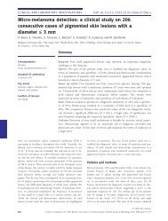
Diagnosis<br />
Because of the wide range of symptoms and its rarity, the diagnosis of GISTs requires a high<br />
index of suspicion. Imaging has become more common in the evaluation of patients with<br />
abdominal symptoms and is often the first modality of assessment. Contrast-enhanced<br />
computed tomography (CT) scans and Magnetic resonance imaging (MRI) are more<br />
frequently able to suggest the diagnosis of GIST. The CT features of GISTs can vary<br />
depending on the size, location, and aggressiveness of tumor. As in the case of other<br />
hypervascular masses, GISTs are visualized as enhancing solid masses and tumor vessels are<br />
often noted on enhanced CT images. Small GISTs typically appear as well defined softtissue,<br />
relatively low-density masses that appear relatively homogeneous on enhanced CT<br />
images. When the masses are large (usually >10 cm), they are often heterogeneous because<br />
of necrosis, hemorrhage, and myxoid degeneration. The classic CT appearance of a GIST is a<br />
well-defined tumor with a heterogeneous rim of soft tissue. On MRI, GISTs are generally<br />
well defined; the solid portions of the masses are typically of low- to intermediate-signal<br />
intensity on T1-weighted images and high signal intensity on T2- weighted images. <br />
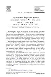
(A) CT image shows the cephalad extent of an extremely large gastric GIST.<br />
(B) CT image shows the heterogeneous nature of the GIST and its mass-producing effect,<br />
as it is pushing the stomach anteriorly, pancreas (and splenic vein) posteriorly ,and left<br />
lateral segment of the liver laterally.<br />
Management of Gastrointestinal Stromal Tumors; Matthew T. Hueman, Richard D.<br />
Schulick
Once the diagnosis of GIST has been suggested on axial imaging, endoscopy can be useful in<br />
further evaluation of a gastric or colorectal submucosal mass. Endoscopic ultrasound scan<br />
can delineate the full depth of the tumor, although it is not able to predict tumor behavior.<br />
Preoperative biopsy generally is not indicated for several reasons. First, GISTs are fragile<br />
and can theoretically rupture and spread tumor cells when biopsied. Second, given their<br />
hypervascular nature, a biopsy can cause intra-tumor hemorrhage. Perhaps most importantly,<br />
pathologists often cannot diagnose GISTs from fine-needle aspirates, especially when the<br />
area sampled is necrotic. Endoscopic biopsy is useful only to confirm the diagnosis, exclude<br />
the diagnosis of lymphoma, which can have a similar radiologic appearance and allow for<br />
neo-adjuvant therapy for a marginally resectable tumor.<br />
Adjuvant Therapy<br />
The approval of imatinib mesylate as (neo) adjuvant therapy has revolutionized the<br />
treatment of GISTs. As a specific tyrosine kinase inhibitor (TKI), imatinib has<br />
shown efficacy in patients with both KIT and PDGFRα mutations. Imatinib is dosed orally<br />
once or twice a day and is generally well tolerated, with rash, diarrhea, and abdominal pain<br />
being the most commonly reported side effects.<br />
Primary resectable disease<br />
Surgery traditionally has been the cornerstone of treatment for<br />
resectable GISTs. In the post-imatinib era, surgery shares a role in a multidisciplinary<br />
treatment plan. In primary disease, complete surgical resection provides the chance of cure.<br />
There are several important principles that guide surgical resection. Because GIST can be<br />
fragile with extensive necrosis or hemorrhage, meticulous dissection is vital to avoid<br />
tumor rupture during the procedure, which can increase the risk of intraperitoneal recurrence.<br />
GISTs are normally surrounded by a pseudo-capsule, that should remain intact after<br />
resection. Given the normal exophytic growth pattern of GISTs within the gastrointestinal<br />
tract, wedge or segmental resections often are possible. GISTs adhere to surrounding<br />
structures; so additional organ resections may be required for complete resection. Margins of<br />
resection should be microscopically negative. To achieve <strong>this</strong>, a 1-cm gross margin is<br />
generally sufficient. Laparoscopy is a useful modality to resect small gastric GISTs,<br />
especially because lymphadenectomy is rarely required. <br />
Primary unresectable GISTs<br />
When a primary GIST appears to be unresectable or borderline, imatinib is the<br />
treatment of choice. A CT scan 1 month after initiation of imatinib is useful to judge<br />
tumor response and thus potential resectability. CT scans are used to judge the effectiveness<br />
of the imatinib, and surgical resection generally is performed within 6 to 9 months. An
additional benefit may be decreased blood loss, given the hypervascularity of large GIST.<br />
One of the pitfalls of neo-adjuvant therapy is that a needle biopsy is necessary to<br />
begin treatment. Given that 15% of patients have primary resistance to imatinib, there is also<br />
a risk that the tumor will become unresectable because of the delay in surgical intervention.<br />
Recurrent and metastatic GISTs<br />
The first line of treatment in patients with recurrent and metastatic disease is imatinib.<br />
Lifelong treatment with imatinib is recommended in patients with imatinibresponsive<br />
GISTs, given that it decreases the likelihood of disease progression.<br />
Approximately 45% of patients with metastatic GISTs have a partial response to imatinib,<br />
whereas 30% maintain stable disease. The success of imatinib is evident in that median<br />
Survival was only 15 months after resection of recurrent GISTs in the pre-imatinib era,<br />
whereas the median overall survival with metastatic disease is now 5 years. The normal<br />
starting dose of imatinib is 400 mg/day. Although there is no difference in overall survival<br />
between 400 and 800 mg/day doses, one study does suggest there may be longer progressionfree<br />
survival with twice-daily imatinib. Given that imatinib is not curative, there often is a<br />
role for surgery in addition to tyrosine kinase inhibition. Combining the two modalities may<br />
delay imatinib resistance and potentially be curative.<br />
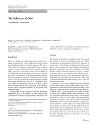
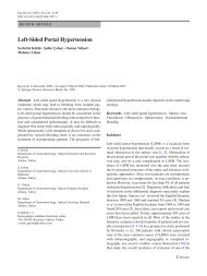
Disease-‐specific survival after complete resection of primary GIST
Imatinib resistant GISTs<br />
In a patient who has a GIST that progresses while taking imatinib at a dose of 400 mg/day,<br />
the dose can be escalated to 800 mg/day. Five percent of patients who progress at 400<br />
mg/day will respond to the elevated dose of imatinib and achieve at least partial remission.<br />
Additionally, 30% of patients will have stable disease. In patients who have imatinibresistant<br />
GISTs or who do not tolerate imatinib, sunitinib is the next line of<br />
therapy. It is a multitargeted tyrosine kinase inhibitor that has both anti-tumor and antiangiogenic<br />
abilities.<br />
Several other treatment modalities are available, including radiation, radio-frequency<br />
ablation, and some novel agents. GISTs are considered radiation-resistant<br />
tumors and thus the only role for radiotherapy is in palliation.<br />
Radiofrequency ablation (RFA) is a technique that is being used more frequently with<br />
metastatic GISTs in the liver.<br />
Vatalanib has a similar mechanism of action to sunitinib and has been shown to have an<br />
effect on imatinib-resistant GISTs. Dasatinib is another tyrosine kinase inhibitor that may<br />
have utility in a GIST with an exon 17 mutation and imatinib resistance. It also inhibits the<br />
src kinase, which is a downstream kinase that can be activated after KIT activation.<br />
Everolimus is another agent currently being studied. It inhibits a mammalian target of the<br />
rapamycin pathway that is activated by the KIT receptor tyrosine kinase.
Algorithm in The Management of GISTs<br />
Gold JS,DeMatteo RP.Combined surgical and molecular therapy: the gastrointestinal<br />
stromal tumor model. Ann Surg 2006;244:176
Summary<br />
A GIST is a rare tumor with a complex natural history. Surgical management is the<br />
mainstay of therapy, with margin-negative resection being the desired surgical outcome.<br />
Targeted therapy with tyrosine kinase inhibitors has revolutionized the care of these<br />
patients, providing improved outcomes for patients who have completely resected tumors<br />
and resulting in prolonged responses in patients who have advanced disease. With ongoing<br />
advancements in the field, it is possible that targeted therapy may be selected in the future<br />
based on the specific mutation exhibited in each GIST and the resulting expected response<br />
rates.<br />
Surgical resection of primary, recurrent, and metastatic GISTs, in combination with tyrosine<br />
kinase inhibition is the standard of care for treating patients with GISTs. A multidisciplinary<br />
approach to diagnosis and treatment is essential for successful outcomes.<br />
References<br />
1. Richard Quek, Suzanne; George<br />
GastrointestinalStromal Tumor :A Clinical<br />
overview. Hematol Oncol Clin N Am 23<br />
(2009) 69–78.<br />
2. Neeta Somaiah, MD,Margaret von<br />
Mehren; New Therapeutic Approaches for<br />
Advanced GastrointestinalStromal<br />
Tumors:, Hematol Oncol Clin N Am 23<br />
(2009) 139–150<br />
3.Matthew T. Hueman, MD,Richard D.<br />
Schulick, MD Management of<br />
Gastrointestinal Stromal Tumors Surg Clin<br />
N Am 88 (2008) 599–614<br />
4. Bernadette Liegl, MD, Jason L.<br />
Hornick, MD, PhD, Alexander J.F. Lazar,<br />
MD, PhD; Contemporary Pathology of<br />
Gastrointestinal Stromal Tumors : Hematol<br />
Oncol Clin N Am 23 (2009) 49–68<br />
5. T. Peter Kingham, MD, Ronald<br />
P.DeMatteo, MD; Multidisciplinary<br />
Treatment of Gastrointestinal Stromal<br />
Tumors:Surg Clin N Am 89 (2009) 217–<br />
233<br />
6. Zubin M. Bamboat, MD, Ronald P.<br />
DeMatteo, MD; Updates on the<br />
Management of Gastrointestinal Stromal<br />
Tumors: Surg Oncol Clin N Am 21 (2012)<br />
301–316<br />
7. Piyaporn Boonsirikamchai, MD,<br />
Donald A. Podoloff, MD,Haesun Choi,<br />
MD; Imaging of Gastrointestinal Stromal<br />
Tumors and Assessment of Benefit from<br />
SystemicTherapy: Hematol Oncol Clin N<br />
Am 23 (2009) 35–48.<br />
8.Demetri GD, Baker LH, Benjamin RS,<br />
et al. Soft tissue sarcoma. J Natl Compr<br />
Canc Netw 2007;5(4):364–99.<br />
9.Steigen SE, Eide TJ. Trends in incidence<br />
and survival of mesenchymal neoplasmof<br />
the digestive tract within a defined<br />
population of northern Norway. APMIS<br />
2006; 114(3):192–200.<br />
10.Tran T, Davila JA, El-Serag HB. The<br />
epidemiology of malignant gastrointestinal<br />
stromal tumors: an analysis of 1,458 cases<br />
from 1992 to 2000. Am J Gastroenterol<br />
2005;100(1):162–8.<br />
11.Prakash S, Sarran L, Socci N, et al.<br />
Gastrointestinal stromal tumors in children<br />
and young adults: a clinicopathologic,<br />
molecular, and genomic study of 15 cases<br />
and review of the literature. J Pediatr<br />
Hematol Oncol 2005;27(4):179–87.<br />
12.DeMatteo RP, Lewis JJ, Leung D, et al.<br />
Two hundred gastrointestinal stromal<br />
tumors: recurrence patterns and prognostic<br />
factors for survival. Ann Surg<br />
2000;231(1):51–8.
HEREDITARY SPHEROCYTOSIS<br />
Case Report<br />
Dr. MI Hampton, Registrar, General Surgery<br />
GSH / UCT<br />
A 29-year-old previously well Xhosa<br />
female was seen in casualty at GF Jooste<br />
Hospital. She presented with a brief<br />
history of fever and severe right upper<br />
quadrant pain associated with nausea and<br />
vomiting. She had become jaundiced and<br />
had started to feel short of breath.<br />
Her abdomen was tender in the right upper<br />
quadrant and a diagnosis of acute<br />
An abdominal ultrasound was<br />
requested <br />
Examination revealed her to be<br />
apyrexial, clinically anaemic and<br />
moderately jaundiced. She was not<br />
diaphoretic or tachycardic and appeared<br />
stable.<br />
Blood results:<br />
Hb 5.3 Total Bilirubin 72<br />
MCV 90 Conj Bilirubin 33<br />
MCH 34 ALP 28<br />
WCC 10.0 GGT 64<br />
Ultrasound showed multiple gall bladder<br />
stones with features of acute cholecystitis<br />
including gall bladder distension, wall<br />
thickening and pericholecystic fluid. The<br />
common bile duct was not distended.<br />
PLT 137 ALT 24<br />
AST 25
The patient was admitted and treated with<br />
intravenous antibiotics and a blood<br />
transfusion.<br />
At <strong>this</strong> point the patient’s jaundice was<br />
unexplained and the cause for the<br />
normocytic anaemia was also unclear.<br />
(HS) was made. The patient’s jaundice<br />
could then be explained as being the result<br />
of haemolysis, and the cholelithiasis could<br />
be explained as being the result of<br />
pigment stone formation in the gall<br />
bladder secondary to haemolysis.<br />
Over the next 3 days the patient became<br />
progressively more ill. She had persistent<br />
sepsis with an elevated white cell count,<br />
spiking temperature and progressive<br />
abdominal pain. She also developed<br />
symptomatic anaemia and required further<br />
blood transfusions.<br />
The patient became even more ill, which<br />
prompted abdominal imaging by means of<br />
a CT scan.<br />
The cause of the anaemia was further<br />
investigated with a peripheral smear<br />
which showed numerous spherocytes.<br />
A diagnosis of Hereditary spherocytosis<br />
This demonstrated massive splenomegaly<br />
in keeping with the diagnosis of HS, and<br />
features of gall bladder empyema with a<br />
gall bladder perforation.<br />
The patient was then taken for an<br />
emergency laparotomy.
Findings at<br />
laparotomy<br />
demonstrating a<br />
thickened pus-filled<br />
gall bladder.<br />
A subtotal<br />
cholecystectomy<br />
was performed and<br />
the patient did<br />
extremely well.<br />
Tip of spleen visible <br />
She has<br />
subsequently been<br />
referred to the<br />
Haematology Clinic<br />
at Groote Schuur<br />
Hospital where she<br />
is undergoing<br />
further<br />
investigation and<br />
management.<br />
Black pigment stones found in gall bladder
Discussion<br />
Red blood cells are flexible biconcave<br />
discs. At any one time there are 2 – 3 x<br />
10 13 red cells present within the<br />
circulation. Senescent red cells are<br />
phagocytosed within the<br />
reticuloendothelial system, which consists<br />
of the spleen, bone marrow and liver.<br />
Red cell membranes are highly<br />
sophisticated structures that allow<br />
conformability, asymmetry and<br />
flexibility of the cells.<br />
They consist of three layers: An outer<br />
glycocalyx layer containing carbohydrate<br />
chains, a middle phospholipid bilayer<br />
which also contains transmenbrane<br />
proteins and an inner cytoskeletal layer.<br />
The integrity of the proteins in the red cell<br />
membrane is essential for effective red<br />
cell functioning.<br />
Hereditary spherocytosis (HS) is an<br />
autosomal dominant condition<br />
more common in the Japanese<br />
and Northern European<br />
population. It results in red cell<br />
membrane defects caused by mutations in<br />
the genes coding for various red cell<br />
transmembrane proteins. 25% of cases<br />
arise due to spontaneous mutations.<br />
There are 5 subtypes, each characterized by a<br />
distinct genetic mutation. Mutation of the<br />
ANK1 gene (type 1 HS) located on<br />
chromosome 8p11.2 which codes for the<br />
protein Ankyrin, is the most common<br />
subtype.<br />
Other mutations result in defects of the<br />
proteins Band 3 and spectrin.<br />
In HS the red cells lose their biconcave<br />
shape and become spherical. This<br />
results in a massive decrease in total red cell<br />
surface area. The cells lose their<br />
conformability and are unable to pass<br />
through sinusoidal channels within the<br />
spleen (which are approximately half the<br />
diameter of a normal red cell) where they<br />
become trapped. They are then<br />
destroyed by the phagocytic<br />
splenic cells and extravascular<br />
haemolysis ensues.<br />
The clinical presentation includes anaemia<br />
and splenomegaly. Haemolytic crises may<br />
arise precipitated by events such as sepsis
and result in jaundice and unconjugated<br />
hyperbilirubinaemia.<br />
electrophoresis is needed to<br />
confirm it.<br />
To make a diagnosis of HS once a<br />
haemolytic anaemia has been recognized<br />
there are various laboratory tests which<br />
should be performed. These include a<br />
peripheral blood smear, which will<br />
demonstrate spherocytes, although<br />
these can also be present in pregnancy and<br />
after blood transfusions.<br />
Osmotic fragility testing exposes the<br />
spherocytic red cells to a hypo osmolar<br />
solution and compares the extent of cell<br />
lysis to a sample of normal red cells.<br />
Although <strong>this</strong> test is useful to suggest a<br />
diagnosis of HS, membrane protein<br />
The options for treatment are haematinic<br />
treatment with blood transfusions as<br />
necessary and splenectomy.<br />
Splenectomy is generally<br />
reserved for those patients with<br />
recurrent haemolytic crises and<br />
who are dependent of frequent<br />
blood transfusion.
Case Report:<br />
Pancreatic mucinous cystadenoma in a young female <br />
Faizel Kimmie, Registrar, General Surgery <br />
GSH/ UCT <br />
Introduction<br />
A young female was recently admitted to<br />
Groote Schuur hospital with a large<br />
cystic abdominal mass.<br />
Histological examination after complete<br />
excision confirmed a pancreatic mucinous<br />
cystadenoma.<br />
Case report<br />
A 29-year-old female presented with a 6-<br />
month history of a progressively<br />
enlarging left upper quadrant mass. This<br />
was associated with intermittent<br />
abdominal pain. There was no associated<br />
weight loss, no loss of appetite or change<br />
in bowel habit. Apart from having had an<br />
uneventful caesarean section 5 years ago,<br />
she had no known co-morbidities. There<br />
was also no history of alcohol abuse or<br />
previous episodes of acute pancreatitis.<br />
On clinical examination she was<br />
found to have a large, round, nontender<br />
left upper quadrant mass<br />
extending from below the left<br />
costal margin, +/- 15 x 15 cm in size.<br />
Abdominal CT scan revealed a septated
cystic mass with foci of calcification<br />
measuring 160 x 127 x 175 mm related<br />
to the tail of the pancreas.<br />
As <strong>this</strong> patient was young and otherwise<br />
healthy, the next step was surgical<br />
excision. A distal pancreatectomy<br />
and splenectomy was done via<br />
an extended left subcostal<br />
incision. The intra-operative finding<br />
was a well-demarcated mass in the left<br />
upper quadrant with the tail of the<br />
pancreas draped over from the medial<br />
side.<br />
Discussion<br />
White arrow indicates the spleen. Black<br />
arrow indicates the tail of pancreas.<br />
Histological examination confirmed a<br />
mucinous cystadenoma with ovarian<br />
stroma. It was multiloculated without any<br />
evidence of stromal invasion.<br />
Pancreatic cystic neoplasms<br />
(PCNs) are usually<br />
asymptomatic and present as an<br />
incidental finding on crosssectional<br />
imaging. Symptoms arise<br />
from compression on surrounding<br />
structures. These patients can then present<br />
with abdominal pain, jaundice or recurrent<br />
episodes of pancreatitis.<br />
It is important to exclude pancreatic<br />
pseudocysts, which are the most<br />
common cystic lesions of the pancreas. In<br />
these cases, clinical history would reveal<br />
previous documented attacks of acute<br />
pancreatitis or risk factors such as alcohol<br />
abuse, gallstones or pancreatic trauma.<br />
The CT scan findings would include a<br />
well-circumscribed cyst and other features<br />
of pancreatitis (gland atrophy, duct<br />
dilation, calcifications).
The most common PCNs include serous<br />
cystadenomas (SCA), mucinous cyst<br />
neoplasms (MCN) and intraductal<br />
papillary mucinous neoplasms (IPMN).<br />
Serous cystadenomas are lined by<br />
glycogen-rich cuboidal epithelium and<br />
have a very low malignant potential.<br />
MCNs differ from IPMNs by the<br />
histological finding of mesenchymal<br />
ovarian-like stroma in the former.<br />
A key feature of IPMN is pancreatic<br />
ductal communication, which MCNs lack.<br />
The important characteristic shared by<br />
IPMNs and MCNs, besides mucin<br />
production, is their definite malignant<br />
potential.<br />
those who are fit for surgery.<br />
MCNs are mostly found in the pancreatic tail<br />
and distal pancreatectomy is usually<br />
sufficient. IPMNs are more frequently found<br />
in the head of the pancreas and require<br />
pancreatoduodenectomy. SCAs have an even<br />
distribution and require resection with the<br />
appropriate segment of pancreas.<br />
The prognosis for patients with MCNs,<br />
without transmural invasion, approaches<br />
100%. No post-operative surveillance is<br />
necessary where there is no transmural<br />
invasion. Patients do, however, require<br />
surveillance after resection of IPMN due<br />
high recurrence rates.<br />
SCAs occur more commonly in women in<br />
the sixth decade of their lives. MCNs are<br />
significantly more common in females<br />
and tend to occur a decade earlier than<br />
SCAs. IPMN tends to occur a bit later<br />
with a mean age of 65 years.<br />
CT is an excellent investigation for cystic<br />
pancreatic lesions. A central scar on<br />
CT is highly suggestive of SCA. MCNs<br />
can be unilocular or multilocular<br />
and may contain peripheral calcifications<br />
on CT. MRI can provide more detail on<br />
morphological appearance. Endoscopic<br />
ultrasound (EUS) is a useful adjunct and<br />
can aid in aspiration and cytology. A<br />
raised fluid amylase indicates pancreatic<br />
ductal communication and CEA is raised<br />
in mucinous lesions. In patients who are<br />
fit for surgery there is no need for preoperative<br />
tissue diagnosis.<br />
Conservative management is considered<br />
in asymptomatic patients who are poor<br />
surgical candidates.<br />
Surgical resection is indicated in<br />
symptomatic patients, where definitive<br />
radiological diagnosis is unclear, and<br />
References<br />
1. Brugge, W.R. et al. 2004. Cystic<br />
Neoplasms of the Pancreas. N Engl J<br />
Med, 351(12):1218-26, September 16<br />
2. Tran Cao, H.S. et al. 2010. Cystic<br />
Neoplasms of the Pancreas. Surg<br />
Oncol Clin N Am, 19: 267-295<br />
3. Verbesey, J.E. et al. 2010. Pancreatic<br />
Cystic Neoplasms. Surg Clin N Am,<br />
90: 411-425<br />
4. Degen, L. Et al. 2008. Cystic and solid<br />
lesions of the pancreas. Best Practice<br />
& Research Clinical<br />
Gastroenterology, 22(1): 91-103
Endoscopic Stenting for Malignant Gastric Outlet<br />
Obstruction <br />
Dr. Galya Chinnery, Consultant, HPB Surgery <br />
GSH / UCT <br />
Summary of the review article “Endoscopic stenting versus<br />
gastrojejunostomy for palliation of malignant gastric outlet<br />
obstruction. Zheng B, Wang X, Ma B, Tian J, Jiang L, Yang K. Dig<br />
Endosc 2012 Mar;24(2):71-8”<br />
Brief overview of the technique of duodenal stenting<br />
Zheng et al have undertaken a systematic<br />
review of the literature analyzing clinical<br />
trials on malignant gastric outlet obstruction<br />
(GOO). Their findings confirm the clinical<br />
impression of patient outcome during the<br />
management of malignant GOO with<br />
endoscopic stents.<br />
While gastrojejunostomy (GJ) has always<br />
been the traditional palliative treatment of<br />
choice, endoscopic stenting (ES) has in<br />
recent years gained in popularity. Suggested<br />
benefits to stenting include a procedure,<br />
which is less invasive and initially<br />
more cost-effective with earlier<br />
relief of symptoms and<br />
resumption of oral intake. Stent<br />
disadvantages are technical failure<br />
during insertion and late stent<br />
migration or tumour ingrowth. The<br />
authors reviewed six RCT studies in their<br />
meta-analysis.<br />
The following aspects were included in<br />
their review:<br />
1. Technical and clinical<br />
success: Meta-analysis showed<br />
significantly higher technical<br />
success in the GJ group (99%)<br />
when compared to the ES group<br />
(82%). No significant difference<br />
was found regarding clinical<br />
success.<br />
2. Time to oral intake: The mean
time to commencing oral intake<br />
was 3.6 days earlier for ES than for<br />
the GJ group.<br />
3. Length of survival: Mean<br />
survival after stenting was 78 (56 –<br />
94) days and 81 (72 – 99) days after<br />
GJ. The majority of the trials<br />
reviewed by the authors indicated<br />
no statistical significance between<br />
the two groups.<br />
4. Quality of Life (QoL):<br />
Different questionnaires were used<br />
to assess QoL and therefor the<br />
results could not be combined to<br />
carry out the meta-analysis.<br />
However stenting appeared to be<br />
associated with significant<br />
improvements in dysphagia, eating<br />
restrictions, dry mouth and reflux,<br />
whilst GJ patients commented on<br />
significant improvements mainly<br />
with dysphagia and eating<br />
restrictions.<br />
reported to be 4.2- 28.6% whilst the<br />
GJ group mortality ranged from 21.4<br />
– 26.7%. This difference was not<br />
significant.<br />
7. Hospital stay: ES patients have a<br />
significantly shorter hospital stay.<br />
8. Medical costs: Costs were<br />
lower in the stented patient<br />
group.<br />
Conclusions:<br />
Zheng et al concluded that ES is a safe and<br />
effective, minimally invasive and costeffective<br />
option for the palliation of<br />
malignant GOO and should be considered<br />
the gold standard. A larger well-designed<br />
RCT was recommended by the authors to<br />
validate the findings of their meta-analysis.<br />
5. Complications: The morbidity<br />
for stenting ranged between 0 –<br />
40%; the GJ group had a morbidity<br />
range of 22.2 – 57.1%. The<br />
differences between the two groups<br />
for major complications was not<br />
significant, however stented<br />
patients were significantly less<br />
likely to develop minor<br />
complications than those<br />
undergoing surgery.<br />
6. Mortality: The ES mortality was<br />
Summary:<br />
ES GJ<br />
Higher technical success +<br />
Rapid oral intake +<br />
Shorter hospital stay +<br />
Lower minor complications +<br />
Improved QoL +<br />
Lower cost +<br />
Clinical success = =
Technique of duodenal stenting<br />
Figure 1<br />
Endoscopic identification of possible residual lumen<br />
Figure 2<br />
Guidewire passed through a co-axial and across the stricture under screening into the<br />
distal lumen
Figure 3<br />
Co-axial passed over the guidewire into distal lumen. Guidewire removed, contrast<br />
injected down co-axial to confirm position beyond stricture and distal patency<br />
Figure 4<br />
Guidewire placed back down the co-axial into the distal lumen. Co-axial removed leaving<br />
guidewire behind. Stent delivery system passed over guidewire across the stricture and<br />
deployment started under screening.
Figure 5<br />
Once fully deployed, guidewire and stent introducer removed. Care must be taken to have<br />
an adequate length of proximal stent within the antrum. Contrast may be injected through<br />
the stent on completion to confirm position. Full expansion on the stent will occur over 24<br />
– 48 hours.
MUST READ HPB <br />
ARTICLES <br />
Dr Stefan Hofmeyr <br />
General Surgery <br />
Consultant, HPB Fellow <br />
GSH
Obstructive jaundice due to pancreatic<br />
carcinoma is a common<br />
presentation/referral to Tertiary Hospitals.<br />
Data from Western centers suggest that<br />
only 20% of patients have resectable<br />
disease at first presentation. Due to<br />
late presentation and delayed referral, the<br />
situation in South Africa is similar, with<br />
most patients deemed unresectable due to<br />
locally advanced or metastatic disease.<br />
The first paper, on the pre-treatment<br />
assessment of these patients, published in<br />
the Annals of Surgical Oncology in 2009,<br />
is a consensus statement on accurate<br />
radiological staging of pancreatic cancer<br />
and the criteria used to determine<br />
resectability. Pretreatment staging creates,<br />
in the absence of distant metastases, three<br />
distinct patient groups. The first group<br />
have locally advanced disease with no<br />
possibilty of R0 resection and require<br />
palliation. Experience with neoadjuvant<br />
chemotherapy and/or radiation in <strong>this</strong><br />
setting, with the aim of converting patients<br />
to resectable status, has been a failure.<br />
well as the management and outcome of<br />
these patients is addressed in the second<br />
paper, published in the Surgical Clinics of<br />
North America in 2010. The paper<br />
describes the borderline resectability<br />
criteria as described by Katz et al and<br />
discusses the rationale for neoadjuvant<br />
therapy in <strong>this</strong> specific subset of patients.<br />
As mean survival after R0 pancreatic<br />
resection and adjuvant therapy has<br />
plateaued at 24 months at best,<br />
neoadjuvant therapy, with many<br />
theoretical benefits, has been investigated<br />
as a treatment strategy in patients with<br />
resectable/borderline resectable disease.<br />
Data is however limited to single center<br />
reports. The third paper, also published in<br />
the Surgical Clinics of North America in<br />
2010, reviews the best available data on<br />
neoadjuvant therapy and upcoming trials<br />
to look out for.<br />
The second group of patients have<br />
resectable disease with little concern<br />
regarding vascular involvement and in<br />
practice 80% of these are resected<br />
successfully. The third group consists of<br />
patients with borderline resectable disease.<br />
The criteria for borderline resectability, as
British Journal of Surgery 2011; 98, 1446-‐1454 <br />
Annals of surgery; Vol 254, Number 6, December 2011 <br />
The timing of cholecystectomy after<br />
various gallstone related biliary conditions<br />
has been the focus of many publications.<br />
With few exceptions, the message is: the<br />
sooner the better. Adherence to <strong>this</strong> policy<br />
is unfortunately inconsistent, the major<br />
stumbling block in the South African<br />
context being the pressure on emergency<br />
and elective lists.<br />
The paper by the Dutch Pancreatic Study<br />
Group on the timing of cholecystectomy<br />
after an episode of mild acute biliary<br />
pancreatitis, published in the British<br />
Journal of Surgery in 2011, reports the<br />
adverse outcome after omission of early<br />
cholecystectomy. While most society<br />
guidelines, though variable, advise early<br />
cholecystectomy, <strong>this</strong> paper reports poor<br />
adherence to these guidelines. In the<br />
interval between recovery from mild acute<br />
biliary pancreatitis and delayed<br />
cholecystectomy (mean = 6 weeks), 13,7%<br />
and 10% were readmitted for biliary events<br />
and recurrent pancreatitis respectively. The<br />
protective effect of endoscopic<br />
sphincterotomy is also mentioned.<br />
Laparoscopic cholecystectomy was<br />
achieved with a conversion rate of 8%<br />
and without bile duct injury.<br />
Early (same admission), rather than<br />
delayed (6 weeks) laparoscopic<br />
cholecystectomy for acute calculous<br />
cholecystitis is a well-established standard<br />
of care, although not always possible due<br />
to pressure on emergency lists. The paper<br />
by the Swiss Association of Laparoscopic<br />
and Thoracoscopic Surgery, published in<br />
the Annals of Surgery in 2011, is one of<br />
the largest reports (4113 patients)<br />
evaluating the outcome of early<br />
laparoscopic cholecystectomy for acute<br />
cholecystitis, illustrating the deleterious<br />
effect of delaying surgery past 48 hrs after<br />
admission. Significant increases in<br />
conversion rates, complications and<br />
hospital stay are reported for every day<br />
surgery is delayed after 48 hrs.
British Journal of surgery 1998, 85, 904-‐906 <br />
The difficult cholecystectomy will<br />
regularly confront a practicing General<br />
Surgeon. Dissection of the hepatocystic<br />
triangle is often hazardous due to<br />
inflammation or chronic fibrosis; and if<br />
persisted in, may lead to serious injury of<br />
structures within the hepatoduodenal<br />
ligament and/or porta hepatis. This classic<br />
paper, reported in the BJS in 1998,<br />
illustrates in text and graphics, the<br />
technique of subtotal laparoscopic<br />
cholecystectomy as a means to avoid<br />
disastrous complications.<br />
www.thelancet.com Vol 379 March 31, 2012 <br />
This is an excellent review of the staging<br />
and management of Hepatocellular<br />
carcinoma by the Barcelona Clinic Liver<br />
Cancer group. Options such as resection,<br />
transplantation, ablation, transarterial<br />
therapy and Sorafenib are reviewed.
CROSSWORD COMPETITION <br />
Dr. David Thomson, General Surgery Consultant <br />
Transplant and Trauma, GSH <br />
T <br />
C
CLUES <br />
Competition entries close May 31 st 2012 <br />
Send your solution to Shreya.r@hotmail.com <br />
Solution format: scan and send to email address. <br />
Prize: Gift voucher: R500 Woolworths
HISTORICAL FACTS<br />
DID YOU KNOW?<br />
Dr S Rayamajhi, Registrar, <br />
General Surgery, UCT <br />
Erich MÜhe, a German surgeon was the first person to perform<br />
laparoscopic cholecystectomy (LC) in 1985, a hundred years<br />
after the first open cholecystectomy.<br />
Erich Muhe <br />
The German surgical society initially rejected his work. In 1992 he<br />
received their highest award. In 1999 SAGES recognized him as the<br />
first surgeon to perform LC.<br />
MÜhe’s fascination with laparoscopy was inspired by Semm, a<br />
gynecologist who reported a laparoscopic appendectomy performed<br />
with a suture technique.<br />
Prof. MÜhe learned laparoscopy from another gynecologist, Willi-<br />
Rinehard Braumann.<br />
His description of LC as recorded in Endoscopy is as follows:<br />
A Galloscope <br />
“The first endoscope constructed and used by ourselves (‘Galloscope’) had side-viewing<br />
optics, and an instrumentation channel with valves, a light conductor and a duct for the<br />
establishment of continuous pneumoperitoneum by the Veress needle technique; the<br />
endoscope was introduced through the umbilicus into the peritoneal cavity. For the insertion,<br />
we used a sharp mandrin within a trocar sleeve. After removal of the mandrin, a trap valve<br />
was ejected from the inner wall of the tube to seal off escaping CO 2 . When the gallbladder<br />
was removed under optical control through the endoscope, the top of the endoscope had to be<br />
taken off. However, the gallbladder could also be removed through the trocar sleeve. When<br />
<strong>this</strong> access route from the umbilicus or suprapubic abdominal wall to the gallbladder was<br />
used, a pneumoperitoneum was indispensable. Therefore, after the first six operations, we<br />
changed the method, and the remaining 88 patients were operated on using a simplified<br />
approach, namely laparoscopic cholecystectomy without pneumoperitoneum and without<br />
optical guidance. Using an access channel at the costal margin <strong>this</strong> served as a firm bone roof<br />
above the gallbladder, and neither a pneumoperitoneum nor optical guidance was necessary.
NEXT ISSUE <br />
LOWER GIT <br />
COMING SOON