Thermodesulfatator indicus gen. nov., sp. nov., a novel thermophilic ...
Thermodesulfatator indicus gen. nov., sp. nov., a novel thermophilic ...
Thermodesulfatator indicus gen. nov., sp. nov., a novel thermophilic ...

You also want an ePaper? Increase the reach of your titles
YUMPU automatically turns print PDFs into web optimized ePapers that Google loves.
H. Moussard and others<br />
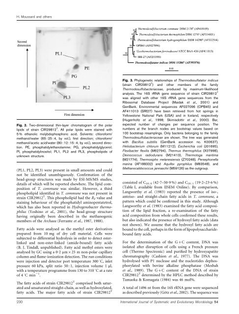
Fig. 2. Two-dimensional thin-layer chromatogram of the polar<br />
lipids of strain CIR29812 T . All polar lipids were stained with<br />
5 % ethanolic molybdopho<strong>sp</strong>horic acid. Solvents: chloroform/<br />
methanol/water (65 : 25 : 4, by vol.), first direction; chloroform/<br />
methanol/acetic acid/water (80 : 12 : 15 : 4, by vol.), second direction.<br />
PE, pho<strong>sp</strong>hatidylethanolamine; PG, pho<strong>sp</strong>hatidylglycerol;<br />
PI, pho<strong>sp</strong>hatidylinositol; PL1, PL2 and PL3, pho<strong>sp</strong>holipids of<br />
unknown structure.<br />
(PL1, PL2, PL3) were present in small amounts and could<br />
not be identified unambiguously. Confirmation of the<br />
head-group structures was made by ESI-MS/MS studies,<br />
details of which will be reported elsewhere. The lipid composition<br />
of T. commune was similar. However, a third<br />
pho<strong>sp</strong>holipid identified in T. commune was not present in<br />
strain CIR29812 T . This pho<strong>sp</strong>holipid had the R F value and<br />
staining behaviour of the pho<strong>sp</strong>hatidyl aminopentatetrol,<br />
which has also been reported in Hydro<strong>gen</strong>obacter thermophilus<br />
(Yoshino et al., 2001), the head-group structure<br />
having originally been described in the methano<strong>gen</strong>ic<br />
members of the Archaea (Ferrante et al., 1987, 1988).<br />
Fig. 3. Phylo<strong>gen</strong>etic relationships of <strong>Thermodesulfatator</strong> <strong>indicus</strong><br />
(strain CIR29812 T ) and other members of the family<br />
Thermodesulfobacteriaceae, produced by maximum-likelihood<br />
analysis. The 16S rRNA <strong>gen</strong>e sequence of strain CIR29812 T<br />
was aligned with other 16S rRNA <strong>gen</strong>e sequences from the<br />
Ribosomal Database Project (Maidak et al., 2001) and<br />
GenBank. Environmental sequences AF027096 (OPB45) and<br />
AF411013 (SRI27) have been retrieved from hot <strong>sp</strong>rings in<br />
Yellowstone National Park (USA) and in Iceland, re<strong>sp</strong>ectively<br />
(Hu<strong>gen</strong>holtz et al., 1998; Skirnisdottir et al., 2000). Bar,<br />
expected number of changes per sequence position. The<br />
numbers at the branch nodes are bootstrap values based on<br />
100 bootstrap resamplings. Only bacteria belonging to the family<br />
Thermodesulfobacteriaceae are shown. The tree was <strong>gen</strong>erated<br />
with Bacillus subtilis (GenBank accession no. K00637),<br />
Heliobacterium chlorum (M11212), Escherichia coli (J01695),<br />
Flexibacter flexilis (M62794), Thermus thermophilus (X07998),<br />
Deinococcus radiodurans (M21413), Thermotoga maritima<br />
(M21774), Thermosipho melanesiensis (Z70248), Persephonella<br />
marina (AF188332) and Aquifex pyrophilus (M83548), and<br />
Methanocaldococcus jannaschii (M59126) as the outgroup.<br />
consisted of C 18 : 0 (42?7–50?9 %) and C 18 : 1 (19?2–23?6%)<br />
(Table I, available from IJSEM Online). By comparison,<br />
Langworthy et al. (1983) reported the presence of iso-,<br />
anteiso- and straight-chain fatty acids in T. commune, a<br />
pattern which could be confirmed in this study. Although<br />
Langworthy et al. (1983) examined the fatty acid composition<br />
of the lipid fraction, a re-examination of the fatty<br />
acid composition from whole cells confirmed these results,<br />
but also indicated the presence of hydroxyl fatty acids (data<br />
not shown). We assume that the hydroxyl fatty acids are<br />
Fatty acids were analysed as the methyl ester derivatives bound to the cell, perhaps in the form of lipopolysaccharidebound<br />
fatty acids.<br />
prepared from 10 mg of dry cell material. Cells were<br />
subjected to differential hydrolysis in order to detect esterlinked<br />
and non-ester-linked (amide-bound) fatty acids For the determination of the G+C content, DNA was<br />
(B. J. Tindall, unpublished). Fatty acid methyl esters were isolated after disruption of cells using a French pressure<br />
analysed by GC using a 0?2 mm625 m non-polar capillary cell (Thermo Spectronic) and purified by hydroxyapatite<br />
column and flame-ionization detection. The run conditions chromatography (Cashion et al., 1977). The DNA was<br />
were injection and detector port temperature 300 uC, inlet hydrolysed with P1 nuclease and the nucleotides depho<strong>sp</strong>horylated<br />
with bovine alkaline pho<strong>sp</strong>hatase (Mesbah<br />
pressure 60 kPa, <strong>sp</strong>lit ratio 50 : 1, injection volume 1 ml,<br />
with a temperature programme from 130 to 310 uC at a rate et al., 1989). The G+C content of the DNA of strain<br />
of 4 uC min 21 .<br />
CIR29812 T determined by the HPLC method described by<br />
Tamaoka & Komagata (1984) was 46 mol%.<br />
The fatty acids of strain CIR29812 T comprised both saturated<br />
and unsaturated straight-chain, as well as hydroxylated,<br />
fatty acids. The major fatty acids of strain CIR29812 T<br />
A total of 1496 nt from the 16S rRNA <strong>gen</strong>e were sequenced<br />
as described previously (Götz et al., 2002). The sequence was<br />
230 International Journal of Systematic and Evolutionary Microbiology 54