View - Biogeosciences
View - Biogeosciences
View - Biogeosciences

You also want an ePaper? Increase the reach of your titles
YUMPU automatically turns print PDFs into web optimized ePapers that Google loves.
<strong>Biogeosciences</strong>, 5, 1295–1310, 2008<br />
www.biogeosciences.net/5/1295/2008/<br />
© Author(s) 2008. This work is distributed under<br />
the Creative Commons Attribution 3.0 License.<br />
<strong>Biogeosciences</strong><br />
Iron oxide deposits associated with the ectosymbiotic bacteria in the<br />
hydrothermal vent shrimp Rimicaris exoculata<br />
L. Corbari 1 , M.-A. Cambon-Bonavita 2 , G. J. Long 3 , F. Grandjean 4 , M. Zbinden 5 , F. Gaill 5 , and P. Compère 1<br />
1 Université de Liège, Laboratoire de Morphologie fonctionnelle et Evolutive, Unité de Morphologie ultrastructurale et Cellule<br />
d’Appui Technologique en Microscopie (Catµ), allée de la chimie, 3, 4000 Liège, Belgium<br />
2 Laboratoire de Microbiologie et Biotechnologie des Extrêmophiles, Ifremer, centre de Brest, BP 70, 29280 Plouzané, France<br />
3 Department of Chemistry, Missouri University of Science and Technology, University of Missouri-Rolla, Rolla, Missouri<br />
65409-0010, USA<br />
4 Department of Physics, B5, University of Liège, 4000 Sart-Tilman, Belgium<br />
5 UMR CNRS 7138 “Systématique, Adaptation et Evolution”, Université Pierre et Marie Curie, 7 Quai St Bernard, Bâtiment<br />
A, 75252 Paris Cedex 05, France<br />
Received: 12 February 2008 – Published in <strong>Biogeosciences</strong> Discuss.: 24 April 2008<br />
Revised: 1 August 2008 – Accepted: 14 August 2008 – Published: 10 September 2008<br />
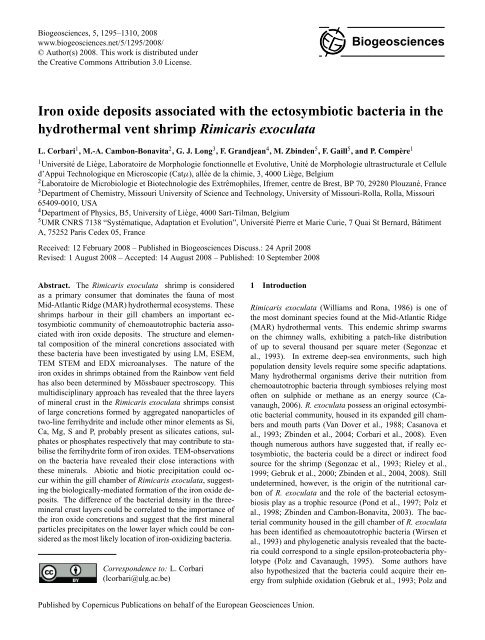
Abstract. The Rimicaris exoculata shrimp is considered<br />
as a primary consumer that dominates the fauna of most<br />
Mid-Atlantic Ridge (MAR) hydrothermal ecosystems. These<br />
shrimps harbour in their gill chambers an important ectosymbiotic<br />
community of chemoautotrophic bacteria associated<br />
with iron oxide deposits. The structure and elemental<br />
composition of the mineral concretions associated with<br />
these bacteria have been investigated by using LM, ESEM,<br />
TEM STEM and EDX microanalyses. The nature of the<br />
iron oxides in shrimps obtained from the Rainbow vent field<br />
has also been determined by Mössbauer spectroscopy. This<br />
multidisciplinary approach has revealed that the three layers<br />
of mineral crust in the Rimicaris exoculata shrimps consist<br />
of large concretions formed by aggregated nanoparticles of<br />
two-line ferrihydrite and include other minor elements as Si,<br />
Ca, Mg, S and P, probably present as silicates cations, sulphates<br />
or phosphates respectively that may contribute to stabilise<br />
the ferrihydrite form of iron oxides. TEM-observations<br />
on the bacteria have revealed their close interactions with<br />
these minerals. Abiotic and biotic precipitation could occur<br />
within the gill chamber of Rimicaris exoculata, suggesting<br />
the biologically-mediated formation of the iron oxide deposits.<br />
The difference of the bacterial density in the threemineral<br />
crust layers could be correlated to the importance of<br />
the iron oxide concretions and suggest that the first mineral<br />
particles precipitates on the lower layer which could be considered<br />
as the most likely location of iron-oxidizing bacteria.<br />
Correspondence to: L. Corbari<br />
(lcorbari@ulg.ac.be)<br />
1 Introduction<br />
Rimicaris exoculata (Williams and Rona, 1986) is one of<br />
the most dominant species found at the Mid-Atlantic Ridge<br />
(MAR) hydrothermal vents. This endemic shrimp swarms<br />
on the chimney walls, exhibiting a patch-like distribution<br />
of up to several thousand per square meter (Segonzac et<br />
al., 1993). In extreme deep-sea environments, such high<br />
population density levels require some specific adaptations.<br />
Many hydrothermal organisms derive their nutrition from<br />
chemoautotrophic bacteria through symbioses relying most<br />
often on sulphide or methane as an energy source (Cavanaugh,<br />
2006). R. exoculata possess an original ectosymbiotic<br />
bacterial community, housed in its expanded gill chambers<br />
and mouth parts (Van Dover et al., 1988; Casanova et<br />
al., 1993; Zbinden et al., 2004; Corbari et al., 2008). Even<br />
though numerous authors have suggested that, if really ectosymbiotic,<br />
the bacteria could be a direct or indirect food<br />
source for the shrimp (Segonzac et al., 1993; Rieley et al.,<br />
1999; Gebruk et al., 2000; Zbinden et al., 2004, 2008). Still<br />
undetermined, however, is the origin of the nutritional carbon<br />
of R. exoculata and the role of the bacterial ectosymbiosis<br />
play as a trophic resource (Pond et al., 1997; Polz et<br />
al., 1998; Zbinden and Cambon-Bonavita, 2003). The bacterial<br />
community housed in the gill chamber of R. exoculata<br />
has been identified as chemoautotrophic bacteria (Wirsen et<br />
al., 1993) and phylogenetic analysis revealed that the bacteria<br />
could correspond to a single epsilon-proteobacteria phylotype<br />
(Polz and Cavanaugh, 1995). Some authors have<br />
also hypothesized that the bacteria could acquire their energy<br />
from sulphide oxidation (Gebruk et al., 1993; Polz and<br />
Published by Copernicus Publications on behalf of the European Geosciences Union.
1296 L. Corbari et al.: Bacteriogenic iron oxides<br />
Cavanaugh, 1995), but this hypothesis has never been confirmed<br />
by any culture experiment. Recently, Zbinden et<br />
al. (2008) demonstrated that the bacterial community in the<br />
shrimp gill chamber is composed of more than one phylotype<br />
and revealed from molecular analysis that three metabolic<br />
bacterial types (iron, sulfide and methane oxidation) may cooccur<br />
within the symbiotic community associated with Rimicaris<br />
exoculata.<br />
In the absence of cultivated bacteria, studies have focussed<br />
on the description of both the bacteria and the mineral deposits<br />
in R. exoculata. Zbinden et al. (2004) described three<br />
bacterial morphotypes, individual rods with an approximate<br />
size of 0.5×1.5 µm, and two types of multicellular filaments,<br />
i.e. thick filaments with 2 to 3 µm diameters and thin filaments<br />
with 0.5 to 1 µm diameters, found within the entire gill<br />
chamber. They mapped the location of these bacteria and divided<br />
their associated minerals into three functional compartments,<br />
that which were considered to represent distinct microenvironments.<br />
One of these compartments, the upper prebranchial<br />
chamber, houses the highest density of both bacteria<br />
and minerals. Recently, Corbari et al. (2008) focussed on<br />
this compartment and delineated the shrimp-bacteria-mineral<br />
association throughout the shrimp moult cycle. This study<br />
performed on about 300 specimens from two vent sites, TAG<br />
and Rainbow, indicated that the bacterial community restarts<br />
after each exuviation and gradually colonises the gill chamber<br />
in five moult stage-correlated steps. Moreover, the presence<br />
of red-brown mineral deposits in the gill chamber, including<br />
the mouth parts and branchiostegites, of the R. exoculata<br />
has already been described (Gloter et al., 2004; Zbinden<br />
et al., 2004). These deposits have been identified as hydrous<br />
iron oxide in the form of ferrihydrite (Gloter et al.,<br />
2004). The extent and density of iron oxide deposits within<br />
the gill chamber are both responsible for the external colour<br />
of the shrimp, a colour that may be macroscopically observed<br />
by transparency through the branchiostegites (Zbinden et al.,<br />
2004). The shrimp external colour ranges from white, indicative<br />
of no mineral deposits, to dark-red, indicative of a heavily<br />
mineralised crust; the colour appears to be highly correlated<br />
with the moult stages (Corbari et al., 2008). The fullyformed<br />
mineral crust is roughly organised in three step-levels<br />
that illustrate the time-related formation and growth of the<br />
mineral particles (Corbari et al., 2008). The shrimp-bacteria<br />
ectosymbiosis is characterised by the presence of iron oxide<br />
deposits suggesting that iron oxidation may represent the major<br />
energy-pathway for the bacterial community, especially<br />
in shrimps found at the Rainbow site (Gloter et al., 2004;<br />
Zbinden et al., 2004, 2008; Schmidt et al., 2008). Moreover,<br />
the simultaneous occurrence of the bacteria and the iron oxide<br />
deposits in the gill chamber of R. exoculata may be interpreted<br />
as biologically-mediated or biogenic (Zbinden et al.,<br />
2004; Gloter et al., 2004, Anderson et al., 2008).<br />
The definition of “biogenic iron oxides” (Fortin and<br />
Châtellier, 2003) refers to iron oxides formed in the presence<br />
of bacteria and includes iron oxides formed as a direct<br />
result of microbial activities (i.e. from enzymatic reactions)<br />
or of passive mechanisms whereby bacterial exudates<br />
trigger the formation and precipitation of iron oxides<br />
minerals. Several studies have documented the formation<br />
and occurrence of iron oxides formed as a result of biotic<br />
pathways in natural environments (Fortin et al., 1998; Fortin<br />
and Châtellier, 2003; Fortin and Langley, 2005; Banfield et<br />
al., 2000; Kennedy et al., 2003, 2004). In this context, the<br />
bacteria-hydrous ferric oxide interactions have also been investigated<br />
to determine the direct and/or indirect bacterial influence<br />
on the hydrous ferric oxide formation (see reviews in<br />
Fortin and Langley, 2005; Klapper and Straub, 2005). Some<br />
authors (Mavrocordatos and Fortin, 2002; Rancourt et al.,<br />
2005) have indicated that bacteria, either iron-metabolizing<br />
or non-metabolizing, could influence the mineral deposition.<br />
They found evidence of the biogenic origin of the hydrous<br />
ferric oxide, identified the presence of poorly crystallized<br />
iron oxides, and determined the typical Fe/O ratios and particle<br />
size ranges. Natural biogenic iron oxides generally contain<br />
impurities, such as adsorbed or structural Si, and phosphate,<br />
sulphate, and manganese and aluminium ions, impurities<br />
that may influence the spatial organization and the morphology<br />
of the mineral particles (Fortin and Châtellier, 2003,<br />
Châtellier et al., 2004). The properties of the iron oxide deposits<br />
and the influence of any impurities on these properties<br />
have been studied in samples from natural environments<br />
(Fortin and Langley, 2005) but have never been investigated<br />
in the case of a bacterial ectosymbiosis.<br />
Because the iron oxide deposition could be actively or<br />
passively promoted by R. exoculata ectosymbiotic bacteria,<br />
the main goal of this study is to investigate in detail<br />
the structure and the composition of the bacteria-associated<br />
mineral particles by using various imaging techniques, such<br />
as back-scattered electron imaging, transmission electron<br />
microscopy, energy dispersive EDX microanalysis, and<br />
Mössbauer spectroscopy. These investigations have been<br />
performed on the fully formed mineral crust of premoult<br />
shrimps, a crust that is divided into three layers related with<br />
the successive steps of formation and growth of the mineral<br />
particles.<br />
2 Materials and methods<br />
2.1 Shrimp selection and samples treatment<br />
Specimens of Rimicaris exoculata were collected during the<br />
French cruise “EXOMAR” (August 2005) at the MAR hydrothermal<br />
vent site Rainbow (36 ◦ 14 ′ N, 33 ◦ 54 ′ W, 2300 m<br />
depth) by using the suction sampler of the ROV “Victor<br />
6000” operating from the RV “Atalante.” Immediately after<br />
retrieval, entire living specimens were either frozen at<br />
−80 ◦ C or dissected into body parts, branchiostegite and tail,<br />
and fixed in a 2.5% glutaraldehyde in seawater 7/10 at pH 7.2<br />
medium.<br />
<strong>Biogeosciences</strong>, 5, 1295–1310, 2008<br />
www.biogeosciences.net/5/1295/2008/
L. Corbari et al.: Bacteriogenic iron oxides 1297<br />
Observations and analyses were performed on preecdysial<br />
specimens, in moult stages D 1 ”’ and D 2 , in agreement<br />
with the moult-staging method of Drach and Tchernigovteff<br />
(1967), based on the development of setae matrices along<br />
the uropods borders. The six frozen and four glutaraldehydefixed<br />
specimens all exhibited an important red-mineral crust<br />
on the inner side of the gill chamber (Fig. 1a and b) in agreement<br />
with the colour categorisation by Corbari et al. (2008).<br />
All the observations were performed on the dorsal median<br />
zone of the branchiostegite of R. exoculata (Fig. 1a) because<br />
it exhibits a regular bacterial and mineral cover that lines<br />
the antero-dorsal compartment of the gill chamber (Zbinden<br />
et al., 2004). The complete branchiostegite and some portions<br />
were photographed with an Olympus SZ40 stereo microscope.<br />
In order to determine the structure and elemental composition<br />
of the bacteria-associated minerals, samples were prepared<br />
for study by transmission and scanning electron microscopy,<br />
EDX microanalysis, and Mössbauer spectroscopy.<br />
During theses preparations, contact between air and the samples<br />
was avoided to prevent alteration in the oxidation and/or<br />
hydration states of the iron oxide minerals. To avoid this aircontact,<br />
frozen specimens were used for compositional analyses<br />
and compared with glutaraldehyde-fixed specimens. For<br />
analytical electron microscopy measurements, the samples,<br />
dissected from frozen specimens, were directly dehydrated<br />
in absolute ethanol and embedded in epoxy resin (Epofix,<br />
Struers) through propylene oxide. Glutaraldehyde-fixed<br />
samples, conserved in seawater with NaN 3 , were quickly<br />
rinsed in distilled water and dehydrated through an ethanolpropylene<br />
oxide series of rinses before embedding in the<br />
EpoFix resin (Struers).<br />
2.2 Scanning Electron Microscopy (SEM) and Energy-<br />
Dispersive X-ray (EDX) microanalysis<br />
Polished thin slices of 20 to 50 µm thickness were obtained<br />
for the branchiostegites of two glutaraldehyde-fixed and two<br />
frozen specimens. The specimens were cut as vertical crosssections<br />
through the mineral crust, i.e. perpendicular to the<br />
branchiostegite cuticle. They were polished by abrasion<br />
on diamond disks and finally mirror polished with a nonaqueous<br />
1 µm diamond suspension (ESCIL, PS-1MIC). The<br />
polished-thin slices were surrounded with a conductive silver<br />
paint to make contact on the surface, carbon-coated in<br />
a Balzers BAF-400 rotary evaporator, and then maintained<br />
in desiccators to prevent air-contact before analysis. Structural<br />
mineral observations and elemental energy-dispersive<br />
X-ray microanalysis were rapidly performed within two days<br />
of preparation in an environmental scanning electron microscope<br />
(FEI XL30 ESEM-FEG), operating at 15 to 20 kV<br />
and a working distance of 10 mm. A total of 15 polishedthin<br />
slices were imaged by back-scattered electrons (BSE)<br />
and analysed for the elemental composition of the minerals<br />
present.<br />
a<br />
b<br />
1 cm<br />
30 µm<br />
Fig. 1. Rimicaris exoculata. (a) Inner side of the branchiostegite<br />
(left side) of premoult specimen in moult stage D1”’ exhibiting a<br />
dense and uniform coating of mineral deposits. The dashed lines<br />
delimit the observed area, the median zone. (b) Polished thin cross<br />
section slices of the mineral crust observed under a light microscope<br />
and exhibiting three different layers of mineral density. Note that<br />
the bacterial filaments are distinguishable on the upper level of the<br />
picture.<br />
Elemental analyses have been carried out on the surface<br />
of 2 µm size mineral concretions. EDX microanalyses<br />
with an acquisition time of 60 s have been obtained for<br />
both the glutaraldehyde-fixed and the frozen samples in order<br />
to determine whether any mineral transformation took<br />
place through chemical reactions during sample preparation.<br />
The elemental quantitative analysis used an automatic background<br />
subtraction and a ZAF correction matrix has been<br />
used to calculate the elemental composition in weight percents<br />
and atomic percents. For quantitative analysis of the<br />
mineral concretions, the contributions of the C-coating and<br />
the embedding resin containing C, O and trace of Cl were<br />
subtracted from the quantitative data of each spectrum. The<br />
contribution of C-coating was evaluated to 25 At % of C from<br />
a pure mineral sample (hydroapatite) C-coated in the same<br />
conditions. The remaining C was attributed to the resin and<br />
www.biogeosciences.net/5/1295/2008/ <strong>Biogeosciences</strong>, 5, 1295–1310, 2008
1298 L. Corbari et al.: Bacteriogenic iron oxides<br />
an amount of O (At %) was subtracted in the same proportion,<br />
deduced from reference spectra of pure resin (C/O ratio<br />
8:1).<br />
2.3 Transmission Electron Microscopy (TEM)<br />
Four glutaraldehyde-fixed specimens were post-fixed in osmium<br />
1%, dehydrated in an ethanol-propylene oxide series<br />
and then embedded in epoxy resin (SPI-PON 812). Ultrathin<br />
sections were obtained with a Reichert-Jung Ultramicrotome<br />
(Ultracut E) by using a diamond knife; uranium acetate<br />
and lead citrate were used as contrast agents. The specimens<br />
were studied with a Jeol (JEM 100-SX) transmission electron<br />
microscope operating at 80 kV. In order to provide a threedimensional<br />
view of the mineral crust organisation, vertical<br />
cross-sections were cut perpendicular to the branchiostegite<br />
cuticle and the mineral surface and horizontal sections were<br />
cut at the three levels of mineral crust as the previously defined.<br />
2.4 Scanning Transmission Electron Microscopy (STEM)<br />
and Energy-Dispersive X-ray (EDX) microanalysis<br />
Ultrathin sections of samples from two frozen shrimps were<br />
cut as previously described, placed on a formvar-coated titanium<br />
grid and carbon-coated in a Balzers BAF-400 rotary<br />
evaporator. They were then imaged without any additional<br />
contrast in a FEI Tecnai G2 Twin scanning-transmission<br />
electron microscope operating at 200 kV.<br />
In order to determine the elemental composition of the<br />
strata observed in the mineral concretions, scanning transmission<br />
electron imaging has been carried out in a both direct<br />
bright-field and a high-angle annular dark-field (HAADF)<br />
imaging modes. The energy-dispersive EDX nanoanalyses<br />
were performed with a nanoprobe spot size of 1nm in diameter.<br />
Profile spectra have been determined on 1 to 1.5 µm<br />
length of ten various mineral concretions.<br />
2.5 Mössbauer spectroscopy<br />
The Mössbauer spectra have been obtained on samples from<br />
frozen shrimps. Two different types of Mössbauer spectral<br />
absorbers have been used. The first contained boron nitride<br />
mixed with 14 mg/cm 2 of lyophilized powder of crust minerals<br />
obtained by scrapings from three shrimps. The second<br />
consisted in the superimposed branchiostegites of one<br />
shrimp. The spectra were measured between 4.2 and 295 K<br />
on a constant-acceleration spectrometer that utilised a room<br />
temperature rhodium matrix cobalt-57 source and was calibrated<br />
at 295 K with α-iron powder. The estimated relative<br />
errors are ±0.005 mm/s for the isomer shifts, ±0.01 mm/s for<br />
the quadrupole splittings and line widths, and ca. ±0.5 T for<br />
the hyperfine field. The absolute errors are estimated to be<br />
approximately twice as large.<br />
2.6 Statistical analysis<br />
The mineralogical compositions and quantifications are reported<br />
as mean values ± standard deviation. Comparisons<br />
of the mineral composition between glutaraldehyde-fixed<br />
and frozen samples have been evaluated by using a Mann–<br />
Whitney U-test, a two-tailed Student’s t-test, a Fisher test,<br />
and/or analysis of the variance. P
g-2008-0022<br />
L. Corbari et al.: Bacteriogenic iron oxides 1299<br />
Corbari et al., Figure 2<br />
a<br />
b<br />
2 µm<br />
c<br />
d<br />
5 µm<br />
e<br />
f<br />
5 µm<br />
Fig. 2. The three levels of the mineral crust. Comparison between the electron back scattering images of the polished-thin sections (vertical<br />
sections) revealing the mineral densities and structures (left column) and the TEM micrographs of horizontal cross-sections exhibiting<br />
mineral associated with bacterial community (right column) at the lower level (a and b), the median level (c and d) and the upper level (e<br />
and f).<br />
The median level of the crust exhibits larger mineral concretions,<br />
globular in shape, (Fig. 2c and d). These globular<br />
concretions often meet to form larger ones that exhibit<br />
a botryoidal structure. In the horizontal cross-sections, the<br />
aggregate density appears rather heterogeneous and consists<br />
of highly mineralised patches interspersed with bacteria rich<br />
areas. The bacterial density in the median layer is always<br />
smaller than in the lower layer; there are fewer rod-shaped<br />
bacteria. Moreover, ghosts of bacteria are also observed<br />
(Fig. 3e and f) in TEM images. These ghosts have bacterial<br />
shapes that are completely enclosed in a heavy mineral<br />
sheath. Sometimes the bacteria are still present but appear<br />
either to be damaged or as membrane remain (Fig. 3e and f).<br />
In other cases, the mineral sheath appears to be empty or to<br />
have been recolonised by other rod-shaped bacteria. These<br />
observations suggest that mineral formation may influence<br />
the survival rate of the bacteria.<br />
www.biogeosciences.net/5/1295/2008/ <strong>Biogeosciences</strong>, 5, 1295–1310, 2008
g-2008-0022<br />
1300 L. Corbari et al.: Bacteriogenic iron oxides<br />
Corbari et al., Figure 3<br />
a<br />
b<br />
c<br />
d<br />
e<br />
500 nm<br />
f<br />
500 nm<br />
Fig. 3. TEM views illustrating the different ways the minerals may be deposited on the rod-shaped bacteria in Rimicaris exoculata. (a)<br />
Mineral deposition in direct contact with the bacteria cell walls. (b) Mineral deposition on secreted bacterial substance. (c) and (d) Methanotrophic<br />
bacteria surrounded by mineral deposits. (e) and (f) Bacterial ghosts coated with minerals and bacterial recolonization of the mineral<br />
sheaths.<br />
The upper level of the mineral crust contains very large<br />
concretions with diameters of up to 2 µm (Fig. 2e and f). The<br />
grape-like concretion shapes with deep indentations suggest<br />
that they result from the aggregation of several smaller ones.<br />
TEM images reveal that the bacteria become very rare in this<br />
upper level. As may be observed in the horizontal sections,<br />
almost the only bacteria present are a few large, thin, bacterial<br />
filaments that perforate throughout the mineral crust.<br />
Moreover, the minerals are not in direct contact with the filament<br />
cell walls but form large sheaths at some distance from<br />
the cell walls (Fig. 2e). Even though the three step-levels<br />
of the mineral crust have been arbitrarily defined, they are<br />
<strong>Biogeosciences</strong>, 5, 1295–1310, 2008<br />
www.biogeosciences.net/5/1295/2008/
Corbari et al., Figure 4<br />
L. Corbari et al.: Bacteriogenic iron oxides 1301<br />
a<br />
Percent Transmission<br />
100<br />
99<br />
98<br />
97<br />
96<br />
95<br />
94<br />
100<br />
98<br />
96<br />
295 K<br />
85 K<br />
Cor<br />
94<br />
2 µm<br />
b<br />
92<br />
-4 -3 -2 -1 0 1 2 3 4<br />
Velocity, mm/s<br />
100.0<br />
99.0<br />
Fig. 4. Bacteria-mineral interactions in Rimicaris exoculata. TEM<br />
view of the lower level (vertical cross-section) where rod-shape bacteria<br />
are very abundant. Note that the bacteria seem to produce some<br />
substance which prevents any direct mineral deposition close to this<br />
layer.<br />
98.0<br />
97.0<br />
96.0<br />
95.0<br />
100.0<br />
99.0<br />
60 K<br />
representative of the different phases of the mineral formation<br />
and are characterised by an inverse correlation, from<br />
low to upper levels, between the amount of mineral deposits<br />
present and the bacterial density.<br />
3.2 Iron oxides identified by Mössbauer spectroscopy<br />
Mössbauer spectroscopy has been used to identify both the<br />
nature and oxidation states of the iron oxides found in the<br />
native minerals through the measurements of the iron-57 isomer<br />
shift and quadrupole splitting. The Mössbauer spectra,<br />
obtained at 85 and 295 K and between 4.2 and 60 K are<br />
shown in Figs. 5a and b, respectively. At 85 and 295 K the<br />
spectra consist of broadened quadrupole doublets, whereas<br />
below 60 K they consist of a superposition of broadened doublets<br />
and sextets. The observed temperature dependence of<br />
the Mössbauer spectra is typical of small superparamagnetic<br />
particles. The spectra have been fit with two symmetric<br />
quadruople doublets and one to three magnetic sextets; the<br />
average hyperfine parameters are given in Table 1.<br />
The weighted average isomer shift, , is typical of<br />
iron(III) (Shenoy et al., 1978) and spectral analysis indicates<br />
that at least 98 % of the iron in the mineral crust<br />
must be present as iron(III); two percent by spectral area<br />
is the approximate detection limit for the presence of any<br />
iron(II). The average hyperfine parameters observed at 295<br />
and 4.2 K are typical (Murad et al., 1987) of two-line ferrihydrite.<br />
Two-line ferrihydrite, Fe 5 HO 8·4H 2 O, is a poorly<br />
crystalline mineral that forms spherical nanoparticles with<br />
Percent Transmission<br />
98.0<br />
97.0<br />
96.0<br />
100.0<br />
99.5<br />
99.0<br />
98.5<br />
98.0<br />
97.5<br />
97.0<br />
100.0<br />
99.6<br />
99.2<br />
98.8<br />
98.4<br />
100.0<br />
99.5<br />
99.0<br />
98.5<br />
50 K<br />
45 K<br />
40 K<br />
4.2 K<br />
98.0<br />
-12 -10 -8 -6 -4 -2 0 2 4 6 8 10 12<br />
Velocity, mm/s<br />
Fig. 5. (a) The 85 and 295 K iron-57 Mössbauer spectra of minerals<br />
from the crust collected on the branchiostegites of three shrimps.<br />
(b) The iron-57 Mössbauer spectra of two superimposed branchiostegites<br />
parts of one shrimp obtained at the indicated temperatures.<br />
www.biogeosciences.net/5/1295/2008/ <strong>Biogeosciences</strong>, 5, 1295–1310, 2008
1302 L. Corbari et al.: Bacteriogenic iron oxides<br />
Table 1. Mössbauer spectral parameters obtained for the Rimicaris exoculata hydrothermal shrimp.<br />
Compound T , , , , , Area, Abs. Area, Assignment<br />
K mm/s ∗ mm/s T mm/s % (%ε) (mm/s)<br />
Rainbow 295 0.358 0.78 0 0.46 100 7.302 Superparamagnetic iron(III)<br />
225 0.403 0.78 0 0.38 100 10.106 Superparamagnetic iron(III)<br />
155 0.439 0.79 0 0.39 100 11.010 Superparamagnetic iron(III)<br />
85 0.465 0.80 0 0.42 100 11.064 Superparamagnetic iron(III)<br />
60 0.464 0.80 0 0.42 82 6.196 Superparamagnetic iron(III)<br />
0.461 −0.04 39.6 1.13 18 1.314 Partially blocked iron(III)<br />
50 0.464 0.80 0 0.42 36 3.6935 Superparamagnetic iron(III)<br />
Corbari et al., Figure 6<br />
0.463 −0.05 21.6 4.35 64 6.693 Partially blocked iron(III)<br />
45 0.464 0.80 0 0.42 23 2.332 Superparamagnetic iron(III)<br />
0.463 −0.06 28.1 3.54 77 7.753 Partially blocked iron(III)<br />
40 0.464 0.80 0 0.42 8 0.901 Superparamagnetic iron(III)<br />
0.463 −0.06 24.0 3.75 92 10.075 Partially blocked iron(III)<br />
4.2 0.481 −0.045 47.8 0.56 100 10.727 Blocked iron(III)<br />
bg-2008-00<br />
∗ The isomer shifts are given relative to room temperature α-iron powder.<br />
Table 2. Typical elemental EDX microanalysis of the mineral crust<br />
of Rimicaris exoculata. Data are expressed as both weight and<br />
atomic percents.<br />
Element Wt % At %<br />
O Ka<br />
C Ka<br />
Fe Ka<br />
C K 37.5 57.3<br />
O K 25.5 29.2<br />
MgK 0.4 0.3<br />
SiK 1.7 1.1<br />
P K 0.6 0.3<br />
S K 0.3 0.2<br />
CIK 0.3 0.2<br />
CaK 0.6 0.3<br />
FeK 32.7 10.8<br />
a diameter of between 2 and 7 nm, a diameter that depends<br />
upon both the crystallinity of the material and the presence<br />
of impurities. Also for the same reasons, the average hyperfine<br />
field observed at 4.2 K may be reduced from 50 to<br />
46.5 T. In the specimens understudy, the average hyperfine<br />
field of 47.8 T corresponds to a reasonably well crystallised<br />
sample. The blocking temperature, i.e. the temperature at<br />
which the absorption areas of the doublets and sextets are<br />
equal, is 55±2 K. By using the anisotropy constant (Murad<br />
et al., 1987) of 4×10 4 J/m 3 for 5 nm Fe 5 HO 8·4H 2 O particles,<br />
an average particle diameter of 5.6 nm is obtained.<br />
3.3 Quantitative EDX microanalyses<br />
BSE images of polished thin slices of frozen specimens provide<br />
a detailed map of the mineral particles in the crust.<br />
EDX microanalyses (n=14) performed in ESEM give accurate<br />
qualitative and quantitative determinations of the ele-<br />
0<br />
Si Ka<br />
Fe La<br />
P Ka<br />
S Ka<br />
Mg Ka Cl Ka<br />
Ca Ka<br />
2.0 4.0 6.0<br />
KeV<br />
Fe Kb<br />
Fig. 6. Elemental EDX microanalyses of the mineral crust of Rimicaris<br />
exoculata. (a) A typical spectrum obtained on mineral particles<br />
of up to 2 µm diameter. The peaks are labelled with the EDX<br />
line of the corresponding element.<br />
mental composition of the mineral deposits. These microanalyses<br />
reveal the predominance of iron, with a K α peak<br />
at 6.400 keV and a K β peak at 7.059 keV, and oxygen with<br />
a K α peak at 0.5425 keV, in the mineral crust (Fig. 6). Minor<br />
amounts of silicon with a K α peak at 1.740 keV, calcium<br />
with a K α peak at 3.690 keV and a K β peak at 4.012 keV,<br />
phosphorus with a K α peak at 2.013 keV, magnesium with<br />
a K α peak at 1.253 keV, and sulphur with a K α peak at<br />
2.307 keV, have also been detected. Elemental quantitative<br />
analyses yield the weight and atomic percentages of the elements<br />
present, see Table 2. In order to determine the relative<br />
amount of the iron oxides and other minerals in the concretions,<br />
we subtract the C of the C-coating and the C, O and Cl<br />
of the embedding resin. It was also assumed that the minor<br />
elements such as Si, Ca, Mg, S and P are present as silicate<br />
8.0<br />
<strong>Biogeosciences</strong>, 5, 1295–1310, 2008<br />
www.biogeosciences.net/5/1295/2008/
L. Corbari et al.: Bacteriogenic iron oxides 1303<br />
Table 3. Mineralogical characterisation of the mineral crust in Rimicaris exoculata. Data are expressed in atomic percentages and results<br />
from EDX quantitative analyses (n=14) of polished thin slices of minerals from two frozen specimens. Mean values have been calculated<br />
after removing the peripheral elements inherent to the resin and the carbon coating. The amount of available oxygen for each mineral or<br />
ligands is calculated by assuming the presence of SO 2−<br />
4 and PO 3−<br />
4 anionic groups as well as silicate with at least two O atoms associated<br />
to each Si atom. The percentages of mineral constituents and ligands have been calculated under the assumption of their most probable<br />
occurrence. All values are mean ± standard deviation.<br />
Elements O Fe Si S P Ca Mg<br />
Mean 67.8±1.4 27.3±0.7 2.6±0.4 0.5±0.1 0.5±0.2 0.7±0.1 0.5±0.3<br />
Available O − 58.5±3.1 5.3±0.8 2.1±0.4 2.0±0.9 − −<br />
Minerals and Fe x O y (OH) z + vH 2 O Si (O 2 ) SO 2−<br />
4 PO 3−<br />
4 Ca, MG Total<br />
Inorganic ligands 85.8±2.9 7.9±1.2 2.6±0.5 2.5±1.2 1.2±0.49 9.9±0.2<br />
cations and anions (sulphate and phosphate) respectively as<br />
they appear as the most probable ligand forms, as suggested<br />
by Châtellier et al. (2001, 2004) and Fortin and Langley<br />
(2005). The associated oxygen was calculated according to<br />
the stoichiometric ratio in the forms of SO 2−<br />
4<br />
and PO 3−<br />
4<br />
and<br />
reported as available O in Table 3. For silicate, a minimum<br />
of two oxygen atoms arbitrary attributed to each Si atom that<br />
appears as Si(O 2 ) in Table 3. The remaining O was considered<br />
as available for Fe and taken into account in the calculation<br />
of the Fe/O ratio. The results of these calculations give<br />
the relative contribution of all the mineral constituents of the<br />
concretions (Table 3). They show that iron(III) oxides correspond<br />
to ca. 85 At % (ca. 90 Wt %) of the minerals present<br />
in the crust, as confirmed by Mössbauer spectroscopy. This<br />
value does not include hydrogen atoms (not measurable) that<br />
may represent 25–50% of the atoms in the mineral, according<br />
to the general Ferrihydrite formula and hydration state<br />
but does never exceed 2 Wt%. The contribution of the inorganic<br />
ligands in the concretions is approximately evaluated at<br />
8% for Si(O 2 ), 2.5% for SO 2−<br />
4<br />
and 2.5% of PO 3−<br />
4<br />
and 1.2%<br />
for Ca and Mg. It is very probable that Ca 2+ and Mg 2+ are<br />
combined in salts with the anions SO 2−<br />
4<br />
and PO 3−<br />
4<br />
because<br />
of the perfect accordance with the stoichiometric ratio in (Ca,<br />
Mg) SO 4 and (Ca, Mg) 3 (PO 4 ) 2 . Ca, Mg salts could thus represent<br />
approximately 6% in the concretions.<br />
Comparative elemental EDX microanalyses carried out on<br />
12 polished thin slices of glutaraldehyde-fixed specimens reveal<br />
very similar proportions of the minor ligands suggesting<br />
that they are not solubilised or removed by the aqueous<br />
preparation procedure. However, these analyses differ from<br />
those of the frozen sample by the Fe/O ratio in the iron oxide,<br />
i.e. after the subtraction of the oxygen linked to minor<br />
elements, see Table 4. The Fe/O ratio is 0.47 in the frozen<br />
specimens while it reaches 0.60 in the glutaraldehyde-fixed<br />
specimens. These measured Fe/O ratios are statistically different<br />
as is indicated by a paired t-test which yields t=7.1,<br />
d.f.=11, and P =0.00002. Hence, the atomic percentage of<br />
oxygen is lower in the glutaraldehyde-fixed specimens, in<br />
which the glutaraldehyde could act as a reducing agent and<br />
modify the iron oxidation state in the mineral particles.<br />
Table 4. Comparison of the mineralogical composition between<br />
frozen and glutaraldehyde-fixed specimens. EDX elemental<br />
quantitative analysis of fourteen frozen samples and twelve<br />
glutaraldehyde-fixed samples. The data have been obtained with<br />
the same experimental procedure. All values are mean ± standard<br />
deviation.<br />
Frozen sp.<br />
Glutharaldehyde-fixed sp.<br />
Elements O Fe O Fe<br />
Mean 67.8±1.4 27.3±0.7 64.5±1.4 28.3±1.3<br />
Available O − 58.5±3.1 − 47.8±3.0<br />
Ratio Fe/O 0.47±0.03 0.60±0.05 a<br />
a Significantly different.<br />
3.4 Structure and composition of the crust minerals<br />
TEM images obtained on U/Pb contrasted ultra-thin vertical<br />
sections indicate that most of the mineral concretions, which<br />
have diameters ranging from 200 to 600 nm, exhibit layered<br />
features in the lower level of the crust (Fig. 7a and b). Most<br />
of the specimens exhibit a multilayered pattern with a periodicity<br />
of a few nanometers, a pattern that suggests that these<br />
concretions are composed of ca. 5 to 10 successive strata. All<br />
of these strata appear as concentric growth layers originating<br />
from a unique nucleation centre, as multiglobular particles<br />
that change their shape and, for the outer particles, follow<br />
the outer particle border (Fig. 7b). Neighbouring concretions<br />
also exhibit layered patterns that may correspond to similar<br />
mineral deposition sequences (Fig. 7a). Mineral nucleation<br />
and deposition occur either close to the rod-shaped bacteria<br />
walls or in their near-neighbour environment. Several nucleation<br />
centres are located close to the same bacteria and the<br />
accumulation of strata leads to the aggregation of mineral<br />
particles that, as a consequence, exhibit a grape-like shape<br />
(Fig. 7b). STEM-HAADF images of particles from frozen<br />
specimens give an inverted mass contrast of the strata. These<br />
images reveal the reality of the strata in terms of the changing<br />
aggregate density and/or the composition within the particles<br />
(Fig. 7c and d).<br />
www.biogeosciences.net/5/1295/2008/ <strong>Biogeosciences</strong>, 5, 1295–1310, 2008
g-2008-0022<br />
Corbari et al., Figure7<br />
1304 L. Corbari et al.: Bacteriogenic iron oxides<br />
a<br />
1 µm<br />
b<br />
200 nm<br />
B<br />
c<br />
B<br />
d<br />
Fig. 7. (a) and (b) TEM views of the mineral particles in the lower level of the crust. The occurrence of contrasted strata is clearly visible.<br />
(c) and (d) STEM images of ultrathin sections of non-contrasted mineral particles revealing the different nature of the mineral strata. B<br />
indicates the bacteria.<br />
In order to determine the exact nature of these strata, EDX<br />
nanoanalyses have been carried out by STEM along a direction<br />
perpendicular to the strata of some particles observed<br />
in the ultrathin sections. The experimental procedure for<br />
obtaining these line profiles of 1 to 1.5 µm length is based<br />
on the acquisition of a sequence of ca. 700 EDX spectra<br />
per line profile. Subsequent data analysis can differentiate<br />
different elements through the number of counts that correspond<br />
to a given element found along the line. Figure 8 illustrates<br />
a typical elemental profile for iron, oxygen, and silicon<br />
along a 1.2 µm line through a stratified mineral particle. The<br />
clear lighter coloured strata in Fig. 8a correspond to a higher<br />
iron and oxygen content than is found in the dark strata, see<br />
Fig. 8b. In spite of a low number of counts, the silicon profile<br />
seems to correlate well with those of iron and oxygen. In order<br />
to more accurately characterise the nature of the mineral<br />
strata, quantitative elemental analyses have been performed<br />
at specific points, i.e. points 1, 2, and 3 in Fig. 8a, at well<br />
separated strata along the line profile. The results (Table 5)<br />
confirm that the iron and oxygen atomic percentages are high<br />
at the lighter strata, points 1 and 3, and very low in the dark<br />
strata, position 2. The strata result thus rather from changes<br />
in the aggregation density than from real compositional differences.<br />
4 Discussion<br />
A multifacitated analysis carried out on the mineral crust of<br />
R. exoculata reveals a mineral content of 85% iron(III) oxide<br />
and associated with 15 % of minor inorganic ligands, possibly<br />
present as silicate and (Ca, Mg) sulphate and phosphate.<br />
The Mössbauer spectral results indicate that the iron(III)<br />
oxide corresponds to two-line ferrihydrite, Fe 5 HO 8·4H 2 O,<br />
which is present in nanometric particles of less than 5 nm diameter.<br />
A transmission and scanning electron microscopic<br />
study of the mineral crust reveals the concretions exhibit<br />
<strong>Biogeosciences</strong>, 5, 1295–1310, 2008<br />
www.biogeosciences.net/5/1295/2008/
L. Corbari et al.: Bacteriogenic iron oxides 1305<br />
Table 5. Elemental quantitative analyses expressed in atomic percent<br />
along the line profile of the iron oxide particle shown in Fig. 8a.<br />
Positions (1), (2), and (3) correspond to the positions indicated in<br />
Fig. 8a.<br />
a<br />
3<br />
Corbari et a<br />
% At Fe(K) O(K) Si(K)<br />
(1) 4.62 10.95 0.55<br />
(2) 1.07 4.57 0.34<br />
(3) 1.99 6.54 0.36<br />
1<br />
2<br />
stratified layers suggesting a sequential deposition with alternating<br />
layers of differing elemental density and composition.<br />
The description of the bacteria-mineral interactions suggests<br />
that both biotic and abiotic process could influence the mineral<br />
formation in the R. exoculata ectosymbiosis.<br />
4.1 Iron oxides<br />
Mössbauer spectral results have confirmed that the mineralbacteria<br />
associated crust which coats the branchiostegite<br />
of Rimicaris exoculata is mainly composed of a hydrous<br />
iron(III) oxide identified as two-line ferrihydrite,<br />
Fe 5 HO 8·4H 2 O (Jambor and Dutrizac, 1998). These results<br />
are in agreement with the conclusions based on transmission<br />
electron microscopy-electron energy loss spectroscopy<br />
(TEM-EELS) of minerals of R. exoculata mouthparts (Gloter<br />
et al., 2004). Further, the particle diameter of 2 to 7 nm obtained<br />
from the Mössbauer spectral results is in complete<br />
agreement with that obtained by high-resolution electron microscopic<br />
images or structural analyses (Gloter et al., 2004;<br />
Michel et al., 2007). However, although an earlier TEM-<br />
EELS analysis (Gloter et al., 2004) indicated a mixture of 55<br />
to 66% iron(III) and 45 to 34% iron(II), only iron(III) has<br />
been detected in the Mössbauer spectra. Hence, if it is assumed<br />
that limit of detection for iron(II) is 2%, iron(III) represents<br />
more than 98% of the total iron present. This difference<br />
in the oxidation state of iron may result from the experimental<br />
procedure used herein and the glutaraldehyde-fixation<br />
used for the TEM-EELS analysis as is discussed below.<br />
Mössbauer spectroscopy utilises bulk samples of the mineral<br />
crust and hence, the results are averaged over a rather<br />
large number of concretions, in contrast with the results obtained<br />
from electron microscopic techniques that are representative<br />
of small portions of the samples. Further, there is<br />
neither radiation damage nor preparative damage of the absorbers<br />
in the Mössbauer spectral experiments. In contrast,<br />
exposure to an electron beam can result in atomic displacement,<br />
electronic reduction, electron-beam sputtering and/or<br />
heating, electrostatic charging, and radiolysis (Egerton et al.,<br />
2004). In a recent study using EELS to evaluate the effects<br />
of electron beam damage to ferrihydrite, Pan et al. (2006)<br />
observed the reduction of iron(III) to iron(II). These results<br />
b<br />
Counts<br />
4 e +7<br />
2 e +7<br />
0<br />
4 e +7<br />
2 e +7<br />
0<br />
4 e +7<br />
2 e +7<br />
1<br />
200 nm<br />
2<br />
0<br />
0 0.4 0.8 1.2<br />
Position (µm)<br />
Fe<br />
Fig. 8. (a) STEM pictures of the analysed particle exhibiting contrasted<br />
strata. The red line corresponds to the spectral profile line<br />
of 1.2 µm length. The numbers 1 to 3 indicate the points where the<br />
elemental quantitative analysis given in Table 5 was carried out. (b)<br />
Element specific spectra acquired along the red profile line. Points<br />
1 to 3 are also indicated at their corresponding positions on the iron<br />
spectrum.<br />
3<br />
O<br />
Si<br />
www.biogeosciences.net/5/1295/2008/ <strong>Biogeosciences</strong>, 5, 1295–1310, 2008
1306 L. Corbari et al.: Bacteriogenic iron oxides<br />
highlight how investigations carried out under high vacuum<br />
in a transmission electron microscope may cause substantial<br />
and perhaps unsuspected changes to a mineral sample.<br />
Ferrihydrite is thus an essential mineral component of the<br />
mineral-bacteria associated crust in R. exoculata. The most<br />
common extracellular biogenic iron oxides include oxyhydroxides,<br />
e.g. goethite, lepidocrocite, akaganeite, and poorly<br />
ordered phases, e.g. two-line and six-line ferrihydrite (Cornell<br />
and Schwertmann, 2003; Fortin and Langley, 2005).<br />
Furthermore, it is commonly accepted that the product of microbial<br />
micro-aerobic iron(II) oxidation is often identified as<br />
a poorly crystalline ferrihydrite. The observation of two-line<br />
ferrihydrite thus supports the hypothesis of the presence of<br />
iron-oxidisers among the ectosymbiotic bacterial community<br />
of R. exoculata (Zbinden et al., 2004) and validates the first<br />
observations on bacterial cultures (Cambon-Bonavita, pers.<br />
com.). In hydrothermal environments, ferrihydrite has previously<br />
been identified because it is commonly intermixed<br />
with lithoautotrophic iron(II) oxidizing bacteria, that act as a<br />
causative agent in the formation of the ferrihydrite (Emerson<br />
and Moyer, 2002; Kennedy et al., 2003, 2004; Little<br />
et al., 2004). Very few vent animals have been discovered<br />
to live in close association with iron oxide deposits. To the<br />
best of our knowledge, only the scaly-foot gastropod found<br />
in the hydrothermal vents at the Indian Ridge has been shown<br />
to exhibit scale-shaped structures, mineralised with iron sulphides<br />
on its foot (Goffredi et al., 2003; Waren et al., 2003).<br />
These structures are associated with bacteria but an iron isotopic<br />
analysis indicates that sulphur and iron in the sclerites<br />
originate from hydrothermal fluids rather than from bacteria<br />
(Suzuki et al., 2006). Herein, the presence of ferrihydrite<br />
in close interaction with hydrothermal metazoan has been<br />
discovered for the first time, in the specific case of the vent<br />
shrimp R. exoculata.<br />
Elemental quantitative analyses have been performed to<br />
determine the Fe/O ratio. Because of the influence of the<br />
sample preparation for ultrastructural and elemental analyses,<br />
an alternative experimental procedure has been adopted<br />
in this study to reduce damage to the samples containing both<br />
bacteria and minerals. The samples for mineralogical analyses<br />
were frozen until used and were directly dehydrated by<br />
ethanol to avoid both air-contact and any aqueous chemical<br />
fixation. This procedure is based on the work of Mavrocordatos<br />
and Fortin (2002), who investigated the influence<br />
of sample preparation on poorly ordered biotic hydrous iron<br />
oxide. To assess the impact of sample fixation on the composition<br />
of the R. exoculata minerals, Fe/O ratios determined<br />
through identical procedures have been compared between<br />
frozen and glutaraldehyde-fixed samples. The Fe/O ratio<br />
of the glutaraldehyde-fixed minerals is significantly different<br />
from that of the frozen minerals. Thus sample preparation<br />
may modify the oxidation state of iron. The use of glutaraldehyde,<br />
a reducing agent, in addition to electron beam<br />
damage in TEM-EELS studies can explain the higher percentage<br />
of iron(II) reported by Gloter et al. (2004) in their<br />
mineral samples. The Fe/O ratio in frozen minerals has been<br />
measured to be 0.47, after subtraction of the oxygen associated<br />
with inorganic elements. This value cannot be compared<br />
with the Fe/O ratio of 0.33 obtained by Gloter et al. (2004)<br />
because both their analytical approach and mineralogical interpretations<br />
are quite different from those used herein. In<br />
contrast, the calculated Fe/O ratio of 0.47 may be compared<br />
with the 0.417 ratio obtained for abiotic ferrihydrite (Mavrocordatos<br />
and Fortin, 2002 and references therein). Moreover,<br />
the calculated Fe/O ratio of 0.47 in frozen minerals is similar<br />
to the Fe/O ratio of 0.48 (Mavrocordatos and Fortin, 2002).<br />
In this study, quantitative TEM-EELS analyses have been<br />
performed on ferrihydrite sample experimentally obtained<br />
by the precipitation of hydrous ferric oxides in the presence<br />
of bacteria exhibiting extracellular polymers. Despites our<br />
slightly different methodologies, the similar Fe/O ratio determined<br />
in the presence of bacteria, suggest the influence of<br />
the bacterial ectosymbiosis on the iron oxide formation.<br />
In conclusion, for future research on R. exoculata minerals,<br />
sample preparation and, more specifically, the drying<br />
process and fixation of the biological samples must be accurately<br />
delineated because they both influence the surface<br />
properties of ferrihydrite and hence, the iron oxidation state<br />
and the Fe/O ratio.<br />
4.2 Intrinsic inorganic constituents<br />
Even though ferrihydrite represents the main component of<br />
the mineral crust in R. exoculata, elemental and quantitative<br />
analyses of the mineral particles have revealed the presence<br />
of minor elements, such as Si, P, Ca, S, and Mg. These elements<br />
are not considered as impurities (Gloter et al., 2004)<br />
but rather to form intrinsic inorganic constituents or ligands<br />
as suggested by Châtellier et al. (2004) because these authors<br />
found that their presence during the oxidation process<br />
can affect the mineralogy as well as the size and structure<br />
of the iron oxide particles. Because of their large surface<br />
areas, small particles of natural biogenic iron oxides generally<br />
contain adsorbed elements and ions, such as silicate,<br />
sulphate, phosphate, and manganese and aluminium cations<br />
(Fortin and Châtellier, 2003; Fortin and Langley, 2005). For<br />
example, nanoparticles of iron oxyhydroxide formed during<br />
the mineralisation process, can adsorb phosphate and<br />
silicate ions (Gilbert and Banfield, 2005). The adsorption<br />
of compounds on ferrihydrite from the surrounding aqueous<br />
milieu can affect its subsequent mineral ordering processes.<br />
Specifically, adsorption of silicates has been found<br />
to inhibit the conversion of ferrihydrite to more crystalline<br />
iron oxides, such as hematite and goethite (Kennedy et al.,<br />
2003; Châtellier et al., 2004). EDX nanoanalyses and elemental<br />
profiles performed on stratified R. exoculata mineral<br />
particles have revealed that silicon is already present in the<br />
early stages of mineral development. Thus, in R. exoculata,<br />
it is evident that inorganic ligands co-precipitate with iron<br />
oxide and are closely associated with it in the nanoparticles<br />
<strong>Biogeosciences</strong>, 5, 1295–1310, 2008<br />
www.biogeosciences.net/5/1295/2008/
L. Corbari et al.: Bacteriogenic iron oxides 1307<br />
because the Si portion cannot be located separately from the<br />
iron oxide even on the nanometric scale reached in STEM. In<br />
the global characterisation of the mineral particles, we have<br />
assumed that they correspond to separated compounds such<br />
as silicate, Ca 2+ , Mg 2+ , SO 2−<br />
4<br />
, and PO3−<br />
4<br />
, even though the<br />
authors consider that silicate, sulphate, phosphate, and magnesium<br />
and calcium cations are substituted or adsorbed to<br />
ferrihydrite. Their presence could also influence or stabilise<br />
this poorly crystalline form of iron oxide. Considering the<br />
stoichiometric ratio of Ca, Mg and P, it is probable that SO 2−<br />
4<br />
and PO 3−<br />
4<br />
are associated with Ca and Mg in (Ca, Mg) salts.<br />
In order to further assess the distribution of the intrinsic<br />
inorganic constituents, EDX spectra acquired at line profiles<br />
have been performed on mineral particles exhibiting stratified<br />
features. At the atomic level, the results indicate that<br />
both iron and the other inorganic ligands are already associated<br />
within nanosized mineral particles. These observations<br />
reveal that iron, oxygen, and silicon proportions are constant<br />
but that the stratification is mainly the consequence of parallel<br />
variations of the concentration of all the constituting elements.<br />
Bacteria-associated mineral concretions appear to be<br />
identical and stratification seems to follow the same sequence<br />
within neighbouring mineral particles. These observations<br />
suggest that micro-environmental variations can occur in the<br />
nearby environment during the formation of the mineral particles.<br />
Uniform patterns of stratification on neighbouring<br />
particles can be explained by either environmental variations<br />
found at the shrimp growth level or by variations in the bacterial<br />
metabolism or activities. Similar patterns of stratification<br />
have been previously described on sediment microfacies<br />
and delineated as microstromatolite (Boulvain et al., 2001).<br />
The “microstromatolite” aspect of the mineral particles in R.<br />
exoculata provides is in favour of the biologically-mediated<br />
origin of iron oxides.<br />
4.3 Bacteria-mineral interactions<br />
In R. exoculata, iron oxide is complexed with intrinsic inorganic<br />
ligands in the presence of an important bacterial community.<br />
Both bacteria and the intrinsic inorganic ligands may<br />
play a role in mineral deposition. The term of biogenic iron<br />
oxides is commonly used to refer to iron oxide formed in the<br />
presence of bacteria. It also includes iron oxides formed as<br />
a direct result of microbial metabolism, i.e. through enzyme<br />
activities, or by passive mechanisms through which bacterial<br />
secretions trigger the formation and precipitation of iron<br />
oxides minerals (Fortin and Châtellier, 2003).<br />
In R. exoculata, mineral deposition only occurs when the<br />
bacterial community is well-developed on the inner side of<br />
the branchiostegite (Corbari et al., 2008). This observation<br />
both contradicts the idea that iron oxide deposition could<br />
only result from a passive chemical-induced precipitation<br />
and supports the idea that bacteria must participate in mineral<br />
formation. Ferrihydrite formed in the absence of bacteria<br />
may be metastable and, typically after few days, transforms<br />
into a more structurally ordered iron oxide, such as<br />
hematite or goethite (Cornell and Schwertmann, 2003). Experiments<br />
performed on bacteriogenic ferrihydrite minerals<br />
obtained from the hydrothermal vents in the Axial Volcano<br />
(Pacific Ridge) demonstrated that even if they were subjected<br />
to heating of up to 80 ◦ C, these minerals did not undergo a<br />
phase transition, and therefore suggested that the presence of<br />
bacteria inhibited the ferrihydrite transformation (Kennedy<br />
et al., 2004). Hence, inside the gill chamber of R. exoculata,<br />
ferrihydrite deposits appear to result from the presence of an<br />
abundant bacterial community and their formation is without<br />
any doubt biologically-mediated. If the moult cycle is used<br />
as a time-scale, the first ferrihydrite deposits are only observed<br />
when the bacterial density reaches a maximum in the<br />
early preecdysial individuals, i.e. the light-red or medium-red<br />
individuals, in stages D 0 to D 1 (Corbari et al., 2008). During<br />
the ten day moult cycle of the vent shrimp, the first mineral<br />
particles appear as ferrihydrite (Corbari and Compère, unpublished<br />
data) as early as the second post-moult day and<br />
continue to deposit until the tenth day, just before exuviation.<br />
Hence, we conclude that the bacterial community could<br />
contribute to the stabilisation of the iron oxide in the form<br />
of ferrihydrite. However, the bacteria organic moieties can<br />
hinder its transformation into a more crystallized iron oxide<br />
(Kennedy et al., 2004). In R. exoculata, such bacteria organic<br />
moieties can act together with the minor ligands and/or<br />
favour their incorporation in the concretions.<br />
TEM observations of the bacterial morphotypes and<br />
their mineral interactions help to elucidate the biologicallymediated<br />
origin of the ferrihydrite deposits inside the gill<br />
chamber of R. exoculata. Different ways of mineral deposition<br />
have been identified, based on the recurrent observations<br />
of both bacterial morphotypes and mineral morphologies.<br />
Two association modes between minerals and rod shaped<br />
bacteria have been observed. In the first, iron oxide deposition<br />
occurs in close contact to the rod cell walls and, in the<br />
second, the iron oxide precipitates on polysaccharide or proteinaceous<br />
extracellular secretions at a significant distance<br />
from the bacterial cells. Such bacterial-mineral relationships<br />
have previously been described in R. exoculata, suggesting<br />
that rods are mainly involved in iron oxide formation (Anderson<br />
et al., 2008). The precipitation of iron oxides on or<br />
near the bacterial cell walls raises the question of the passive<br />
or active implication of the bacteria in their deposition.<br />
Passive production of biogenic iron oxide is related to the<br />
reactivity of the bacterial cell walls. Mineral formation on<br />
the bacteria is generally not controlled by the organism but,<br />
rather results from the chemistry of the cell environment and<br />
the physicochemistry of the bacterial surface, (Fortin and<br />
Langley, 2005). This implies the adsorption and/or nucleation<br />
of iron oxide particles on bacterial cell walls that simply<br />
acts as a passive deposition template (Konhauser, 1997;<br />
Fortin and Châtellier, 2003; Klapper and Straub, 2005,).<br />
Whatever the type of surface structure the cell may have,<br />
the main charged chemical groups found at neutral pH are<br />
www.biogeosciences.net/5/1295/2008/ <strong>Biogeosciences</strong>, 5, 1295–1310, 2008
1308 L. Corbari et al.: Bacteriogenic iron oxides<br />
carboxyl, phosphoryl, and amino groups (Douglas and Beveridge,<br />
1998). We thus suspect that similar mineral-bacteria<br />
associations take place in R. exoculata and that passive deposition<br />
of iron oxides could occur. However, the observation<br />
of bacterial ghost surrounded by dense mineral deposits<br />
suggest that the precipitation on iron (III) oxide in<br />
the vicinity of the cell or at the cell surface is harmful for<br />
the cells, probably by limiting substrate diffusion and uptake<br />
as assumed by Hallberg and Ferris (2004). Moreover,<br />
these bacteria encrusted in iron oxides are probably not ironoxidisers<br />
because iron oxides seem to improve their survival<br />
rates. Indeed, the most studied iron-oxidising bacteria,<br />
the neutrophilic aerobic iron(II) oxidisers from Gallionella<br />
and Leptothrix genus, are known to produce extracellular organic<br />
polymers that nucleate iron(III) precipitates at some<br />
distance of the bacterial envelope, avoiding encrustation that<br />
could cause cell death (Hallberg and Ferris, 2004; Kappler<br />
et al., 2005). Thus this active production of iron(III) oxide<br />
by metabolic oxidation of environmental iron(III) followed<br />
by bacterial-induced iron(III) oxide deposition on specific<br />
bacterial secretions must be distinguished from passive<br />
biologically-induced iron oxide precipitation. Hence, if ironoxidizing<br />
bacteria are present among the ectosymbiotic community<br />
in R. exoculata, they could use this strategy to keep<br />
their metabolism active and only those with extracellular secretion<br />
would be involved in iron oxide formation. The presence<br />
of a mineral boundary between the dense rod population,<br />
located at the lower layer and the mineralised area suggests<br />
a strategy involving the production of an extracellular<br />
organic material in order to prevent mineral deposition directly<br />
on the bacterial cell walls. This could also be true for<br />
the sheathed large filaments.<br />
Interestingly, the appearance of methanotrophic bacteria<br />
clusters also support the above described mechanism in<br />
which bacteria exude organic substances to prevent any mineral<br />
deposition directly on their cell walls. Further, the presence<br />
of methanotrophic bacteria (Zbinden et al., 2008) suggests<br />
a more diversified bacterial community than previously<br />
mentioned (Segonzac et al., 1993; Zbinden et al., 2004; Corbari<br />
et al., 2008). Herein, methanotrophic bacteria show intact<br />
internal structures, i.e. stalks, and are distributed in clusters<br />
that indicate an active metabolism.<br />
The three step-levels in the mineral crust formation previously<br />
described (Corbari et al., 2008) indicates that iron oxide<br />
particle growths are continuously initiated from the lower<br />
level, in close association with growing bacteria and subsequently<br />
grow into the median and upper levels. The mineralisation<br />
within the gill chamber could be described as a<br />
dynamic process in which particles increase in size and are<br />
simultaneously pushed upward by the formation of new particles.<br />
As has been illustrated herein, the lower level exhibits<br />
the highest bacterial density and is mainly composed of rod<br />
bacteria. This level may be considered as a bacterially active<br />
layer and its evolution in time, based on the moult cycle,<br />
shows continuous growth (Corbari et al., 2008). Nevertheless,<br />
the concretions in their final state may results from biotic<br />
as well as from abiotic iron oxide precipitation, owing<br />
that, if present, iron-oxidising bacteria have to compete with<br />
abiotic oxidation of iron (Schmidt et al., 2008).<br />
Finally, the weak mineral deposition, maximal rod-shaped<br />
bacterial density, and presence of extracellular secretions<br />
suggest that iron-oxidising bacteria may be located in this<br />
layer, a layer that may act as a potential reserve for active<br />
ectosymbiotic bacteria.<br />
5 Conclusions<br />
The multidisciplinary approach used in the present study<br />
provides new details about the iron oxide deposits associated<br />
with ectosymbiotic bacteria in Rimicaris exoculata. The<br />
mineral crust has been identified as a dense layer of two-line<br />
ferrihydrite nanoparticles associated with other intrinsic inorganic<br />
ligands. Ultrastructural observations and analytical<br />
data give evidences of the biologically induced deposition<br />
of iron oxides and support the role of bacterial morphotypes<br />
in active production of iron oxides. The process of mineralisation<br />
in the gill chambers of R. exoculata remains complex<br />
(co-occurrence of biotic and abiotic processes) because<br />
the combined effects of the intrinsic inorganic constituents<br />
and the bacterial influence are difficult to disentangle. But<br />
the evolution of the bacterial density in the three levels of<br />
the mineral crust is closely related to the amount of iron deposited<br />
and it is proposed that the lower level is the likely region<br />
where the iron-oxidising bacteria could be located. But<br />
the presence of a more diversified bacterial community raises<br />
the question on the metabolic or genetic diversity of these<br />
bacteria.<br />
Because the main studies on R. exoculata ectosymbiosis<br />
have been performed on shrimps from the vent site Rainbow,<br />
the influence of the chemical vent environment should be<br />
studied in the future by comparing ectosymbiosis and its associated<br />
minerals in R. exoculata specimens collected at different<br />
vent sites, for instance TAG, Logatchev, and Snake Pit.<br />
This indirect approach could be used to evaluate how representative<br />
is the R. exoculata ectosymbiosis and, thus to determine<br />
whether iron oxidation represents the most favourable<br />
energetic-pathways for ectosymbiotic bacteria (Schmidt et<br />
al., 2008).<br />
Acknowledgements. The authors thank A. Godfroy, the chief<br />
scientist of the EXOMAR cruise, as well as the captain and crew<br />
of the RV “Atalante” and the ROV “Victor” team. The authors also<br />
wish to express their appreciation to N. Decloux for her excellent<br />
technical assistance with transmission and scanning electron<br />
microscopy. This work was partly funded with the help of the<br />
MOMARNET program. The fellowship of L. Corbari and a part<br />
of this work were supported by the Belgian Fund for Joint Basic<br />
Research FNRS (F.R.F.C Belgium, conventions no. 2.4594.07.F).<br />
The authors also thank the centre of Microscopy of Liège (CATµ;<br />
dir. R. Cloots) for giving access to high performance equipment<br />
<strong>Biogeosciences</strong>, 5, 1295–1310, 2008<br />
www.biogeosciences.net/5/1295/2008/
L. Corbari et al.: Bacteriogenic iron oxides 1309<br />
EM, funded by F.R.F.C and FEDER. F. Grandjean acknowledges,<br />
with thanks, the financial support of the FNRS, Belgium, through<br />
grants 9.456595 and 1.5.064.05.<br />
Edited by: K. Küsel<br />
References<br />
Anderson, L., Halary, S., Lechaire, J.-P., Boudier, T., Frebourg,<br />
G., Marco, S., Zbinden, M., and Gaill, F.: Tomography of<br />
bacteria-mineral associations within the deep-sea hydrothermal<br />
vent shrimp Rimicaris exoculata, CR Chim., 11, 268–280, 2008.<br />
Banfield, J. F., Welch, S. A., Zhang, H., Ebert, T. T., and Penn, L.<br />
P.: Aggregation-based crystal growth and microstructure development<br />
in natural iron oxyhydroxide biomineralization products,<br />
Science 289, 751–754, 2000.<br />
Boulvain, F., De Ridder, C., Mamet, B., Préat, A., and Gillan,<br />
D.: Iron microbial communities in belgian frasnian carbonate<br />
mounds, Facies, 44, 47–60, 2001.<br />
Casanova, B., Brunet, M., and Segonzac, M.: L’impact d’une<br />
épibiose bactérienne sur la morphologie fonctionnelle de<br />
crevettes associées à l’hydrothermalisme médio-atlantique, Cah.<br />
Biol. Mar., 34, 573–588, 1993.<br />
Cavanaugh, C., McKiness, Z., Newton, I., and Stewart, F.: Marine<br />
chemosynthetic symbioses, in: The prokaryotes, edited by:<br />
Dworkin, M., Falkow, S., Rosenberg, E., Schleifer, K., and<br />
Stackebrandt, E., Springer, New York, 475–507, 2006.<br />
Châtellier, X., Fortin, D., West, M., Leppard, G., and Ferris, F.:<br />
Effect of the presence of bacterial surfaces during the synthesis<br />
of fe-oxides by oxidation of ferrous ions, Eur. J. Miner., 13, 705–<br />
714, 2001.<br />
Châtellier, X., West, M. M., Rose, J., Fortin, D., Leppard, G. G.,<br />
and Ferris, F. G.: Characterization of iron-oxides formed by oxidation<br />
of ferrous ions in the presence of various bacterial species<br />
and inorganic ligands, Geomicrobiol. J., 21, 99–112, 2004.<br />
Corbari, L., Zbinden, M., Cambon-Bonavita, M.-A., Gaill, F., and<br />
Compère, P.: Bacterial symbionts and mineral deposits in the<br />
branchial chamber of the hydrothermal vent shrimp Rimicaris<br />
exoculata: Relationship to moult cycle, Aquatic Biology, 1, 225–<br />
238, 2008.<br />
Cornell, R. and Schwertmann, U.: The iron oxides – structure, properties,<br />
occurrences and uses 2nde edition, Weinheim ed., Wiley-<br />
VCH Verlag, 664 pp., 2003.<br />
Desbruyeres, D., Biscoito, M., Caprais, J.-C., Colaco, A., Comtet,<br />
T., Crassous, P., Fouquet, Y., Khripounoff, A., Le Bris, N., and<br />
Olu, K.: Variations in deep-sea hydrothermal vent communities<br />
on the Mid-Atlantic Ridge near the Azores plateau, Deep-sea<br />
Res. PtI, 48, 1325–1346, 2001.<br />
Douglas, S. and Beveridge, T. J.: Mineral formation by bacteria in<br />
natural microbial communities, FEMS Microbiol. Ecol., 26, 79–<br />
88, 1998.<br />
Drach, P. and Tchernigovtzeff, C.: Sur la méthode de détermination<br />
des stades d’intermue et son application générale aux crustacés,<br />
Vie et Milieu, 18, 595–609, 1967.<br />
Edwards, K. J., Bach, W., McCollom, T. M., and Rogers, D. R.:<br />
Neutrophilic iron-oxidizing bacteria in the ocean: Their habitats,<br />
diversity, and roles in mineral deposition, rock alteration, and<br />
biomass production in the deep-sea, Geomicrobiol. J., 21, 393–<br />
404, 2004.<br />
Egerton, R. F., Li, P., and Malac, M.: Radiation damage in the TEM<br />
and SEM, Micron, 35, 399–409, 2004.<br />
Emerson, D. and Moyer, C. L.: Neutrophilic Fe-oxidizing bacteria<br />
are abundant at the Loihi seamount hydrothermal vents and play<br />
a major role in Fe oxide deposition, Appl. Environ. Microbiol.,<br />
68, 3085–3093, 2002.<br />
Fortin, D., Ferris, G., and Scott, S.: Formation of Fe-silicates and<br />
Fe-oxides on bacterial surfaces in samples collected near hydrothermal<br />
vents on the southern explorer ridge in the northeast<br />
Pacific ocean, American Mineralogist, 83, 1399–1408, 1998.<br />
Fortin, D. and Châtellier, X.: Biogenic iron-oxides, Recent Research<br />
Developments in Mineralogy, 3, 47–63, 2003.<br />
Fortin, D. and Langley, S.: Formation and occurrence of biogenic<br />
iron-rich minerals, Earth-Sci. Rev., 72, 1–19, 2005.<br />
Gebruk, A., Pimenov, N., and Savvichev, A.: Feeding specialization<br />
of bresiliid shrimps in the tag site hydrothermal community, Mar.<br />
Ecol.-Prog. Ser., 98, 247–253, 1993.<br />
Gebruk, A. V., Southward, E. C., Kennedy, H., and Southward, A.<br />
J.: Food sources, behaviour, and distribution of hydrothermal<br />
vent shrimps at the mid-atlantic ridge, J. Mar. Biol. Ass. U.K,<br />
80, 485–499, 2000.<br />
Gilbert, B. and Banfield, J. F.: Molecular-scale processes involving<br />
nanoparticulate minerals in biogeochemical systems, Rev. Mineral.<br />
Geochem., 59, 109-155, 2005.<br />
Gloter, A., Zbinden, M., Guyot, F., Gaill, F., and Colliex, C.: TEM-<br />
EELS study of natural ferrihydrite from geological-biological interactions<br />
in hydrothermal systems, Earth Planet. Sci. Lett., 222,<br />
947–957, 2004.<br />
Goffredi, S., Hurtado, L., Hallam, S., and Vrijenhoek, R.: Evolutionary<br />
relationships of deep-sea vent and cold seep clams (mollusca:<br />
Vesicomyidae) of the “Pacifica” Species complex, Mar.<br />
Biol., 142, 311–320, 2003.<br />
Hallberg, R. and Ferris, F. G.: Biomineralization by Gallionella,<br />
Geomicrobiol. J., 21, 325–330, 2004.<br />
Jambor, J. L. and Dutrizac, J. E.: Occurrence and constitution of<br />
natural and synthetic ferrihydrite, a widespread iron oxyhydroxide,<br />
Chem. Rev., 98, 2549–2586, 1998.<br />
Kappler, A., Schink, B., and Newman, D. K.: Fe(III) mineral<br />
formation and cell encrustation by the nitrate-dependent Fe(II)-<br />
oxidizer strain bofen1, Geobiology, 3, 235–245, 2005.<br />
Kappler, A. and Straub, K. L.: Geomicrobiological cycling of iron,<br />
Rev. Mineral. Geochem., 59, 85–108, 2005.<br />
Kennedy, C. B., Martinez, R. E., Scott, S., and Ferris, F. G.: Surface<br />
chemistry and reactivity of bacteriogenic iron oxidesfrom<br />
axial volcano, juan de fuca ridge, north-east pacific ocean, Geobiology,<br />
1, 59–69, 2003.<br />
Kennedy, C. B., Scott, S. D., and Ferris, F. G.: Hydrothermal phase<br />
stabilization of 2-line ferrihydrite by bacteria, Chem. Geol., 212,<br />
269–277, 2004.<br />
Konhauser, K. O.: Bacterial iron biomineralisation in nature, FEMS<br />
Microbiol. Rev., 20, 315–326, 1997.<br />
Little, C., Glynn, S., and Mills, R. A.: Four-hundred-and-ninetymillion-year<br />
record of bacteriogenic iron oxide precipitation at<br />
sea-floor hydrothermal vents, Geomicrobiol. J., 21, 415–429,<br />
2004.<br />
Mavrocordatos, D. and Fortin, D.: Quantitative characterization of<br />
iron oxides formed on bacterial walls by tem-eels, Am. Miner.,<br />
87, 940–946, 2002.<br />
www.biogeosciences.net/5/1295/2008/ <strong>Biogeosciences</strong>, 5, 1295–1310, 2008
1310 L. Corbari et al.: Bacteriogenic iron oxides<br />
Michel, F. M., Ehm, L., Antao, S. M., Lee, P. L., Chupas, P. J.,<br />
Liu, G., Strongin, D. R., Schoonen, M. A. A., Phillips, B. L.,<br />
and Parise, J. B.: The structure of ferrihydrite, a nanocrystalline<br />
material, Science, 316, 1726–1729, 2007.<br />
Murad, E. and Johnston, J. H.: Mössbauer spectroscopy applied to<br />
inorganic chemistry, G. J. Long, Ed, Plenum Press New York,<br />
507 pp., 1987.<br />
Murad, E. and Schwertmann, U.: Iron oxide mineralogy of some<br />
deep-sea ferromanganese crusts, Am. Miner., 73, 1395–1400,<br />
1988.<br />
Page, H. M., Fisher, C. R. and Childress, J.: The role of suspensionfeeding<br />
in the nutritional biology of a deep-sea mussel with<br />
methanotrophic symbionts, Mar. Biol., 104, 251–257, 1990.<br />
Pan, Y., Brown, A., Brydson, R., Warley, A., Li, A., and Powell,<br />
J.: Electron beam damage studies of synthetic 6-line ferrihydrite<br />
and ferritin molecule cores within a human liver biopsy, Micron,<br />
37, 403–411, 2006.<br />
Polz, M. F. and Cavanaugh, C. M.: Dominance of one bacterial phylotype<br />
at a Mid-Atlantic Ridge hydrothermal vent site, PNAS, 92,<br />
7232–7236, 1995.<br />
Polz, M. F., Robinson, J. J., Cavanaugh, C. M. and Van Dover, C. L.:<br />
Trophic ecology of massive shrimp aggregations at a mid-atlantic<br />
ridge hydrothermal vent site, Limnol. Oceanogr., 43, 1631–1638,<br />
1998.<br />
Pond, D., Bell, M., Dixon, D., and Sargent, J.: Occurrence of<br />
16:2(n-4) and 18:2(n-4) fatty acids in the lipids of the hydrothermal<br />
vent shrimp Rimicaris exoculata: Nutritional and trophic implications,<br />
Mar. Ecol.-Prog. Ser., 156, 167–174, 1997.<br />
Rieley, G., Dover, C. L. V., Hedrick, D. B., and Eglinton, G.:<br />
Trophic ecology of Rimicaris exoculata: A combined lipid abundance/stable<br />
isotope approach, Mar. Biol., 133, 495–499, 1999.<br />
Schmidt, C., Vuillemin, R., Le Gall, C., Gaill, F., and Le Bris, N.:<br />
Geochemical energy sources for microbial primary production in<br />
the environment of hydrothermal vent shrimps, Mar. Chem., 108,<br />
18–31, 2008.<br />
Segonzac, M., de Saint-Laurent, M., and Casanova, B.: L’énigme<br />
du comportement trophique des crevettes Alvinocarididae des<br />
sites hydrothermaux de la dorsale Médio-Atlantique, Cah. Biol.<br />
Mar., 34, 535–571, 1993.<br />
Shenoy, G. K., Wagner, F. E., and Kalvius, G. M.: Mössbauer isomer<br />
shifts, edited by: Shenoy, G. K. and Wagner, F. E., North-<br />
Holland Amsterdam, 49 pp., 1978.<br />
Suzuki, Y., Kopp, R. E., Kogure, T., Suga, A., Takai, K., Tsuchida,<br />
S., Ozaki, N., Endo, K., Hashimoto, J., Kato, Y., Mizota,<br />
C., Hirata, T., Chiba, H., Nealson, K. H., Horikoshi, K., and<br />
Kirschvink, J. L.: Sclerite formation in the hydrothermal-vent<br />
“Scaly-foot” Gastropod-possible control of iron sulfide biomineralization<br />
by the animal, Earth Planet. Sci. Lett., 242, 39–50,<br />
2006.<br />
Van Dover, C., Fry, B., Grassle, F., Humphris, S., and Rona, P. A.:<br />
Feeding biology of the shrimp Rimicaris exoculata at hydrothermal<br />
vents on the mid-atlantic ridge, Mar. Biol., 98, 209–216,<br />
1988.<br />
Warén, A. S., Bengtson, S. K., Goffredi, S., and Van Dover, C. L.:<br />
A hydrothermal-vent gastropod with iron sulfide biomineralized<br />
dermal sclerites, Science, 302, 1007, 2003.<br />
Williams, A. and Rona, P. A.: Two new caridean shrimps (bresiliidae)<br />
from a hydrothermal field on the mid-Atlantic Ridge., J.<br />
Crustac. Biol., 6, 446–462, 1986.<br />
Wirsen, C. O., Jannasch, H. W., and Molyneaux, S.: Chemosynthetic<br />
microbial activity at Mid-Atlantic Ridge hydrothermal<br />
vent sites, J. Geophys. Res., 98, 9693–9703, 1993.<br />
Zbinden, M. and Cambon-Bonavita, M.-A.: Occurrence of deferribacterales<br />
and entomoplasmatales in the deep-sea alvinocarid<br />
shrimp Rimicaris exoculata gut, FEMS Microbiol. Ecol., 46, 23–<br />
30, 2003.<br />
Zbinden, M., Le Bris, N., Gaill, F., and Compère, P.: Distribution<br />
of bacteria and associated minerals in the gill chamber of<br />
the vent shrimp Rimicaris exoculata and related biogeochemical<br />
processes, Mar. Ecol.-Prog. Ser., 284, 237–251, 2004.<br />
Zbinden, M., Shillito, B., Le Bris, N., de Villardi de Montlaur, C.,<br />
Roussel, E., Guyot, F., Gaill, F., and Cambon-Bonavita, M.-A.:<br />
New insigths on the metabolic diversity among the epibiotic microbial<br />
communitiy of the hydrothermal shrimp rimicaris exoculata,<br />
J. Exp. Mar. Biol. Ecol., 359, 131–140, 2008.<br />
<strong>Biogeosciences</strong>, 5, 1295–1310, 2008<br />
www.biogeosciences.net/5/1295/2008/