Brace Yourself: Redesigning the Charleston Back Brace
Brace Yourself: Redesigning the Charleston Back Brace
Brace Yourself: Redesigning the Charleston Back Brace

Create successful ePaper yourself
Turn your PDF publications into a flip-book with our unique Google optimized e-Paper software.
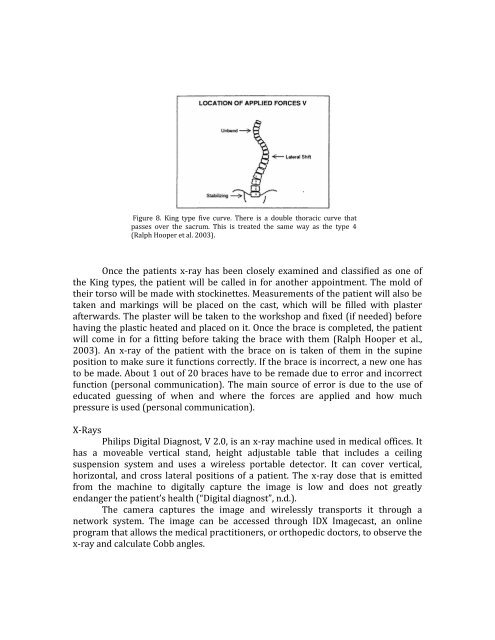
Figure 8. King type five curve. There is a double thoracic curve that<br />
passes over <strong>the</strong> sacrum. This is treated <strong>the</strong> same way as <strong>the</strong> type 4<br />
(Ralph Hooper et al. 2003).<br />
Once <strong>the</strong> patients x-ray has been closely examined and classified as one of<br />
<strong>the</strong> King types, <strong>the</strong> patient will be called in for ano<strong>the</strong>r appointment. The mold of<br />
<strong>the</strong>ir torso will be made with stockinettes. Measurements of <strong>the</strong> patient will also be<br />
taken and markings will be placed on <strong>the</strong> cast, which will be filled with plaster<br />
afterwards. The plaster will be taken to <strong>the</strong> workshop and fixed (if needed) before<br />
having <strong>the</strong> plastic heated and placed on it. Once <strong>the</strong> brace is completed, <strong>the</strong> patient<br />
will come in for a fitting before taking <strong>the</strong> brace with <strong>the</strong>m (Ralph Hooper et al.,<br />
2003). An x-ray of <strong>the</strong> patient with <strong>the</strong> brace on is taken of <strong>the</strong>m in <strong>the</strong> supine<br />
position to make sure it functions correctly. If <strong>the</strong> brace is incorrect, a new one has<br />
to be made. About 1 out of 20 braces have to be remade due to error and incorrect<br />
function (personal communication). The main source of error is due to <strong>the</strong> use of<br />
educated guessing of when and where <strong>the</strong> forces are applied and how much<br />
pressure is used (personal communication).<br />
X-Rays<br />
Philips Digital Diagnost, V 2.0, is an x-ray machine used in medical offices. It<br />
has a moveable vertical stand, height adjustable table that includes a ceiling<br />
suspension system and uses a wireless portable detector. It can cover vertical,<br />
horizontal, and cross lateral positions of a patient. The x-ray dose that is emitted<br />
from <strong>the</strong> machine to digitally capture <strong>the</strong> image is low and does not greatly<br />
endanger <strong>the</strong> patient’s health (“Digital diagnost”, n.d.).<br />
The camera captures <strong>the</strong> image and wirelessly transports it through a<br />
network system. The image can be accessed through IDX Imagecast, an online<br />
program that allows <strong>the</strong> medical practitioners, or orthopedic doctors, to observe <strong>the</strong><br />
x-ray and calculate Cobb angles.