Diaporthe rhusicola - Fungal Planet
Diaporthe rhusicola - Fungal Planet
Diaporthe rhusicola - Fungal Planet

Create successful ePaper yourself
Turn your PDF publications into a flip-book with our unique Google optimized e-Paper software.
134 Persoonia – Volume 26, 2011<br />
<strong>Diaporthe</strong> <strong>rhusicola</strong>
<strong>Fungal</strong> <strong>Planet</strong> description sheets<br />
135<br />
<strong>Fungal</strong> <strong>Planet</strong> 81 – 31 May 2011<br />
<strong>Diaporthe</strong> <strong>rhusicola</strong> Crous, sp. nov.<br />
Diaporthis neotheicolae similis, sed conidiis majoribus, (7–)8–9(–10) ×<br />
3(–3.5) µm, discernitur.<br />
Etymology. Named after the host from which it was collected, Rhus<br />
pendulina.<br />
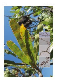
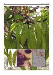
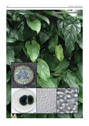
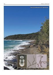
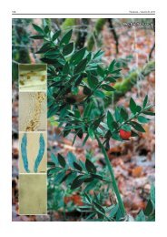
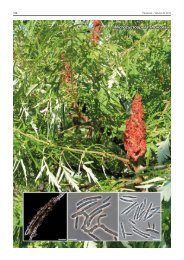
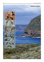
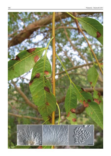
Leaf spots brown, amphigenous, subcircular, up to 1 cm diam,<br />
similar to those associated with Muribasidiospora indica on<br />
this host (Crous et al. 2003), except that in the latter they tend<br />
to be red to red-purple in colour. Pycnidia formed readily on<br />
potato-dextrose agar (PDA), oatmeal agar (OA) and malt extract<br />
agar (MEA); erumpent, flattened, black, multilocular, up<br />
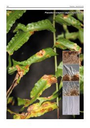
to 600 µm diam. Conidiophores on PDA lining the inner cavity,<br />
hyaline, smooth, 1–3-septate, subcylindrical, unbranched or<br />
branched (below or above), 20–40 × 2–3 µm. Conidiogenous<br />
cells terminal, hyaline, smooth, subcylindrical, 15–25 × 2–3<br />
µm, tapering somewhat towards a truncate apex, 1–1.5 µm,<br />
with a flaring collarette, up to 5 µm wide and long. Paraphyses<br />
intermingled among conidiophores, hyaline, smooth, subcylindrical,<br />
branched or not, up to 80 µm long, 2–3 µm wide. Conidia<br />
(7–)8–9(–10) × 3(–3.5) µm, solitary, hyaline, smooth, guttulate,<br />
subcylindrical to fusoid-ellipsoidal, apex obtuse, widest in middle,<br />
tapering to a truncate base, 1 µm diam.<br />
Culture characteristics — (in the dark, 25 °C, after 2 wk):<br />
Colonies spreading, with moderate to fluffy aerial mycelium and<br />
feathery margins, covering the dish in 2 wk; on MEA surface<br />
dirty white, reverse dirty white with patches of iron-grey; on OA<br />
iron-grey in centre, with patches of olivaceous grey and pale<br />
olivaceous grey; on PDA olivaceous grey in centre, with dirty<br />
white outer region, forming a diffuse yellow pigment in agar.<br />
Typus. South Africa, Western Cape Province, Cape Town, Kirstenbosch<br />
Botanical Garden, on leaves of Rhus pendulina (White Karee), 8 May 2010,<br />
P.W. Crous, holotype CBS H-20589, cultures ex-type CPC 18191 = CBS<br />
129528, ITS sequence GenBank JF951146 and LSU sequence GenBank<br />
JF951166, MycoBank MB560170.<br />
Notes — Based on a megablast search of NCBI’s GenBank<br />
nucleotide database, the closest hits using the ITS sequence<br />
are Phomopsis sp. BLE12 (FN868477; Identities = 574/574<br />
(100 %), Gaps = 0/574 (0 %)), Phomopsis sp. FE 86 (FJ440699;<br />
Identities = 537/537 (100 %), Gaps = 0/537 (0 %)), Phomopsis<br />
sp. 1 (AY485723; Identities = 514/517 (99 %), Gaps = 1/517<br />
(0 %)) and <strong>Diaporthe</strong> neotheicola (anamorph: Phomopsis theicola)<br />
(DQ286286; Identities = 519/523 (99 %), Gaps = 2/523<br />
(0 %)) (Santos & Phillips 2009). As far as we could establish,<br />
this is the first association of <strong>Diaporthe</strong> with a leaf spot disease<br />
on Rhus pendulina.<br />
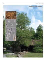
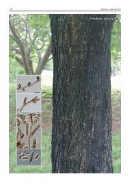
Colour illustrations. Leaf symptoms on Rhus pendulina in Kirstenbosch<br />
Botanical Garden; conidiophores giving rise to conidia. Scale bars = 10 µm.<br />
Pedro W. Crous & Johannes Z. Groenewald, CBS-KNAW <strong>Fungal</strong> Biodiversity Centre, P.O. Box 85167, 3508 AD Utrecht, The Netherlands;<br />
e-mail: p.crous@cbs.knaw.nl & e.groenewald@cbs.knaw.nl<br />
© 2011 Nationaal Herbarium Nederland & Centraalbureau voor Schimmelcultures