PDF 281 kb - Fungal Planet
PDF 281 kb - Fungal Planet
PDF 281 kb - Fungal Planet

Create successful ePaper yourself
Turn your PDF publications into a flip-book with our unique Google optimized e-Paper software.
196 Persoonia – Volume 23, 2009<br />
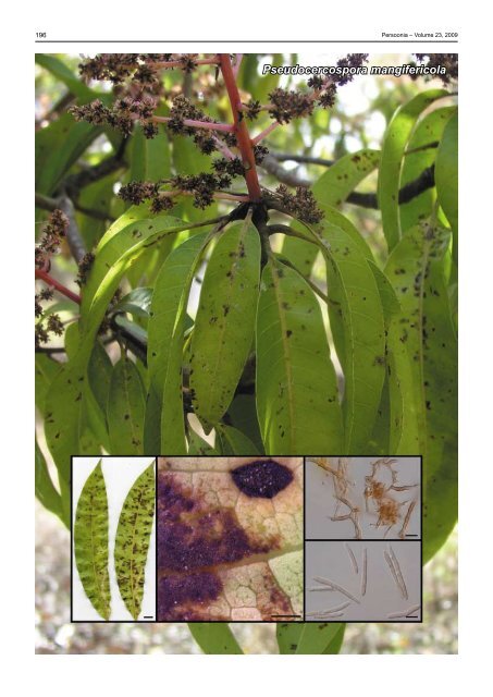
Pseudocercospora mangifericola
Persoonial Reflections<br />
197<br />
<strong>Fungal</strong> <strong>Planet</strong> 42 – 23 December 2009<br />
Pseudocercospora mangifericola R.G. Shivas, A.J. Young & Grice, sp. nov.<br />
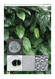
Mycelium internum ad externum, pallidum-brunneum ad brunneum; hyphae<br />
superficiales septatae, leves, ramosae, 2.5−4.5 µm latae. Stromata absunt.<br />
Conidiophora sola vel in fasciculis laxis usque ad sex, orientia ex hyphis<br />
superficialibus, lateralia, erecta, ampulliformia ad subcylindracea, recta vel<br />
interdum geniculata, 8−45 × 3.0−4.5 µm, raro ramosa. Cellulae conidiogenae<br />
terminales, pallidae-brunneae et pallidiores in apice. Conidia sola, pallidabrunnea,<br />
subcylindracea, 10−47 × 2.5−3.5 µm, recta vel partim curvata, apex<br />
rotundatus, basis obconice truncata ad subtruncata, 0–5-septata, interdum<br />
constricta in uno vel pluribus septis, levis.<br />
Etymology. Derived from the name of the host plant Mangifera in the<br />
Anacardiaceae.<br />
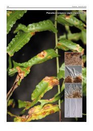
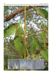
Leaf spots amphigenous; conspicuous on abaxial surface,<br />
polyangular to irregular, often vein-limited, 2–8 mm wide,<br />
margins diffuse or narrowly water-soaked, dark brown in centre<br />
becoming paler towards margin, often widespread over the<br />
entire leaf surface, lesions sometimes covered with abundant<br />
sporulation; inconspicuous on adaxial leaf surface, irregular,<br />
margins diffuse, pale to yellowish brown, always smaller than<br />
the corresponding lesion on the abaxial surface. Mycelium<br />
internal and external, pale to medium brown; superficial hyphae<br />
septate, smooth, branched, 2.5–4.5 µm wide. Stromata<br />
absent. Conidiophores solitary or in loose fascicles of up to 6,<br />
arising from superficial hyphae, lateral, erect, ampulliform to subcylindrical,<br />
straight or occasionally geniculate, 8–45 × 3.0–4.5<br />
µm, rarely branched. Conidiogenous cells terminal, pale brown<br />
becoming paler towards the apex, proliferating percurrently or<br />
occasionally sympodially, smooth or minutely roughened from<br />
torn annulations, rounded at apex. Conidia solitary, pale brown,<br />
subcylindrical, 10–47 × 2.5–3.5 µm, straight or partially curved,<br />
apex rounded, base obconically truncate to subtruncate, 0–5-<br />
septate, sometimes constricted at one or more septa, smooth;<br />
hila inconspicuous.<br />
Culture characteristics — Colonies on malt extract agar<br />
(Difco) circular, up to 30 mm diam after 28 d at 25 °C, grey to<br />
pale olivaceous-grey, reverse olivaceous-black, radially furrowed,<br />
flat and raised in the centre, margin entire, smooth.<br />
Typus. Australia, Queensland, Tolga, 17° 13' S, 145° 28' E, Mangifera<br />
indica cv. Kensington Pride, 28 Aug. 2009, K.R.E. Grice & P. Holt, BRIP<br />
52776b, holotype; cultures ex-type BRIP 52776b, GenBank GU188048,<br />
MycoBank MB515467.<br />
Notes — Pseudocercospora mangifericola parasitised mango<br />
(Mangifera indica) leaves that were also infected at low levels<br />
with Scolecostigmina mangiferae (syn. Cercospora mangiferae,<br />
Stigmina mangiferae), which differs symptomatically by producing<br />
angular, black lesions with chlorotic haloes visible on both<br />
leaf surfaces (see middle photograph in adjacent illustrations)<br />
and is clearly distinguishable based on ITS sequence. The only<br />
other cercosporoid fungus reported on mango is C. mangiferaeindicae,<br />
which differs by having broader conidia measuring 3–6<br />
µm in width 1 .<br />
76<br />
76<br />
59<br />
64<br />
74<br />
Pseudocercospora avicenniae BRIP 52764 (GU188047)<br />
Pseudocercospora mangifericola BRIP 52776 (GU188048)<br />
Pseudocercospora lythracearum voucher KACC 42649 (EF535720)<br />
Pseudocercospora paraguayensis strain CBS 111286 (DQ267602)<br />
Passalora schizolobii strain CBS 120029 (DQ885903)<br />
Pseudocercospora tereticornis CPC:13008 (GQ852769)<br />
Pseudocercospora vitis voucher KACC 42650 (EF535721)<br />
Mycosphaerella heimii strain AGI004D (EU882122)<br />
0.005<br />
An ITS neighbour-joining tree constructed using MEGA4 2 . The<br />
scale bar shows 0.005 changes per site, and bootstrap support<br />
values from 1 000 replicates are shown at the nodes. The species<br />
described here is printed in bold face. The tree was rooted<br />
to Mycosphaerella heimii (GenBank EU882122).<br />
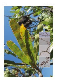
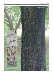
Colour illustrations. Mangifera indica with leaf spots caused by Pseudocercospora<br />
mangifericola at Tolga, Queensland; leaf (upper side left, lower<br />
side right) infected with P. mangifericola; leaf spots caused by P. mangifericola<br />
(left and middle) and Scolecostigmina mangiferae (top right); hyphae and<br />
conidiophores; conidia in vivo. Scale bars (from left to right) = 1 cm, 1 mm,<br />
10 µm, 10 µm.<br />
References. 1 Crous PW, Braun U. 2003. Mycosphaerella and its anamorphs.<br />
1. Names published in Cercospora and Passalora. CBS Biodiversity<br />
Series 1: 1–157. 2 Tamura K, Dudley J, Nei M, Kumar S. 2007. MEGA4:<br />
Molecular Evolutionary Genetics Analysis (MEGA) v4.0. Molecular Biology<br />
and Evolution 24: 1596–1599.<br />
© 2009 Nationaal Herbarium Nederland & Centraalbureau voor Schimmelcultures<br />
Roger G. Shivas & Anthony J. Young, Plant Pathology Herbarium, Queensland Primary Industries and Fisheries,<br />
80 Meiers Rd, Indooroopilly 4068, Queensland, Australia<br />
e-mail: roger.shivas@deedi.qld.gov.au & anthony.young@deedi.qld.gov.au<br />
Kathy R. E. Grice, Centre for Tropical Agriculture, Queensland Primary Industries and Fisheries, 28 Peters Street,<br />
Mareeba 4880, Queensland, Australia; e-mail: kathy.grice@deedi.qld.gov.au