Pseudocercospora nephrolepidicola - Fungal Planet
Pseudocercospora nephrolepidicola - Fungal Planet
Pseudocercospora nephrolepidicola - Fungal Planet

You also want an ePaper? Increase the reach of your titles
YUMPU automatically turns print PDFs into web optimized ePapers that Google loves.
138 Persoonia – Volume 25, 2010<br />
<strong>Pseudocercospora</strong> <strong>nephrolepidicola</strong>
Persoonial Reflections<br />
139<br />
<strong>Fungal</strong> <strong>Planet</strong> 59 – 23 December 2010<br />
<strong>Pseudocercospora</strong> <strong>nephrolepidicola</strong> Crous & R.G. Shivas, sp. nov.<br />
Teleomorph. Mycosphaerella-like.<br />
<strong>Pseudocercospora</strong>e nephrolepidis similis, sed conidiis minoribus, (40–)50–<br />
60(–95) × (2.5–)3.5(–4) µm, distinguitur.<br />
Etymology. Named after the host from which it was collected, Nephrolepis<br />
(Lomariopsidaceae).<br />
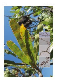
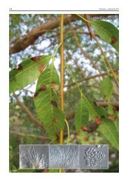
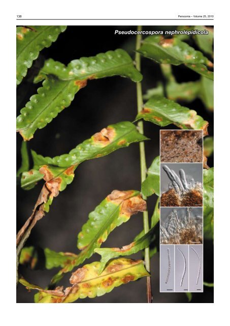
Leaf spots amphigenous, medium brown, with indistinct margins,<br />
2–12 mm diam. Conidiomata pale to medium brown,<br />
amphigenous, fasciculate, arising from a well-developed<br />
subepidermal, medium brown stroma, up to 150 µm wide, and<br />
50 µm high. Mycelium consisting of smooth, septate, brown,<br />
branched, 2–3 µm diam hyphae. Conidiophores subcylindrical,<br />
medium brown, smooth, unbranched or branched below, irregularly<br />
geniculate-sinuous, in loosely aggregated fascicles, or<br />
separate on superficial mycelium, 1–4-septate, 25–45(–90) ×<br />
2.5–3(–3.5) µm. Conidiogenous cells terminal on conidiophore,<br />
integrated, subcylindrical, pale brown, smooth, proliferating 1–2<br />
times percurrently near apex, 15–25(–40) × (2–)2.5(–3) µm.<br />
Conidia medium brown, smooth, guttulate, subcylindrical,<br />
straight to irregularly flexuous, apex obtusely rounded, base<br />
truncate, 3–6(–9)-septate, (40–)50–60(–95) × (2.5–)3.5(–4)<br />
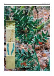
µm; hila not thickened nor darkened. Ascomata globose,<br />
erumpent, brown, up to 80 µm diam, with a central ostiole.<br />
Asci subcylindrical to narrowly obovoid, 35–50 × 8–10 µm.<br />
Ascospores fusoid-ellipsoidal, widest in middle of apical cell,<br />
tapering towards both ends, apex acutely rounded, constricted<br />
at septum, 9–11 × 2.5–3.5 µm.<br />
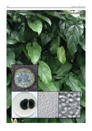
Culture characteristics — (in the dark, 25 °C, after 2 wk):<br />
Colonies spreading, erumpent, with folded surface and even,<br />
lobate margins, reaching up to 15 mm diam. On potato-dextrose<br />
agar surface smoke-grey with patches of grey-olivaceous,<br />
iron-grey in reverse; on malt extract agar pale olivaceous-grey<br />
(surface), iron-grey in reverse; on oatmeal agar olivaceous-grey<br />
with patches of pale olivaceous-grey.<br />
Typus. Australia, Queensland, Brisbane Botanical Garden, on fronds<br />
of Nephrolepis falcata, 14 July 2009, P.W. Crous & R.G. Shivas, CBS-H<br />
20492 holotype, cultures ex-type CPC 17050, 17049 = CBS 128211, ITS<br />
sequence of CPC 17049 GenBank HQ599590 and LSU sequence of CPC<br />
17049 GenBank HQ599591, MycoBank MB517538.<br />
Notes — There are several specimens of <strong>Pseudocercospora</strong><br />
spp. on Nephrolepis in BRIP, which cannot easily be identified<br />
using morphology alone. <strong>Pseudocercospora</strong> <strong>nephrolepidicola</strong><br />
is morphologically and phylogenetically distinct from P. nephrolepidis<br />
(on Nephrolepis cordifolia (as N. auriculata) in Taiwan 1 ;<br />
conidia subcylindrical, (32–)67–101(–113) × 2–3 μm, 2–9<br />
septate; CBS 119121), in that its conidia are shorter, and wider.<br />
Furthermore, <strong>Pseudocercospora</strong> phyllitidis, which was described<br />
from leaves of Nephrolepis sp. from Florida, has smaller<br />
stromata (up to 75 µm diam) with straight to mildly curved<br />
obclavate conidia, 20–80 × 2–3.5 µm 2 , than the Australian<br />
specimen. A megablast search of NCBIs GenBank nucleotide<br />
database using the LSU sequence retrieved as closest sisters<br />
Mycosphaerella quasiparkii (GenBank EU882143; Identities<br />
= 807/808 (99 %), Gaps = 0/808 (0 %)), Rosenscheldiella<br />
brachyglottidis (GenBank GQ355334; Identities = 874/886<br />
(99 %), Gaps = 0/886 (0 %)), Mycosphaerella swartii (GenBank<br />
DQ923536; Identities = 865/888 (98 %), Gaps = 3/888 (0 %))<br />
and <strong>Pseudocercospora</strong> vitis (GenBank GU214483; Identities<br />
= 864/889 (98 %), Gaps = 5/889 (0 %)). A megablast with the<br />
ITS sequence revealed high identity to ‘Mycosphaerella sp. De-<br />
No’ (GenBank HM189290; Identities = 481/482 (99 %), Gaps =<br />
0/482 (0 %)), M. quasiparkii (GenBank EU882127; Identities =<br />
573/597 (96 %), Gaps = 17/597 (2 %)) and <strong>Pseudocercospora</strong><br />
schizolobii (GenBank GQ852765; Identities = 571/610 (94 %),<br />
Gaps = 28/610 (4 %)).<br />
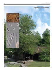
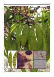
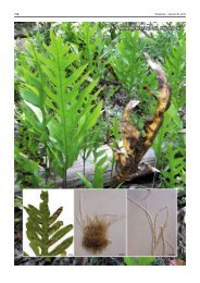
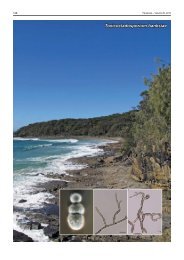
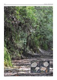
Colour illustrations. Nephrolepis falcata at Brisbane Botanical Gardens;<br />
conidiomata on frond; ascomatum, asci with ascospores; conidiophores,<br />
conidia. Scale bars = 10 µm.<br />
References. 1 Kirschner R, Chen CJ. 2007. Foliicolous hyphomycetes from<br />
Taiwan. <strong>Fungal</strong> Diversity 26: 219–239. 2 Chupp C. 1954. A monograph of the<br />
fungus genus Cercospora. Ithaca, New York. Published by the author.<br />
Pedro W. Crous & Johannes Z. Groenewald, CBS-KNAW <strong>Fungal</strong> Biodiversity Centre, P.O. Box 85167, 3508 AD Utrecht, The Netherlands;<br />
e-mail: p.crous@cbs.knaw.nl & e.groenewald@cbs.knaw.nl<br />
Roger G. Shivas, Agri-Science Queensland, Ecosciences Precinct, Dutton Park 4102, Queensland, Australia;<br />
e-mail: roger.shivas@deedi.qld.gov.au<br />
© 2010 Nationaal Herbarium Nederland & Centraalbureau voor Schimmelcultures