DIFFICULTIES IN THE DIAGNOSIS OF BEZOARS ... - doki.NET Portál
DIFFICULTIES IN THE DIAGNOSIS OF BEZOARS ... - doki.NET Portál
DIFFICULTIES IN THE DIAGNOSIS OF BEZOARS ... - doki.NET Portál
You also want an ePaper? Increase the reach of your titles
YUMPU automatically turns print PDFs into web optimized ePapers that Google loves.
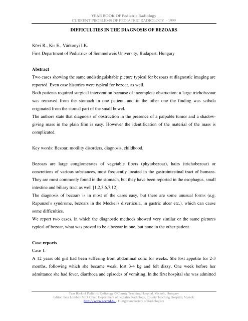
YEAR BOOK <strong>OF</strong> Pediatric Radiology<br />
CURRENT PROBLEMS <strong>OF</strong> PEDIATRIC RADIOLOGY - 1999<br />
<strong>DIFFICULTIES</strong> <strong>IN</strong> <strong>THE</strong> <strong>DIAGNOSIS</strong> <strong>OF</strong> <strong>BEZOARS</strong><br />
Kövi R., Kis E., Várkonyi I.K.<br />
First Department of Pediatrics of Semmelweis University, Budapest, Hungary<br />
Abstract<br />
Two cases showing the same undistinguishable picture typical for bezoars at diagnostic imaging are<br />
reported. Even case histories were typical for bezoar, as well.<br />
Both patients required surgical intervention because of incomplete obstruction: a large trichobezoar<br />
was removed from the stomach in one patient, and in the other one the finding was scibala<br />
originated from the stomal part of the small bowel.<br />
The authors state that diagnosis of obstruction in the presence of a palpable tumor and a shadowgiving<br />
mass in the plain film is easy. However the identification of the material of the mass is<br />
complicated.<br />
Key words: Bezoar, motility disorders, diagnosis, childhood.<br />
Bezoars are large conglomerates of vegetable fibers (phytobezoar), hairs (trichobezoar) or<br />
concretions of various substances, most frequently located in the gastrointestinal tract of humans.<br />
They are most commonly found in the stomach, but they have been reported in the esophagus, small<br />
intestine and biliary tract as well [1,2,3,6,7,12].<br />
The diagnosis of bezoars is in most of the cases easy, but there are some unusual forms (e.g.<br />
Rapunzel's syndrome, bezoars in the Meckel's diverticula, in gastric ulcer etc.), which can cause<br />
some difficulties.<br />
We report two cases, in which the diagnostic methods showed very similar or the same pictures<br />
typical of bezoar, what was proved to be a bezoar in one, but none in the other patient.<br />
Case reports<br />
Case 1.<br />
A 12 years old girl had been suffering from abdominal colic for weeks. She lost appetite for 2-3<br />
months, following which she became weak, lost 3-4 kg and felt dizzy. One week before her<br />
admittance she had fever, diarrhoea and episodes of vomiting. In the first hospital she was admitted<br />
Year Book of Pediatric Radiology © County Teaching Hospital, Miskolc, Hungary<br />
Editor: Bela Lombay M.D. Chief, Department of Pediatric Radiology, County Teaching Hospital, Miskolc<br />
http://www.socrad.hu - Hungarian Society of Radiologists
YEAR BOOK <strong>OF</strong> Pediatric Radiology<br />
CURRENT PROBLEMS <strong>OF</strong> PEDIATRIC RADIOLOGY - 1999<br />
to, upper gastrointestinal contrast study was carried out. Because of its result and the patient's<br />
history, the suspicion of an abdominal tumor arose, so she was sent to our department.<br />
At physical examination a hard abdominal mass, as big as a fist was palpated in the left side of the<br />
abdomen (Fig. 1,2).<br />
Fig. 1: Plain film shows a big mass filling the stomach.<br />
(Remaining contrast material in the large bowel] from the previous barium study.)<br />
Fig. 2: US revealed a solid mass in the projection on the stomach with calcification in its wall and a<br />
wide hypoechoic shadow.<br />
Year Book of Pediatric Radiology © County Teaching Hospital, Miskolc, Hungary<br />
Editor: Bela Lombay M.D. Chief, Department of Pediatric Radiology, County Teaching Hospital, Miskolc<br />
http://www.socrad.hu - Hungarian Society of Radiologists
YEAR BOOK <strong>OF</strong> Pediatric Radiology<br />
CURRENT PROBLEMS <strong>OF</strong> PEDIATRIC RADIOLOGY - 1999<br />
Fig. 3: Surgical finding: the trichobezoar consists of dark hair and trapped food particles, taking the<br />
shape of the stomach.<br />
The plain film showed a big mass filling the stomach. Ultrasound examination revealed a solid mass<br />
in the projection on the stomach with calcification in its wall. CT affirmed the<br />
suspicion of bezoar. The patient's condition deteriorated, blood test revealed severe anaemia (Hb:<br />
74 gll, Hct: 24 %). After stabilization of her condition, the girl underwent laparotomy.<br />
Trichobezoar filling out the stomach was removed by gastrostomy (Fig. 3.)<br />
The postoperative course was uneventful, gastric passage was normal.<br />
Detailed verbal exploration of the girl threw light on her trichotillomania. The girl was referred to a<br />
paediatric psychologist for assessment and therapy.<br />
Case 2.<br />
A 8 months old baby girl was admitted to our department because of exsiccosis and enteritis. The<br />
patient's history included sigmoideostomy because of cloacal malformation and cutaneoureterostomy<br />
because of ureterovesical stenosis in the right solitary kidney.<br />
Physical examination revealed symptoms of exsiccosis. Blood tests showed acidosis (pH: 7,27;<br />
pC02: 22,4 Hgmm; actHCO,: 9,4 mmol/I; BE: -15,6 mmol/l; p02: 63,9 Hgmm; 02 SAT: 88,4 %)<br />
and acetonuria was present (Fig. 4,5.).<br />
Year Book of Pediatric Radiology © County Teaching Hospital, Miskolc, Hungary<br />
Editor: Bela Lombay M.D. Chief, Department of Pediatric Radiology, County Teaching Hospital, Miskolc<br />
http://www.socrad.hu - Hungarian Society of Radiologists
YEAR BOOK <strong>OF</strong> Pediatric Radiology<br />
CURRENT PROBLEMS <strong>OF</strong> PEDIATRIC RADIOLOGY - 1999<br />
Fig. 4: Plain film shows a mottled shadow in projection of sigmoideostoma.<br />
Fig. 5: A solid mass with strong first-interface reflection of sound was seen by US.<br />
In the plain film some dilated bowel loops and a soft-tissue lesion of 5 cm in diameter in projection<br />
on the sigmoidostomy were seen. Contrast enema showed the same findings. Ultrasound revealed a<br />
solid mass with hyperechogenic calcified surface with acoustic shadowing.<br />
At the surgical revision of the stoma and removal of the mass which led to bowel obstruction the<br />
diagnosis of a scibalum was made. Follow-ups indicate that the baby is doing well.<br />
Discussion<br />
Predisposing factors for formation of gastrointestinal bezoars include all the situations with<br />
decreased bowel motility, e.g. operations on the stomach or small intestine, psychotic problems, etc.<br />
[1,2, 10]. Trichobezoars are most commonly seen in the stomach of young girls. Phytobezoars<br />
Year Book of Pediatric Radiology © County Teaching Hospital, Miskolc, Hungary<br />
Editor: Bela Lombay M.D. Chief, Department of Pediatric Radiology, County Teaching Hospital, Miskolc<br />
http://www.socrad.hu - Hungarian Society of Radiologists
YEAR BOOK <strong>OF</strong> Pediatric Radiology<br />
CURRENT PROBLEMS <strong>OF</strong> PEDIATRIC RADIOLOGY - 1999<br />
generally develop after gastrointestinal operations, in case of motility disturbances with decreased<br />
peristalsis and in presence of undigested materials, as medicaments, vegetable fibres, cellulose,<br />
chewing gum, etc. [5,7,12].<br />
Gastrointestinal bezoars clinically manifest in complete or incomplete obstruction in most of the<br />
cases.<br />
The diagnosis of obstruction is easy, but identifying the material causing the obstruction is almost<br />
always impossible.<br />
Diagnosis of a bezoar may be suspected at physical examination at palpation, in plain films and on<br />
ultrasonography [10]. CT and MR may provide help in difficult cases (e.g. Rapulzen's syndrome),<br />
and reveal possible complications [3,9,11].<br />
The therapy of bezoars includes removal of the material and prevention of recurrence. Generally<br />
surgical removal of the bezoar is used [10], but nonsurgical procedures have also been reported.<br />
Small gastric bezoars can be solved by enzymatic therapy (chymopapain, cellulase, acetylysteine)<br />
[6] or may be removed by endoscopy [4,12]. Nasogastric lavage can be effective in the cases of<br />
smaller gastric and intestinal bezoars [12]. Endoscopic fragmentation in the stomach may be<br />
successful, but close observation is required because fragments may drop into the small bowel and<br />
lead to obstruction [4,12].<br />
In cases of complete obstruction the only way for solving the problem is surgery. Laparotomy is<br />
required in the cases of large bezoars and complications [8,9]<br />
Prognosis after the surgery is excellent. However some cases of residual and recurrent bezoars have<br />
been reported. Remains of some parts of the bezoar may be prevented by thorough checking of the<br />
intestine and stomach during operation.<br />
Psychiatric evaluation and therapy is the best method in preventing recurrence of trichobezoars. In<br />
preventing the recurrence of phytobezoars high fiber diet has to be avoided, correction of impaired<br />
teeth and use of prokinetic agents may also be of help. Longstanding obstruction may lead to<br />
dilatation of a part of the bowel, which may cause repeated bezoar formation. Ellaway suggest<br />
reducing the size of the dilated bowel by infolding plication [1].Both of our patients underwent<br />
surgical intervention because of incomplete obstruction. There was no difficulty in diagnosing<br />
obstruction itself. In the patient's history in both of the cases predisposing factors for bezoar-<br />
formation were present (trichotillomania, bowel operation with narrowed stoma). The mass causing<br />
obstruction gave the same picture in both cases on ultrasonography and in the plain film. Howewer,<br />
in the second patient there was no bezoar formation present.<br />
Year Book of Pediatric Radiology © County Teaching Hospital, Miskolc, Hungary<br />
Editor: Bela Lombay M.D. Chief, Department of Pediatric Radiology, County Teaching Hospital, Miskolc<br />
http://www.socrad.hu - Hungarian Society of Radiologists
YEAR BOOK <strong>OF</strong> Pediatric Radiology<br />
CURRENT PROBLEMS <strong>OF</strong> PEDIATRIC RADIOLOGY - 1999<br />
On the basis of morphology it can not always be decided what the cause of the obstruction is. In<br />
presence of an incomplete obstruction knowledge of the material content of the obstructing mass<br />
would be of great help in making a choice for a possible nonsurgical removal (e.g. a bezoar formed<br />
around a piece of chewing gum can not be solved enzimatically, and possibly active substances in<br />
pharmacobezoars should be considered as well).<br />
References<br />
1. Ellaway C, Beasley SW: Bezoar formation and malabsorption secondary to persistent dilata<br />
tion and dysmotility of the duodenum after repair of proximal jejuna) atresia. Pediatr Surg Int<br />
12:190-191, 1997.<br />
2. Escamilla C, Robles CR, Parrilla PP, Lujan MJ, Liron RR, Torralba-Martinet JA: Intestinal<br />
obstruction and bezoars. J Am Coll Surg 179(3), 285-8, 1994.<br />
3. Frazzini VI, English WJ, Bashist B, Moore E: Small bowel) obstruction due to phytobezoar<br />
formation within Meckel diverticulum: CT findings. J Comput Assist Tomogr 20(3), 390-<br />
392,1996.<br />
4. Manbeck MA, Walter MH, Chen YK: Gastric bezoar formation in a patient with Scleroderma:<br />
Endoscopic Removal using the gallstone mechanical lithotripter, AJG 91, 1285, 1996.<br />
5. Milov DE, Andres JM, Erhart NA, Bailey DJ: Chewing gum bezoars of the gastrointestinal<br />
tract. (Electronic article) Pediatr 102(2), e22, 1998.<br />
6. Nomura H, Kitamura T, Takahashi Y: Small bowel] obstruction during enzymatic treatment of<br />
gastric bezoar. Endoscopy 29(5), 424-6, 1997.<br />
7. Razafimahefa H, Mouterde O, Devaux AM: Esophageal bezoar in a sucralfate-treated child.Arch<br />
Pédiatr4:656-661, 1997.<br />
8. Réti Gy, Pályi M, Nagy A: Giant "casting" besoir in stomach which can be removed only<br />
by surgical intervention. Orv Hetil 138(3), 149-151, 1997. (in Hung)<br />
9. Siegel MJ:Pediatric Sonography. Raven Press Second Edition 219-221, 1994.<br />
10. Sinzig M, Umschaden HW, Haselbach H, Illing P: Gastric trichobezoar with gastric ulcer: MR<br />
findings. Pediatr Radio] 28:296, 1998.<br />
11. West WM, Duncan ND: CT appearences of the Rapunzel syndrome: an anusual form of bezoar<br />
and gastrointestinal obstruction. Pediatr Radio) 28:315, 1998.<br />
12. Yin WY, Lin PW, Huang SM, Lee PC, Lee CC, Chang TW, Yang YJ: Bezoar manifested with<br />
digestive and biliary obstruction. Hepatogastroenterology 44:1037-45, 1997.<br />
Year Book of Pediatric Radiology © County Teaching Hospital, Miskolc, Hungary<br />
Editor: Bela Lombay M.D. Chief, Department of Pediatric Radiology, County Teaching Hospital, Miskolc<br />
http://www.socrad.hu - Hungarian Society of Radiologists
Rita Kövi M. D.<br />
YEAR BOOK <strong>OF</strong> Pediatric Radiology<br />
CURRENT PROBLEMS <strong>OF</strong> PEDIATRIC RADIOLOGY - 1999<br />
First Department of Pediatrics of Semmelweis University<br />
H-1083 Budapest, Bokay J. u. 53. Hungary<br />
Tel: (36-1) 334-3186, Fax: (36-1) 303 6077<br />
E-mail: kovi@gyerl.sote.hu<br />
Year Book of Pediatric Radiology © County Teaching Hospital, Miskolc, Hungary<br />
Editor: Bela Lombay M.D. Chief, Department of Pediatric Radiology, County Teaching Hospital, Miskolc<br />
http://www.socrad.hu - Hungarian Society of Radiologists


