PHOIBOS 100/150 - Specs
PHOIBOS 100/150 - Specs
PHOIBOS 100/150 - Specs

Create successful ePaper yourself
Turn your PDF publications into a flip-book with our unique Google optimized e-Paper software.
Surface Analysis<br />
Technology<br />
Vacuum<br />
Components<br />
Surface Analysis<br />
System Software<br />
Computer<br />
Technology<br />
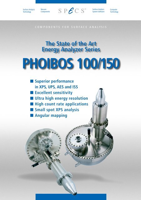
COMPONENTS FOR SURFACE ANALYSIS<br />
The State of the Art<br />
Energy Analyzer Series<br />
<strong>PHOIBOS</strong> <strong>100</strong>/<strong>150</strong><br />
■ Superior performance<br />
in XPS, UPS, AES and ISS<br />
■ Excellent sensitivity<br />
■ Ultra high energy resolution<br />
■ High count rate applications<br />
■ Small spot XPS analysis<br />
■ Angular mapping
<strong>PHOIBOS</strong> <strong>100</strong>/<strong>150</strong><br />
<strong>PHOIBOS</strong> <strong>100</strong>/<strong>150</strong><br />
With the new generation of the well-tried and<br />
proven <strong>PHOIBOS</strong> series of hemispherical<br />
energy analyzers SPECS sets a new standard.<br />
The <strong>PHOIBOS</strong> analyzer<br />
is a main part of the SPECS<br />
multi-technique surface<br />
analysis systems.<br />
New approaches and technical solutions lead<br />
to an instrument that combines excellent performance<br />
and highest reliability for the largest<br />
possible variety of experimental conditions.<br />
<strong>PHOIBOS</strong><br />
The Greek deity<br />
Apollo was often<br />
called<br />
<strong>PHOIBOS</strong> Apollo,<br />
an epithet that means<br />
"bright".<br />
Most advanced and<br />
sophisticated computer<br />
simulations were used to fully<br />
characterize and optimize the<br />
electron optical properties of<br />
the analyzer and transfer lens.<br />
Analyzer<br />
■<br />
■<br />
■<br />
True 180 ° hemispherical energy<br />
analyzer with <strong>100</strong> or <strong>150</strong> mm<br />
mean radius<br />
Exclusive use of non-magnetic<br />
materials inside µ-metal shielding<br />
Sophisticated Slit Orbit mechanism<br />
for external setting of<br />
8 entrance and 3 exit slits<br />
The <strong>PHOIBOS</strong> analyzer is available with <strong>100</strong> and<br />
<strong>150</strong> mm mean radius.<br />
Due to the modular concept of the construction<br />
the analyzer can be easily adapted to meet special<br />
requirements.<br />
Highly effective fringe field corrections in the entrance<br />
and exit areas result in excellent energy<br />
resolution at very low kinetic energies as demonstrated<br />
in various benchmark tests (see Xe 5p 3/2<br />
gas phase UPS spectrum).<br />
With the new sophisticated Slit Orbit the user can<br />
independently select one of 8 pairs of entrance<br />
slits and one of 3 exit slits via one rotary drive<br />
from outside the vacuum. Entrance and exit slits<br />
can be operated independently.<br />
In each pair of entrance slits one slit defines the<br />
energy resolution while the other slit serves to<br />
match the angular spread for the analyzer. This<br />
arrangement allows optimum transmission for<br />
all chosen slit sizes and resolution settings.<br />
For ultimate performance the analyzer and the<br />
lens system are constructed entirely from nonmagnetic<br />
materials inside the µ-metal shielding.<br />
Additionally, a mesh-covered hole is placed in the<br />
outer hemisphere in line-of-sight of the lens axis<br />
in order to reduce the scattered intensity. A view<br />
port is provided for through the lens (optical)<br />
alignment.<br />
C o m p e t e n c e i n S u r f a c e A n a l y s i s
<strong>PHOIBOS</strong> <strong>100</strong>/<strong>150</strong><br />
Transfer Lens<br />
■<br />
■<br />
■<br />
■<br />
■<br />
High étendue for XPS and UPS<br />
High point transmission for<br />
synchrotron, AES and ISS<br />
applications<br />
Multimode transfer lens for angular<br />
and spatially resolved studies<br />
Iris diffraction plane aperture<br />
Small spot analysis down to<br />
<strong>100</strong> µm resolution<br />
Ag 3d 5/2 Peak Area (Normalized Units)<br />
1.0<br />
0.8<br />
0.6<br />
0.4<br />
0.2<br />
0.0<br />
SSXPS<br />
<strong>PHOIBOS</strong> <strong>100</strong> MCD-5<br />
Ag/Cu edge<br />
Mg K α<br />
Ø Slit 6 mm<br />
Ø Iris 10 mm<br />
Ø Slit 1 mm<br />
Ø Iris 5 mm<br />
High Magnification<br />
FWHM = 458µm<br />
FWHM = 96 µm<br />
-750 -500 -250 0 250 500 750<br />
Lateral Displacement (µm)<br />
Ag<br />
Cu<br />
lateral displacement<br />
Experimental XPD<br />
photoelectron hologram<br />
of bulk 2s emission<br />
from Si(111) excited<br />
by Mg Kα using a<br />
<strong>PHOIBOS</strong> <strong>150</strong> MCD-9<br />
analyzer. The angular<br />
resolution was set with<br />
the Iris to 1°<br />
(data with courtesy of<br />
T. Matsushita, A. Agui<br />
and A. Yoshigoe,<br />
Spring-8, Japan)<br />
ÉTENDUE<br />
The product of area<br />
and solid angle of the<br />
electrons accepted by<br />
the spectrometer.<br />
This specification<br />
is required to determine<br />
XPS sensitivity.<br />
The multi element two-stage transfer lens was<br />
designed to yield ultimate transmission and<br />
well-defined optical properties. It may be operated<br />
in several different modes for angular and<br />
spatially resolved studies to adapt the analyzer<br />
to different tasks. All lens modes can be set<br />
electronically.<br />
The standard working distance of 40 mm and<br />
the 44° conical shape of the front part of the<br />
lens provide optimum access to the sample for<br />
all types of excitation sources.<br />
For small spot analysis a lateral resolution down<br />
to <strong>100</strong> µm is available using the High Magnification<br />
Mode and the novel Iris aperture.<br />
In the Magnification Modes angle-resolving is<br />
accomplished with an Iris aperture in the diffraction<br />
plane of the lens system. Using this Iris<br />
the angular resolution can be continuously adjusted<br />
between ±1° and ±9° while keeping the<br />
acceptance area on the sample constant.<br />
Small Spot XPS at a Ag/Cu edge with a broad illuminating<br />
source using the Iris aperture. The typical<br />
broadening of the acceptance area due to lens aberrations<br />
is eliminated.<br />
The Area Modes were optimized to allow very<br />
high transmissions for different spot sizes of the<br />
source.<br />
In the Angular Dispersion Modes electrons<br />
leaving the sample within a given angular<br />
range are focussed on the same location on the<br />
analyzer entrance independent of their position<br />
on the sample. The angular modes allow the<br />
user to optimize the angular resolution down to<br />
±0.05° with the Slit Orbit.<br />
With a 2-D detection system high angular<br />
resolution can be achieved in the nondispersion<br />
direction of the analyzer without restricting the<br />
acceptance angle (Angular Mapping). These<br />
modes are the ideal choice for angular dependent<br />
studies.<br />
BROADENING<br />
The FWHM for a Gaussian<br />
broadening at an edge is<br />
equivalent to a 12 to 88% intensity<br />
change across the<br />
step edge.<br />
A 20 to 80% rule defines a<br />
value which is only 0.71 of<br />
the Gaussian broadening.<br />
ACCEPTANCE AREA<br />
A focused electron beam<br />
rastered over the sample area<br />
could be used to determine<br />
the FWHM of the acceptance<br />
area without restricting the<br />
acceptance angle with an<br />
aperture.<br />
With a broad illuminating<br />
X-ray source also small intensities<br />
from outside the analysis<br />
area can contribute considerably<br />
to the signal. The<br />
Iris aperture eliminates these<br />
contributions.<br />
POINT TRANSMISSION<br />
For a point source, defined<br />
as the solid angle<br />
over which charged particles<br />
are accepted by the<br />
spectrometer and transmitted<br />
to the dispersing<br />
element.<br />
This specification<br />
is required to determine<br />
point source sensitivity.<br />
Intensity (Mcps)<br />
10<br />
8<br />
6<br />
4<br />
Cut-off<br />
2<br />
0<br />
20<br />
UPS<br />
<strong>PHOIBOS</strong> <strong>150</strong> MCD-9<br />
Ag<br />
He I (hν = 21.23 eV)<br />
15<br />
10<br />
Binding Energy (eV)<br />
FWHM = 130 meV<br />
Fermi Edge at T = 300 K<br />
5<br />
× 50<br />
0<br />
The Ag 3d peak obtained with the <strong>PHOIBOS</strong> <strong>150</strong><br />
MCD-9 demonstrates the high étendue of the analyzer<br />
and the transfer lens.<br />
With the high brightness SPECS ultraviolet excitation<br />
sources the <strong>PHOIBOS</strong> analyzer series guarantees<br />
superior performance in ultraviolet photoelectron<br />
spectroscopy.<br />
C o m p e t e n c e i n S u r f a c e A n a l y s i s
<strong>PHOIBOS</strong> <strong>100</strong>/<strong>150</strong><br />
Mode Acceptance area Acceptance angle Typical Applications<br />
Spatially resolved Entrance slit size divided by M Continuously adjustable down Small area XPS, UPS<br />
independent of analyzer settings. to ±1° using Iris aperture standard ARXPS and ARUPS<br />
Slit sizes 7 ✕ 20, 3 ✕ 20, 1 ✕ 20,<br />
0.5 ✕ 20, 0.2 ✕ 20, ∅7, 3 and 1 mm<br />
High Magnification Magnification M = 10 Up to ±9°<br />
Medium Magnification Magnification M = 5 Up to ±6°<br />
Low Magnification Magnification M = 2 Up to ±3°<br />
Transmission optimized Optimized for different Large Area XPS<br />
spot sizes of the source<br />
Monochromated XPS<br />
Typical spot size<br />
AES, ISS and<br />
Large Area ∅ 5 mm Up to ±5° synchrotron studies<br />
Medium Area ∅ 2 mm Up to ±7°<br />
Small Area ∅ 0.1 mm Up to ±9°<br />
The table shows the lens<br />
modes of operation. The multielement<br />
transfer lens of the<br />
<strong>PHOIBOS</strong> analyzer may be<br />
operated in several different<br />
modes: spatial resolution,<br />
optimzed transmission or<br />
angular resolution (see table).<br />
Additional acceleration modes<br />
for low kinetic energy applications<br />
are available (HighMagnification2<br />
and SmallArea2).<br />
Angular resolved Slightly decreasing with Entrance slit size divided by D Low kinetic<br />
increasing retarding ratio independent of analyzer settings energy applications.<br />
and independent of slit sizes Slit sizes 7 ✕ 20, 3 ✕ 20, 1 ✕ 20, High angular resolved<br />
0.5 ✕ 20, 0.2 ✕ 20, ∅ 7, 3 and 1 mm ARXPS / ARUPS with a 2D<br />
High Angular Dispersion D = 3.2 mm / ° (±3° acceptance) detection system<br />
Medium Angular Dispersion D = 2.2 mm / ° (±4° acceptance) (Angular Mapping)<br />
Low Angular Dispersion<br />
D = 1.2 mm / ° (±7° acceptance)<br />
Wide Angle Mode<br />
D = 0.5 mm / ° (±13° acceptance)<br />
Power Supply<br />
■<br />
■<br />
■<br />
■<br />
■<br />
■<br />
■<br />
■<br />
One power supply for all analyzer<br />
operation modes<br />
Modular design / architecture<br />
Including detector high voltage<br />
modules<br />
Fully digitized power supply<br />
20 bit high-precision voltage<br />
modules<br />
Truly bipolar<br />
Very short settle times<br />
Super low noise modes<br />
With the HSA 3500 SPECS presents a new versatile<br />
high voltage power supply for electrostatic<br />
field applications. The modular design of the unit<br />
allows independent setting of all voltages - no<br />
voltage dividers are used.<br />
Each module is fully galvanically floating, highly<br />
stable and linear. The voltages are controlled<br />
by high-precision 20-bit digital-to-analog con-<br />
verters. The bipolarity of the modules allows a<br />
maximum settle time of 3 ms.<br />
Each module is equipped with a microcontroller<br />
allowing independent setting of all voltages.<br />
Analog-to-digital converters for output voltages<br />
and output currents facilitate diagnosis and error<br />
localization.<br />
The complete electronics package is contained in<br />
a single 19” standard rack housing with removable<br />
cables.<br />
The power supply can be operated in FAT (Fixed<br />
Analyzer Transmission) or FRR mode (Fixed Retarding<br />
Ratio).<br />
C o m p e t e n c e i n S u r f a c e A n a l y s i s
<strong>PHOIBOS</strong> <strong>100</strong>/<strong>150</strong><br />
Both pass energy and retarding ratio can be continuously<br />
adjusted to fine-tune resolution and intensity.<br />
With an energy span of ±3500 eV the power<br />
supply of the <strong>PHOIBOS</strong> analyzer provides a wider<br />
energy range than most other instruments and<br />
gives access also to the high kinetic energy lines.<br />
For ultra high energy resolution applications the<br />
unit can be operated in a 400 V or a 40 V bipolar<br />
range with extremely low ripple. Step widths<br />
down to 80 µeV are possible. These ranges<br />
guarantee extraordinary stability and low-noise,<br />
allowing ultra high resolution measurements.<br />
The power supply provides the fast and reliable<br />
CAN Bus interface and an internal microprocessor<br />
for fast and reliable processing and remote<br />
control.<br />
Intensity (kcps)<br />
140<br />
120<br />
<strong>100</strong><br />
80<br />
60<br />
BAES<br />
<strong>PHOIBOS</strong> <strong>100</strong> MCD-5<br />
L M M<br />
3 2,3 2,3<br />
L M M<br />
2 2,3 2,3<br />
L M M<br />
3 2,3 4,5<br />
L 2 M 2,3 M 4,5<br />
L M M<br />
3 4,5 4,5<br />
L M M<br />
2 4,5 4,5<br />
Ag LMM<br />
BAES<br />
Bremsstrahlung<br />
induced Auger<br />
Electron Spectroscopy<br />
40<br />
20<br />
Bremsstrahlung induced<br />
Auger electron spectra<br />
L M N<br />
3 4,5 4,5<br />
L M M<br />
1 4,5 4,5<br />
L M N<br />
2 4,5 4,5<br />
0<br />
1600 1800 2000 2200 2400 2600 2800 3000 3200 3400<br />
Kinetic Energy (eV)<br />
The spectrum shows the Fermi edge of Ag at 300 K<br />
measured with monochromated XPS. The width of the<br />
edge is 210 meV (84% to 16 %).<br />
The advantage of BAES is a considerably improved signal-to-noise<br />
ratio compared to AES, because the background<br />
signal from inelastically scattered primary<br />
electrons is eliminated. This makes it unnecessary to<br />
differentiate the spectra.<br />
HSA 3500 MODES OF OPERATION<br />
Range Typical Application Minimum Step Width Ripple Pass Energy<br />
0 ... ±3500 V AES, ISS and XPS 7 meV 9.7 mV 0 ... 660 eV<br />
0 ... -<strong>150</strong>0 V XPS 1.6 meV 2.6 mV 0 ... <strong>100</strong> eV<br />
0 ... ±400 V UPS and LEIS 800 µeV 400 µV 0 ... 200 eV<br />
0 ... ±40 V UPS and LEIS 80 µeV 50 µV 0 ... 20 eV<br />
ULTRA HIGH ENERGY RESOLUTION<br />
ANALYZER RESOLUTION<br />
The data show that broadening<br />
due to the analyzer (
<strong>PHOIBOS</strong> <strong>100</strong>/<strong>150</strong><br />
Detection<br />
■<br />
■<br />
■<br />
Ultra-fast, low-noise<br />
preamplifier and counter<br />
Single channel or multi channel<br />
detection with up to 9 channels<br />
MCD, CCD, Spin or Delay Line Detector<br />
can be retrofitted on-site<br />
the analyzer for band mapping, angular mapping,<br />
high resolution XPS/UPS (see p. 5 Xe 5p 3/2<br />
spectrum), and image state spectroscopy with<br />
2PPE (see data below).<br />
<strong>PHOIBOS</strong> analyzers are equipped with a flangemounted<br />
detector assembly.<br />
The standard detector assembly consists of either<br />
one, five or nine single channel electron multipliers<br />
(SCD, MCD-5 or MCD-9) arranged as a single<br />
block which provides both compactness and<br />
durability. Channel electron multipliers with an<br />
extended dynamic range for extremely high<br />
count rate applications are used as standard.<br />
Upgrades from single to multi channel detection<br />
can be performed on-site.<br />
The design of the detection electronics takes into<br />
account the need for reliable counting results<br />
even in difficult environments and for extended<br />
dynamic ranges.<br />
Other detector types, such as 2D-CCD, 3D-Delay-<br />
Line or Spin Detectors can easily be retrofitted<br />
without modification to the <strong>PHOIBOS</strong> <strong>100</strong> or <strong>150</strong><br />
analyzer.<br />
The 2D CCD detector system simultaneously<br />
uses both the energy and angular resolution of<br />
The system features a 12 bit digital CCD camera<br />
with a dynamic range of <strong>100</strong>0. The detector design<br />
is especially optimized for the detection of<br />
low kinetic energy electrons.<br />
A 3D (one time and two lateral dimensions) segmented<br />
delay-line detector system can be mounted<br />
on a <strong>PHOIBOS</strong> <strong>100</strong> or <strong>150</strong> analyzer. The new<br />
hybrid design (segmented delay-line) combines<br />
high countrates (50 MHz) with extremly high<br />
temporal resolution (125 ps) in one device.<br />
The SPECS Spin detector is based on the established<br />
Rice University micro-Mott design.<br />
It detects two Spin components in addition to six<br />
standard MCD channels.<br />
The image shows the<br />
two-photon photoemission<br />
signal of the imagepotential<br />
states n=1,2<br />
and n=3 from Cu(<strong>100</strong>)<br />
at 300 K. The surface<br />
has been analyzed using<br />
a <strong>PHOIBOS</strong> <strong>150</strong> analyzer<br />
with the 2D CCD Detector.<br />
Frequency-tripled<br />
pulses from a Ti:sapphire<br />
laser system are used to<br />
excite the electrons,<br />
while the fundamental<br />
pulses photoemit them<br />
with a time delay of<br />
130 fs. Data courtesy M.<br />
Rohleder, W. Berthold, J.<br />
Güdde and U. Höfer<br />
(Philipps-University Marburg,<br />
Germany)<br />
HIGH TRANSMISSION FOR AES AND ISS<br />
DEAD TIME<br />
For a detection system<br />
with a dead time τ the<br />
observed count rate N1<br />
and the true count rate<br />
N is related by<br />
N1 = N / ( N τ +1 )<br />
Intensity (Mcps)<br />
130<br />
120<br />
110<br />
<strong>100</strong><br />
90<br />
80<br />
70<br />
60<br />
50<br />
40<br />
AES<br />
<strong>PHOIBOS</strong> <strong>100</strong> MCD-5<br />
Cu MVV<br />
C LMM<br />
O KVV<br />
Corrected for dead time τ = 4.6 ns<br />
High current AES survey spectra<br />
Cu LMM<br />
120 Mcps<br />
200 400 600 800 <strong>100</strong>0 1200<br />
Kinetic Energy (eV)<br />
Intensity (Mcps)<br />
2.0<br />
1.5<br />
1.0<br />
0.5<br />
0.0<br />
ISS<br />
<strong>PHOIBOS</strong> <strong>100</strong> SCD<br />
Ag<br />
+<br />
He , 2 keV, 0.5 µA (90 V biased)<br />
1.6 Mcps<br />
0.5% resolution<br />
<strong>150</strong>0 1600 1700 1800 1900<br />
Kinetic Energy (eV)<br />
The high current AES survey spectrum shows the high<br />
count rate capability of the <strong>PHOIBOS</strong> detection system<br />
with the extended dynamic range CEMs.<br />
Ion scattering spectroscopy (ISS), performed with inverse<br />
polarity at the analyzer electrodes, is included in<br />
the standard package of analyzer and power supply.<br />
C o m p e t e n c e i n S u r f a c e A n a l y s i s
<strong>PHOIBOS</strong> <strong>100</strong>/<strong>150</strong><br />
Data Acquisition & Processing<br />
■<br />
■<br />
■<br />
■<br />
■<br />
Windows operating system<br />
Predefined measurement limited<br />
by computer memory only<br />
All analyzer operating modes<br />
supported<br />
XPS and AES database<br />
CasaXPS for data processing<br />
The SPECS software package combines ease<br />
of operation with powerful data acquisition<br />
and analysis routines. The data acquisition and<br />
data processing software provides computer<br />
control of all analysis methods possible with the<br />
<strong>PHOIBOS</strong> analyzer.<br />
Predefined sequence measurements are limited<br />
only by computer memory and disk space. For<br />
synchrotron applications, including ARUPS, CIS<br />
and CFS experiments, external components such<br />
as stepper motors or monochromators can be<br />
controlled directly or by CORBA (Common Object<br />
Request Brokerage Architecture) interfaces.<br />
These interfaces can be easily programmed in<br />
C++. User defined lens curves are possible for<br />
spectrometer operation.<br />
Performance<br />
For MCD detectors, the user has online access to<br />
the separate channels. A semi-automatic dispersion<br />
calibration ensures optimal resolution and<br />
energy calibration for all analyzer settings. An<br />
MCD ratemeter is included.<br />
Standard data processing tools include background<br />
subtraction (linear, Shirley and Tougaard<br />
background), satellite subtraction, smoothing,<br />
integration, differentiation, numerical operations,<br />
scaling, view options, shift, work function<br />
adjustment and much more.<br />
Peak fitting routines and quantification with easily<br />
configurable files for peak parameters (including<br />
database for XPS and AES) result in comprehensive<br />
surface analysis software for a wide<br />
range of applications.<br />
<strong>Specs</strong>Lab<br />
data acquistion<br />
software<br />
COUNT RATES<br />
All values specified in the table are in cps for the signal above the background.<br />
Resolution SCD MCD-5 MCD-9<br />
XPS<br />
Ag 3d 5/2 , Mg K α , 15 kV, 300 W, distance sample-anode < 15 mm<br />
0.85 eV 300,000 1,700,000 3,000,000<br />
1.00 eV 900,000 4,600,000 9,000,000<br />
1.40 eV 2,000,000 12,000,000 26,000,000<br />
UPS<br />
Ag valence band, He I, (*) Fermi edge width (12 to 88%) at T=300K<br />
140 meV* 2,000,000 10,000,000 20,000,000<br />
AES<br />
Cu LMM, 20 nA sample current (+15 V bias), 5 keV<br />
0.5 % 300,000 1,500,000 3,000,000<br />
ISS Ag , 0.5 µA sample current (+90 V bias), 2 keV, He +<br />
0.5 % 1,200,000 6,000,000 12,000,000<br />
C o m p e t e n c e i n S u r f a c e A n a l y s i s
<strong>PHOIBOS</strong> <strong>100</strong>/<strong>150</strong><br />
Technical Data<br />
Detection electronics PCU 300<br />
Channels 1, 5 or 9<br />
Preamplifier<br />
300 MHz<br />
Input Impedance 50 Ω<br />
Threshold Level 4 to 200 mV<br />
Additional electronic<br />
dead time<br />
6 ns to 160 ns<br />
Counter<br />
160 MHz, 24 bit per channel<br />
Data transfer<br />
fully digital via on-board<br />
CAN bus interface<br />
Mounting<br />
directly on detector flange<br />
single multi-pin feedthrough<br />
Size 69 ✕ 104 ✕ 163 mm 3<br />
Housing<br />
RF-shielded aluminum case<br />
Power supply HSA 3500<br />
Interface<br />
D/A converters<br />
Lens modes<br />
Detector supply<br />
Size<br />
Weight<br />
CAN bus<br />
20 bit, high-precision and<br />
highly stable<br />
12 modes<br />
spatially and angular resolved<br />
0 to 3500 V<br />
19" (W) ✕ 310 mm (H) ✕ 511 mm (D)<br />
19 kg<br />
<strong>PHOIBOS</strong> <strong>100</strong> <strong>PHOIBOS</strong> <strong>150</strong><br />
Viewport for Alignment<br />
Viewport for Alignment<br />
19<br />
65<br />
DN350CF<br />
DN<strong>100</strong>CF<br />
Electrical Feedthrough<br />
Additional Pumping Port (DN38CF)<br />
Weight approx. 70 kg<br />
Dimensions in mm<br />
Variable Slit Drive<br />
R<strong>100</strong><br />
DN<strong>100</strong>CF<br />
DN<strong>100</strong>CF rotatable<br />
Ø 419<br />
Sample<br />
44°<br />
Ø 95<br />
Rotary Drive<br />
for Iris Aperture<br />
(not visible)<br />
40<br />
196<br />
Customized Length<br />
215<br />
185<br />
430<br />
DN500CF<br />
70<br />
Mounting Lug M12<br />
DN<strong>150</strong>CF<br />
Electrical Feedthrough<br />
Additional Pumping Port (DN63CF)<br />
Variable Slit Drive<br />
Weight approx. <strong>100</strong>kg<br />
Dimensions in mm<br />
DN<strong>150</strong>CF<br />
R<strong>150</strong><br />
DN<strong>100</strong>CF tapped<br />
Ø 554<br />
Sample<br />
44°<br />
Ø 95<br />
Rotary Device<br />
for Iris<br />
Aperture<br />
(not visible)<br />
40<br />
430 196 280 19<br />
196<br />
Customized Length<br />
11/06 SPECS reserves the right to alter technical specification without further notice.<br />
SPECS GmbH - Surface Analysis<br />
and Computer Technology<br />
Voltastrasse 5<br />
13355 Berlin<br />
Germany<br />
Tel.: +49 30 467824 - 0<br />
Fax: +49 30 4642083<br />
E-mail: support@ specs.de<br />
http://www.specs.de<br />
ISO 9001 Certificate<br />
Your Representative:<br />
C o m p e t e n c e i n S u r f a c e A n a l y s i s