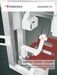
SCANORA® 3Dx / PDF Brochure US - Soredex
SCANORA® 3Dx / PDF Brochure US - Soredex
SCANORA® 3Dx / PDF Brochure US - Soredex

You also want an ePaper? Increase the reach of your titles
YUMPU automatically turns print PDFs into web optimized ePapers that Google loves.
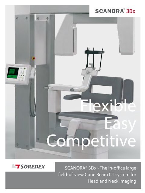
Flexible<br />
Easy<br />
Competitive<br />
SCANORA® <strong>3Dx</strong> - The in-office large<br />
field-of-view Cone Beam CT system for<br />
Head and Neck imaging
SCANORA® <strong>3Dx</strong>. The solution.<br />
Right FOV for each task<br />
SCANORA® <strong>3Dx</strong> makes advanced 3D imaging easy in the head and neck area. The system is ideal<br />
for ENT (Ear, Nose, Throat), dentomaxillofacial and cranial examinations in imaging centers,<br />
ENT offices, total care oral and maxillofacial clinics, hospitals and multispecialty dental practices.<br />
3D imaging raises diagnostic possibilities to a new level compared to 2D imaging. Compared to<br />
medical CT imaging, CBCT imaging offers many benefits: lower dose as the FOV size and location can<br />
be optimized to avoid radiation sensitive organs, better spatial resolution in bony structures and<br />
lower cost of purchase, commissioning, maintenance and use. Examinations are fast and convenient<br />
for the patients.<br />
A suitable imaging protocol can be defined for every diagnostic task by adjusting the field-of-view,<br />
resolution and dose. The FOV can be freely located to any region of interest in the head and neck area.<br />
SCANORA® <strong>3Dx</strong> fields-of-view (H × D in millimeters)<br />
A CBCT system is also a perfect addition to an existing medical CT. Both systems have their applications.<br />
A CBCT system can effectively perform, e.g. sinus studies and postoperative follow-up cases, that<br />
previously were handled with overdimensioned medical CT systems.<br />
Summary of<br />
benefits<br />
Flexible<br />
• Six, plus two optional, fields-of-view<br />
from 50 x 50 mm to 240 x 165 mm<br />
• The FOV can be freely located to<br />
different areas of the head and neck<br />
• Comprehensive software offering<br />
Easy<br />
Small S (50x50)<br />
Small S+ (50x100) Medium M (80x100) Medium M+ (80x165)<br />
• Seated patient, head in normal position<br />
• 12” HD clear touch control panel for<br />
ensuring easy workflow<br />
• Compatible with leading navigation<br />
and surgical guide systems<br />
Competitive<br />
• DICOM®/PACS compatibility<br />
• Optional CCD - RealPAN sensor for high<br />
quality dental panoramic imaging, with<br />
AutoSwitch 2D/3D mode change<br />
• Small footprint<br />
Large L (140x100) Large L+ (140x165) XL (180x165)<br />
Optional<br />
XL+ (240x165)<br />
Optional
Excellent diagnostic<br />
performance<br />
SCANORA® <strong>3Dx</strong> system provides diagnostic tools for a wide area of applications.<br />
The system is very versatile. Special attention has been paid to the needs of ENT clinicians.<br />
The FOV selection, ease of use and the software features offer efficient solutions for diagnostic tasks.<br />
ENT<br />
The imaging tasks of otorhinolaryngology<br />
can be effectively carried out.<br />
Temporal bone<br />
Temporal bone<br />
structures (M).<br />
Axial, coronal and sagittal slice<br />
series are the most common way<br />
of diagnosing the image. The three<br />
basic views clearly show all the<br />
sinuses (L+).<br />
The above case zoomed in for<br />
showing the auditory ossicles<br />
more clearly.<br />
The datasets are compatible with<br />
leading surgical navigation systems.
Clear display of craniofacial conditions.<br />
Craniofacial<br />
An osteoma in<br />
frontal sinus (L+).<br />
The sinonasal cavities<br />
are sound. No further<br />
treatment needed.<br />
The system is effective in surgical planning and follow-up.<br />
Facial surgery<br />
A postoperative trauma<br />
case (L+). Left side blow-out<br />
fracture. Assessment for<br />
orbital floor reconstruction.<br />
3D, coronal and oblique sagittal reformatted<br />
images (L+) of a 30 year old male after<br />
orbital floor and medial wall reconstruction<br />
and zygomatic fracture reposition.
The largest FOV (XL+) shows the whole facial complex.<br />
The FOV can be easily positioned in all the areas of<br />
the head and neck. The cervical area as an example.<br />
Orthognathic surgery<br />
Cervical spine<br />
3D, axial and synthetic panoramic<br />
reformatted images (XL+).<br />
A postoperative study of a 38 year<br />
old male after bimaxillary osteotomy<br />
2 months before. Swelling and pain<br />
in the left mandibular region, but in<br />
CBCT there were no signs of surgical<br />
complications in that area.<br />
Coronal, axial and sagittal reformatted<br />
images of the cervical spine (L ) in a<br />
50 year old patient showing spondylotic<br />
osteophytes in the ventrolateral region at<br />
the level C5 – C6 and C6 – C7 and very small<br />
calcification of the posterior longitudinal<br />
ligament at level C5 – C6. No degeneration<br />
is seen at the level of facet joints.
Dental and TMJ applications<br />
RealPAN Panoramic imaging<br />
Dental<br />
SCANORA® <strong>3Dx</strong> uses a dedicated CCD sensor for high resolution panoramic imaging. With the panoramic<br />
option SCANORA® <strong>3Dx</strong> provides the speed and efficiency of a traditional panoramic unit.<br />
The dental panoramic image provides an overview of the dentition and jaw area.<br />
A temporo-mandibular joint study. Osteoarthritis in the right TMJ. (M+)<br />
The unique patented<br />
AutoSwitch feature changes<br />
detectors automatically between<br />
panoramic and 3D modes.<br />
Odontogenic sinus problem. (L+).<br />
A study of the dental region. Right palatal cleft. (S).
Comprehensive<br />
software offering<br />
Modern dose 3D imaging<br />
SCANORA® <strong>3Dx</strong> produces image data in<br />
DICOM®*) format. With its open architecture<br />
it allows versatile and optimized software<br />
solutions to be tailored for your practice.<br />
The local area network (LAN) with several<br />
viewing stations is the solution for most<br />
practice applications allowing the system to<br />
be linked with the network and system server.<br />
SCANORA® software is the main platform,<br />
including the local patient image database<br />
and panoramic image handling.<br />
The system comes with comprehensive<br />
patient management capabilities, a server<br />
enabled image database and comprehensive<br />
tools for 2D and 3D image processing,<br />
diagnostics, treatment planning and reporting.<br />
Freely distribute clinical cases on CD/DVD<br />
to referring clinicians. Referring clinicians<br />
can utilize the free viewer without investing<br />
in special software or import the images in<br />
DICOM® format into their own 3D software.<br />
* Digital Imaging and Communication<br />
in Medicine<br />
X-ray imaging is a balance between image<br />
quality and x-ray dose by following the<br />
ALARA* principle. With SCANORA® <strong>3Dx</strong> this<br />
tradeoff has been successfully addressed.<br />
Thanks to wide adjustment ranges of<br />
parameters the overall radiation dose<br />
for specific diagnostic indications can be<br />
optimized by selecting the smallest FOV<br />
for each task and adjusting the mA and<br />
resolution accordingly. The key factors<br />
in achieving this are sophisticated x-ray<br />
generation, selectable imaging modes,<br />
a state-of-the-art flat panel detector<br />
and innovative image reconstruction<br />
technology.<br />
SCANORA® <strong>3Dx</strong> gives you the ability to<br />
carefully minimize the dose according to the<br />
diagnostic task, whether it is a question of<br />
detailed primary diagnostic or a follow-up<br />
study.<br />
The software offering can be tailored<br />
for different specialties including for<br />
instance following features:<br />
*) ALARA = As Low As Reasonably Achievable<br />
Modality<br />
workstation<br />
SCANORA®<br />
Image management<br />
Panoramic imaging<br />
PMS integration<br />
DICOM® services<br />
3D visualization software<br />
3D image handling<br />
Implant planning<br />
Reporting<br />
Additional SCANORA®<br />
and 3D visualization clients<br />
ENT<br />
• Virtual endoscopy<br />
• Airway analysis<br />
• Segmentation<br />
Radiology<br />
• Reporting<br />
• DICOM® printing<br />
• PACS connectivity<br />
• Radiology views<br />
• Image fusion<br />
3 rd party<br />
software<br />
Implant planning<br />
Drill and surgical guides<br />
Navigated surgery<br />
3D modeling<br />
Dental<br />
• Implant planning<br />
• 3D orthodontic analysis<br />
• TMJ diagnostics<br />
3 rd party<br />
PACS<br />
DICOM®<br />
printer
SCANORA® <strong>3Dx</strong> imaging programs<br />
Technical<br />
data<br />
FOV(H × D) Voxel sizes (mm) Name Application examples<br />
(mm) Std res High res<br />
50 × 50 0.15 0.1 S Single implants, wisdom tooth, localized problems,<br />
endo, perio<br />
50 × 100 0.4 0.2 S+ Maxilla or mandible, implants, drill guides<br />
80 × 100 0.25 0.15 M Maxilla and mandible, implants, temporal bone<br />
80 x165 0.35 0.15 M+ Both temporal bones, jaws and TMJ’s<br />
140 × 100 0.35 0.25 L Sinuses, cervical spine, airways<br />
140 x 165 0.3 0.2 L+ Sinuses, orthognathic surgery, ENT, ortho<br />
180 x 165<br />
optional<br />
240 x 165<br />
optional<br />
0.5 0.3 XL Trauma, facial and orthognathic surgery<br />
0.5 0.3 XL+ Trauma, facial and orthognathic surgery<br />
3D imaging parameters<br />
Scan time<br />
18 - 34 s<br />
Effective exposure time 2.4 - 6 s<br />
3D image receptor type Flat panel a-Si<br />
(77.7")<br />
Dedicated panoramic imaging (Optional)<br />
Adult panoramic program<br />
Child program<br />
Five partial segments<br />
Lateral TMJ program<br />
X-ray generator<br />
Tube<br />
Fixed anode tube<br />
Focal spot<br />
0.5 mm<br />
Target angle<br />
15 degrees<br />
kV 60-90<br />
mA 4-10<br />
1940 (76.4")<br />
1600 (63")<br />
General<br />
Weight<br />
Dimensions (HxWxD)<br />
310 kg (690 lbs)<br />
1973 mm × 1600 mm<br />
× 1400 mm<br />
(77.7” × 63” × 55.1”)<br />
Power requirements<br />
Line voltage 220-240 VAC (±10 %),<br />
50/60 Hz<br />
1400 (55.1")<br />
1740 (68.5")
Head office and factory:<br />
SOREDEX<br />
Nahkelantie 160, Tuusula<br />
P.O. Box 148, FI-04301 Tuusula<br />
Finland<br />
Tel. +358 10 270 2000<br />
Fax +358 9 701 5261<br />
info@soredex.com<br />
SOREDEX <strong>US</strong>A<br />
1245 W. Canal Street<br />
Milwaukee, WI 53233<br />
U.S.A.<br />
Tel. +1 800 558 6120<br />
Fax +1 414 481 8665<br />
usainfo@soredex.com<br />
SOREDEX Germany<br />
Schutterstrasse 12<br />
77746 Schutterwald<br />
Germany<br />
Tel: +49 (0) 781 28 41 98-0<br />
Fax: +49 (0) 781 28 41 98-30<br />
kontakt@soredex.de<br />
Digital<br />
imaging<br />
made<br />
easy<br />
www.soredex.com • www.soredex.de • www.soredex.com/usa<br />
210298-1 6/13 Printed in Finland<br />
Since 1977 SOREDEX has been a leader in providing innovative imaging solutions for<br />
demanding professionals. Through continuous evolution and refinement we have set<br />
the highest industry standards for Quality, Reliability and Efficiency.<br />
We are committed to following this path today and in the future.<br />
SCANORA®/AutoSwitch/RealPAN/Digital imaging made easy is a registered trademark / a common law<br />
trademark of SOREDEX. Other product names and trademarks are the property of their respective owners.<br />
CE-marked, NB (CE) number 0537. Electrical safety meets the IEC 60601-1 standard. Manufacturing complies<br />
with ISO 13485:2003, ISO 9001:2008, and ISO 14001:2004.<br />
DICOM® is the registered trademark of the National Electrical Manufacturers Association for<br />
its standards publications relating to digital communications of medical information<br />
SOREDEX reserves the right to make changes in specifications and features shown herein at any time<br />
without notice or obligation. Contact your SOREDEX representative for the most up-to-date information.<br />
© 2012 SOREDEX