36598-DFU - CooperSurgical
36598-DFU - CooperSurgical
36598-DFU - CooperSurgical

You also want an ePaper? Increase the reach of your titles
YUMPU automatically turns print PDFs into web optimized ePapers that Google loves.
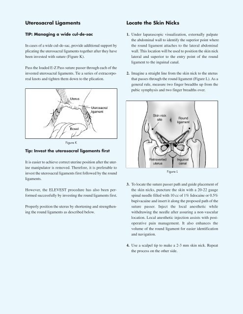
Uterosacral Ligaments<br />
TIP: Managing a wide cul-de-sac<br />
In cases of a wide cul-de-sac, provide additional support by<br />
plicating the uterosacral ligaments together after they have<br />
been invested with suture (Figure K).<br />
Pass the loaded E-Z Pass suture passer through each of the<br />
invested uterosacral ligaments. Tie a series of extracorporeal<br />
knots and tighten them down to the plication.<br />
Locate the Skin Nicks<br />
1. Under laparascopic visualization, externally palpate<br />
the abdominal wall to identify the superior point where<br />
the round ligament attaches to the lateral abdominal<br />
wall. This location will be used to position the skin nick<br />
lateral and superior to the entry point of the round<br />
ligament to the inguinal canal.<br />
2. Imagine a straight line from the skin nick to the uterus<br />
that passes through the round ligament (Figure L). As a<br />
general rule, measure two finger breadths up from the<br />
pubic symphysis and two finger breadths over.<br />
Figure K<br />
Tip: Invest the uterosacral ligaments first<br />
It is easier to achieve correct uterine position after the uterine<br />
manipulator is removed. Therefore, it is preferable to<br />
invest the uterosacral ligaments first followed by the round<br />
ligaments.<br />
However, the ELEVEST procedure has also been performed<br />
successfully by investing the round ligaments first.<br />
Properly position the uterus by shortening and strengthening<br />
the round ligaments as described below.<br />
Figure L<br />
3. To locate the suture passer path and guide placement of<br />
the skin nicks, puncture the skin with a 20-22 gauge<br />
spinal needle filled with 10 cc of 1% lidocaine or 0.5%<br />
bupivacaine and insert it along the proposed path of the<br />
suture passer. Inject the local anesthetic while<br />
withdrawing the needle after assuring a non-vascular<br />
location. Local anesthetic injection assists with postoperative<br />
pain management. It also enhances the<br />
volume of the round ligament for easier identification<br />
and navigation.<br />
4. Use a scalpel tip to make a 2-3 mm skin nick. Repeat<br />
the process on the other side.