

PDF Version - Glidewell Dental Labs
PDF Version - Glidewell Dental Labs
PDF Version - Glidewell Dental Labs

You also want an ePaper? Increase the reach of your titles
YUMPU automatically turns print PDFs into web optimized ePapers that Google loves.
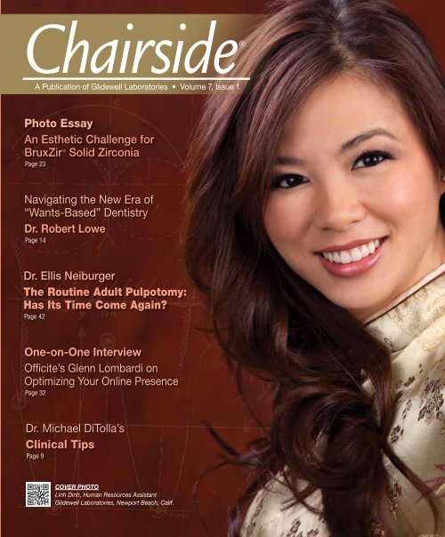
Chairside®<br />
A Publication of <strong>Glidewell</strong> Laboratories • Volume 7, Issue 1<br />
Photo Essay<br />
An Esthetic Challenge for<br />
BruxZir ® Solid Zirconia<br />
Page 23<br />
Navigating the New Era of<br />
“Wants-Based” Dentistry<br />
Dr. Robert Lowe<br />
Page 14<br />
Dr. Ellis Neiburger<br />
The Routine Adult Pulpotomy:<br />
Has Its Time Come Again?<br />
Page 42<br />
One-on-One Interview<br />
Officite’s Glenn Lombardi on<br />
Optimizing Your Online Presence<br />
Page 32<br />
Dr. Michael DiTolla’s<br />
Clinical Tips<br />
Page 9<br />
COVER PHOTO<br />
Linh Dinh, Human Resources Assistant<br />
<strong>Glidewell</strong> Laboratories, Newport Beach, Calif.
Contents<br />
9 Dr. DiTolla’s Clinical Tips<br />
In this issue, I showcase Ultradent’s Opalescence ®<br />
Trèswhite Supreme preloaded teeth-whitening trays,<br />
which provide an easy way to deliver same-day bleaching<br />
trays. Also featured are: NTI ® Superflex Diamond<br />
Discs from Axis <strong>Dental</strong>, my top instrument pick for trimming<br />
and shaping provisionals; the KaVo QUATTROcare<br />
Automatic Handpiece Maintenance System, which I trust<br />
to keep my favorite handpieces in tip-top shape; and<br />
NoMIX ® Temporary Cement from Centrix, a necessity for<br />
short-term cementation.<br />
14 Upgrading Porcelain Veneer Restorations:<br />
A Case Report<br />
Have you had patients come to your office requesting<br />
an upgrade of perfectly serviceable restorations based<br />
solely on esthetics? Dr. Robert Lowe presents one such<br />
case in his article that outlines the process of upgrading<br />
veneers to satisfy a patient’s esthetic demands. He<br />
discusses a new age of “wants-based” dentistry, which<br />
is often purely esthetic in nature, and how to navigate<br />
customers’ wants and perceived needs in this era of<br />
elective dentistry.<br />
23 Photo Essay: BruxZir ® Solid Zirconia<br />
Anterior Esthetic Challenge<br />
This photo essay illustrates our laboratory’s latest<br />
advancements in improving the translucency and esthetic<br />
properties of BruxZir Solid Zirconia. To showcase this<br />
product, we put it to the challenge of replacing old<br />
crowns on tooth #8 and #9. After viewing the case, I<br />
think you will see why we decided to give the BruxZir<br />
Solid Zirconia motto an upgrade as well.<br />
32 One-on-One with Dr. Michael DiTolla:<br />
Interview of Glenn Lombardi<br />
In a day and age when a business’s success and growth<br />
often hinges on online reviews and other social media<br />
standards, some dental practitioners would like to believe<br />
they are safe from this type of information technology, yet<br />
they simply are not. Now, more than ever, it is important<br />
to ensure your practice is up-to-date on its social media<br />
practices. Glenn Lombardi, president of Officite LLC, a<br />
leading national provider of premier websites and turnkey<br />
Internet marketing solutions for the dental community,<br />
talks about what dentists can do to optimize their online<br />
presence.<br />
Contents 1
Contents<br />
42 Is It Time to Do Routine Adult Pulpotomies?<br />
Due to the current dampened state of the economy, a<br />
growing number of patients are unable to afford traditional<br />
endodontic treatment, yet the need for root canal therapy<br />
continues to rise. Dr. Ellis Neiburger discusses the oftenoverlooked<br />
pulpotomy as an alternative to traditional endo,<br />
its long history and how it fits into today’s dental practice<br />
in this must-read article.<br />
52 In Praise of Electric Handpieces<br />
As Dr. Elliot Mechanic explains, dentists have come a long<br />
way from being regarded as “drillers, fillers and billers,” to<br />
now increasingly being seen as artists and healers. With the<br />
advent of “smart” technologies such as the electric handpiece,<br />
dentists can treat their once-fearful patients with a<br />
greater level of ease and increased efficiency. Dr. Mechanic<br />
outlines the use and benefits of electric handpieces, including<br />
the important role they play in crown preparation.<br />
56 The Remake Debate<br />
How do you handle remake cases? Maribeth Marsico, senior<br />
editor at LMT Communications, explores the remake process,<br />
the biggest remake culprit and what can be done to<br />
cut down on these “cases-gone-wrong.” There will always<br />
be remakes in dentistry, but as this report shows, it is the<br />
duty of the lab, the technicians and the dentists to work together<br />
to ensure the patient gets their final desired result.<br />
<strong>Glidewell</strong> Publications for iPad<br />
iPAD APP Chairside is now available on<br />
your iPad. Search “<strong>Glidewell</strong>” in the iTunes<br />
Store and download the free app.<br />
60 Digital Impressions for an<br />
Immediate Denture<br />
Versatility, accuracy and ease of use are just some of the<br />
benefits digital impression technology offers over conventional<br />
impression techniques. This case study from<br />
Dr. Dean Saiki illustrates how digital impressions are not<br />
only equal to conventional impressions, but are in some<br />
instances the only appropriate option.<br />
ALSO IN THIS ISSUE<br />
8 By the Numbers<br />
64 The Chairside Photo Hunt<br />
2<br />
www.chairsidemagazine.com
Publisher<br />
Jim <strong>Glidewell</strong>, CDT<br />
Editor-in-Chief and Clinical Editor<br />
Michael C. DiTolla, DDS, FAGD<br />
Managing Editors<br />
Jim Shuck; Mike Cash, CDT<br />
Creative Director<br />
Rachel Pacillas<br />
Copy Editors<br />
Jennifer Holstein,<br />
Megan Affleck, David Frickman<br />
Statistical Editor<br />
Darryl Withrow<br />
Digital Marketing Manager<br />
Kevin Keithley<br />
Graphic Designers/Web Designers<br />
Jamie Austin, Deb Evans, Joel Guerra, Audrey Kame,<br />
Lindsey Lauria, Phil Nguyen, Kelley Pelton,<br />
Melanie Solis, Ty Tran, Makara You<br />
Photographer<br />
Sharon Dowd<br />
Clinical Videographer<br />
James Kwasniewski<br />
Illustrator<br />
Wolfgang Friebauer, MDT<br />
Coordinator and Ad Representative<br />
Teri Arthur<br />
(teri.arthur@glidewelldental.com)<br />
If you have questions, comments or complaints regarding<br />
this issue, we want to hear from you. Please e-mail us at<br />
chairside@glidewelldental.com. Your comments may be featured<br />
in an upcoming issue or on our website:<br />
www.chairsidemagazine.com.<br />
© 2012 <strong>Glidewell</strong> Laboratories<br />
Neither Chairside magazine nor any employees involved in its publication<br />
(“publisher”), makes any warranty, express or implied, or assumes<br />
any<br />
Neither<br />
liability<br />
Chairside<br />
or responsibility<br />
Magazine<br />
for<br />
nor<br />
the<br />
any<br />
accuracy,<br />
employees<br />
completeness,<br />
involved in its<br />
or<br />
publication<br />
usefulness<br />
of<br />
(“publisher”),<br />
any information,<br />
makes<br />
apparatus,<br />
any warranty,<br />
product,<br />
express<br />
or<br />
or<br />
process<br />
implied,<br />
disclosed,<br />
or assumes<br />
or<br />
represents<br />
any liability<br />
that<br />
or<br />
its<br />
responsibility<br />
use would<br />
for<br />
not<br />
the<br />
infringe<br />
accuracy,<br />
proprietary<br />
completeness,<br />
rights. Reference<br />
or usefulness<br />
herein to<br />
of<br />
any<br />
any information,<br />
specific commercial<br />
apparatus,<br />
products,<br />
product,<br />
process,<br />
or process<br />
or<br />
disclosed,<br />
services by<br />
or<br />
trade<br />
represents<br />
name, trademark,<br />
that its use<br />
manufacturer<br />
would not infringe<br />
or otherwise<br />
proprietary<br />
does<br />
rights.<br />
not<br />
Reference<br />
necessarily<br />
herein<br />
constitute<br />
to any<br />
or<br />
specific<br />
imply its<br />
commercial<br />
endorsement,<br />
products,<br />
recommendation,<br />
process, or services<br />
or favoring<br />
by<br />
by<br />
trade<br />
the<br />
name,<br />
publisher.<br />
trademark,<br />
The views<br />
manufacturer<br />
and opinions<br />
or otherwise<br />
of authors<br />
does not<br />
expressed<br />
necessarily<br />
constitute<br />
herein do not necessarily<br />
or imply its<br />
state<br />
endorsement,<br />
or reflect<br />
recommendation,<br />
those of the publisher<br />
or favoring<br />
and<br />
shall<br />
by<br />
not<br />
the<br />
be<br />
publisher.<br />
used for<br />
The<br />
advertising<br />
views and<br />
or product<br />
opinions<br />
endorsement<br />
of authors expressed<br />
purposes.<br />
CAUTION:<br />
herein do<br />
When<br />
not necessarily<br />
viewing the<br />
state<br />
techniques,<br />
or reflect<br />
procedures,<br />
those of the<br />
theories<br />
publisher<br />
and materials<br />
and<br />
shall<br />
that<br />
not<br />
are<br />
be used<br />
presented,<br />
for advertising<br />
you must<br />
or<br />
make<br />
product<br />
your<br />
endorsement<br />
own decisions<br />
purposes.<br />
about<br />
specific<br />
CAUTION:<br />
treatment<br />
When<br />
for<br />
viewing<br />
patients<br />
the<br />
and<br />
techniques,<br />
exercise<br />
procedures,<br />
personal professional<br />
theories and<br />
judgment<br />
materials<br />
regarding<br />
that are<br />
the<br />
presented,<br />
need for<br />
you<br />
further<br />
must<br />
clinical<br />
make your<br />
testing<br />
own<br />
or<br />
decisions<br />
education<br />
about<br />
and<br />
your<br />
specific<br />
own<br />
treatment<br />
clinical expertise<br />
for patients<br />
before<br />
and<br />
trying<br />
exercise<br />
to implement<br />
personal<br />
new<br />
professional<br />
procedures.<br />
judgment<br />
regarding the need for further clinical testing or education and<br />
your own clinical expertise before trying to implement new procedures.<br />
Chairside is a registered trademark of <strong>Glidewell</strong> Laboratories.<br />
Chairside ® Magazine is a registered trademark of <strong>Glidewell</strong> Laboratories.<br />
Editor’s Letter<br />
Ahh, the Internet. Like many of you, I grew up in an era<br />
where most arguments were never really settled. Unless<br />
one of the arguing parties actually owned an Encyclopedia<br />
Britannica and the argument was about which bird can<br />
hover (answer: the hummingbird), most $20 bets entered a<br />
permanent state of limbo. In 2012, this is no longer true. I<br />
was recently marveling at how nearly any fact can be pulled<br />
up on the Web in less than 10 seconds. (Wikipedia should<br />
be getting a cut of all the bets it’s helping to settle!)<br />
While the power of the Internet to transform our everyday<br />
lives is undeniable and largely positive, as dentists we<br />
are faced with something dentists in the “golden age of<br />
dentistry” never had to deal with. Do you remember the<br />
“golden age”? Dentistry was great back then! The golden<br />
age is loosely defined as the time period 10 years before<br />
you graduated from dental school — for me that’s 1978.<br />
Back then, all patients were independently wealthy, and<br />
they lusted after full-mouth rehabilitations. When they were<br />
happy, they said “thank you.” When they were unhappy,<br />
they told six to eight people.<br />
Today’s patients — happy or unhappy — are increasingly<br />
Internet savvy, and with the increase in social media platforms<br />
and participants, managing these communication<br />
channels becomes necessary. We can’t please every patient,<br />
and we are going to disappoint some. Unless we learn how<br />
to manage social media, we just have to hope that the ones<br />
we disappoint are blood relatives or in-laws.<br />
It’s inevitable that you will eventually have a bad review or<br />
two on Yelp or another social media site that doesn’t even<br />
exist today. Read my recent interview with Glenn Lombardi<br />
to find out how you can use this new form of communication<br />
to your advantage. I know the dentists back in the<br />
“golden age” didn’t have to deal with this, and you never<br />
signed up for this, but it’s here. Appoint a staff member to<br />
lead your team’s social media efforts and follow Glenn’s<br />
well-reasoned advice!<br />
Yours in quality dentistry,<br />
Dr. Michael C. DiTolla<br />
Editor-in-Chief, Clinical Editor<br />
mditolla@glidewelldental.com<br />
Editor’s Letter 3
Letters to the Editor<br />
Dear Dr. DiTolla,<br />
First of all, I want to thank you for helping<br />
me with anterior crown preps! I, too, find<br />
that as soon as I begin preparing, I lose<br />
my frame of reference. I have tried various<br />
depth measurement methods, but your<br />
Reverse Preparation Technique is priceless.<br />
Also, your in-depth video demonstration of<br />
its use is outstanding. Thank you so much<br />
for transforming my technique and for my<br />
newly gained confidence in accurately doing<br />
anterior crown preps in a timely manner.<br />
I have a question for you: From the brief<br />
view I got of your dental unit on your video,<br />
it looks like you have an A-dec unit (Continental<br />
style). Also, the electric handpiece,<br />
if I viewed it correctly, is an A-dec/W&H<br />
electric motor. However, I noticed that you<br />
use the KaVo ELECTROtorque high-speed<br />
attachment. I did not know that KaVo electric<br />
handpieces were capable of connecting<br />
to A-dec/W&H electric motors. Do you<br />
need a special coupling/adaptor, or are you<br />
able to simply snap it on the same way you<br />
would if you used an A-dec/W&H electric<br />
handpiece? Also, I have the same A-dec<br />
unit plus A-dec/W&H electric motor, but<br />
I use the A-dec/W&H electric handpieces. I<br />
find them to be very good, but I must admit<br />
I have never tried the KaVo ELECTROtorque<br />
electric handpiece. Have you used or tried<br />
the A-dec/W&H electric handpiece? If so,<br />
4<br />
www.chairsidemagazine.com<br />
how does it differ from the KaVo electric<br />
handpiece that you use?<br />
Thank you very much for your time. Again, I<br />
enjoy watching and reviewing your instructional<br />
videos!<br />
– Larry Kolar, DDS<br />
Chicago, Ill.<br />
Dear Larry,<br />
Thanks for the kind words!<br />
The KaVo electric handpieces do snap<br />
directly onto the A-dec motor; no<br />
adapter is needed. I have never used<br />
anything but KaVo handpieces, even<br />
going back to my air turbine days, so<br />
I guess that means I’ve been pretty<br />
satisfied with KaVo.<br />
I know KaVo is doing a special promotion<br />
where, if you go to www.trykavo.<br />
com, they will send you an electric<br />
handpiece at no charge that you can<br />
snap on for a few days, prep some<br />
teeth and see which one you like better.<br />
Let me know what you think!<br />
Best,<br />
– Mike<br />
Dear Dr. DiTolla,<br />
I was wondering which is the strongest<br />
anterior bridge material besides monolithic<br />
zirconia? I have had failures with IPS<br />
Empress ® II and IPS e.max ® (Ivoclar<br />
Vivadent). Fractures usually occur when<br />
patients unknowingly bite into a hard bone<br />
(meat that is supposed to be boneless), or<br />
forget they need to be cautious with the<br />
restorations and chomp on something like<br />
a hard baguette. The bridges in these cases<br />
have had solid, broad connectors. Could<br />
you comment on IPS e.max versus zirconia<br />
with layered porcelains (e.g., 3M ESPE <br />
Lava )? Is it possible to make the lingual<br />
occlusion of an anterior maxillary bridge<br />
in zirconia and layer just the facial with<br />
porcelain, using the same concept of metal<br />
occlusion in a PFM? I am sitting on a case,<br />
so a quick response would be appreciated.<br />
Mahalo for your input.<br />
– Todd Okazaki, DDS<br />
Haleiwa, Hawaii<br />
Dear Todd,<br />
Good question! First of all, you are<br />
correct in thinking that monolithic<br />
zirconia, such as BruxZir ® Solid<br />
Zirconia (<strong>Glidewell</strong> Laboratories), is the<br />
strongest all-ceramic bridge material<br />
that we have. This time last year, I would<br />
have hesitated to recommend that a<br />
dentist prescribe BruxZir Solid Zirconia<br />
for an anterior bridge. The esthetic<br />
nature of BruxZir restorations has really<br />
improved over the last year, although<br />
it hasn’t quite caught up with its<br />
monolithic brethren, such as IPS e.max.<br />
An anterior PFM bridge is probably<br />
the strongest solution, although the<br />
ceramic material can certainly chip<br />
off the metal understructure, and the<br />
esthetics can be compromised by the<br />
lack of translucency and possibly<br />
exposed metal margins.<br />
I am not sure I would want to go with<br />
the zirconia-layered-with-ceramic option<br />
you mention, as we have noticed<br />
more chipping with that combination<br />
of materials than we have with porcelain<br />
fused to metal. In fact, porcelain<br />
fused to zirconia would probably be<br />
my last choice after BruxZir Solid Zirconia,<br />
IPS e.max and PFM.<br />
You also asked about making the lingual<br />
of the bridge in solid zirconia,<br />
similar to a metal lingual on a PFM<br />
restoration. While we do fabricate<br />
bridges like that on request from dentists,<br />
we don’t get many requests for it.<br />
Perhaps the reason is that the strength<br />
of BruxZir Solid Zirconia comes from<br />
its monolithic nature — the fact that<br />
it doesn’t have any ceramic material<br />
fused to it. As a result, it fractures and<br />
chips less than any other restoration<br />
in the lab (except cast gold, of course).<br />
When we do make a BruxZir restoration<br />
like that, we typically place the
ceramic material on the facial and<br />
carry it from the gingival down to the<br />
incisal edge, without wrapping the<br />
incisal edge. We want to allow the<br />
patient to function in protrusion on<br />
the zirconia, rather than the porcelain.<br />
But doing this takes it from being a<br />
monolithic BruxZir restoration to a<br />
bilayered restoration, which is more<br />
prone to chipping.<br />
While there are no absolutes, my first<br />
choice today is IPS e.max for a 3-unit<br />
bridge in the anterior on a patient<br />
who does not show a lot of wear. For<br />
that same bridge in a patient who<br />
does show signs of wear, my choice is<br />
BruxZir Solid Zirconia, especially if the<br />
patient has previously chipped a PFM<br />
restoration. As the size of the bridge<br />
increases beyond 3 units, I begin to<br />
consider PFM as my choice because<br />
of the superior strength of the metal<br />
connectors when compared to any allceramic<br />
system, especially when there<br />
is a lack of room for the connectors.<br />
As always, your mileage may vary.<br />
Hope that helps!<br />
– Mike<br />
CONNECT WITH CHAIRSIDE<br />
FOLLOW US ON TWITTER<br />
Find us @<strong>Glidewell</strong><strong>Dental</strong><br />
FIND US ON FACEBOOK<br />
Search “<strong>Glidewell</strong>” to see<br />
what’s new.<br />
ITUNES WATCH AND<br />
LEARN<br />
Go the iTunes Store and search<br />
“<strong>Glidewell</strong>.” Plus, download the<br />
free <strong>Glidewell</strong> Publications app<br />
for iPad.<br />
SHARE YOUR THOUGHTS<br />
Visit www.chairsidemagazine.com<br />
and select “Contact Us.” Or write to:<br />
<strong>Glidewell</strong> Laboratories<br />
ATTN: Chairside magazine<br />
4141 MacArthur Blvd.<br />
Newport Beach, CA 92660<br />
ACCESS OUR RESOURCES<br />
Clinical videos, product information<br />
and patient resources are a click<br />
away at www.glidewelldental.com.<br />
ADVERTISE/SUBMIT AN<br />
ARTICLE<br />
Call 888-303-4221<br />
Letters should include writer’s full name,<br />
address and daytime phone number. All<br />
correspondence may be published and<br />
edited for clarity and length.<br />
Dear Dr. DiTolla,<br />
I haven’t talked with my wife for days. She can’t stop!<br />
– Tom Novak, DDS<br />
Weatherford, Texas<br />
Letters to the Editor 5
Contributors<br />
Michael C. DiTolla, DDS, FAGD<br />
Dr. Michael DiTolla is a graduate of University of the Pacific Arthur A. Dugoni School of Dentistry. As<br />
director of clinical education and research at <strong>Glidewell</strong> Laboratories in Newport Beach, Calif., he performs<br />
clinical testing on new products in conjunction with the company’s R&D department. <strong>Glidewell</strong> dental<br />
technicians have the privilege of rotating through Dr. DiTolla’s operatory and experiencing his commitment<br />
to excellence through his prepping and placement of their restorations. He is a CR evaluator and lectures<br />
nationwide on both restorative and cosmetic dentistry. Dr. DiTolla has several clinical programs available<br />
on DVD through <strong>Glidewell</strong> Laboratories. For more information on his articles or to receive a free copy of<br />
Dr. DiTolla’s clinical presentations, call 888-303-4221 or e-mail mditolla@glidewelldental.com.<br />
Glenn Lombardi<br />
Glenn Lombardi is president of Officite LLC, a leading national provider of premier websites and<br />
turnkey Internet marketing solutions for the dental community. Since 2002, Officite has built thousands<br />
of websites for healthcare practices around the world, which have generated hundreds of thousands of<br />
new patient appointment requests. Glenn is a frequent speaker at national and state dental association<br />
meetings, including the AGD and D.C. <strong>Dental</strong> Society annual meetings. His presentations focus on<br />
professional website development, search engine optimization, and how to seamlessly integrate the Internet<br />
into your practice to attract new patients and increase case acceptance. Contact him at 800-908-2483 or<br />
glombardi@officite.com.<br />
Robert A. Lowe, DDS, FAGD, FICD, FADI, FACD, FIADFE<br />
Dr. Robert Lowe graduated magna cum laude from Loyola University School of Dentistry in 1982 and was<br />
a clinical professor of restorative dentistry at the school until its closure in 1993. Since January 2000,<br />
Dr. Lowe has maintained a private practice in Charlotte, N.C. He lectures internationally and his work is<br />
frequently published in well-known dental journals on esthetic and restorative dentistry. Dr. Lowe received<br />
fellowships in the AGD, ICD, ADI, ACD and IADFE, received the 2004 Gordon Christensen Outstanding<br />
Lecturers Award and, in 2005, Diplomat status on the American Board of Aesthetic Dentistry. Contact him<br />
at 704-364-4711 or boblowedds@aol.com.<br />
6<br />
www.chairsidemagazine.com
Elliot Mechanic, DDS<br />
Dr. Elliot Mechanic has been practicing general and esthetic dentistry in Montreal, Quebec, since 1979.<br />
Dr. Mechanic serves as Oral Health’s editorial board member for esthetics, and is a member of numerous<br />
professional organizations, including the International Academy for <strong>Dental</strong>-Facial Esthetics, Academy of<br />
Laser Dentistry and the AACD. Dr. Mechanic takes great pride in his work, which has afforded him the<br />
pleasure to work with executives, professionals, celebrities, international stars and everyone in between. He<br />
can be reached at 514-769-3939.<br />
Ellis J. Neiburger, DDS<br />
Dr. Ellis “Skip” Neiburger graduated from the University of Illinois at Chicago College of Dentistry in 1968,<br />
where he did postgraduate work on pulp histology in the Department of Oral Pathology. Dr. Neiburger<br />
currently practices general dentistry in Waukegan, Ill. A former vice president of the American Association<br />
of Forensic Dentists, Dr. Neiburger has been the association’s journal editor since 1978. He also was<br />
publisher/editor for <strong>Dental</strong> Computer Newsletter (the journal that introduced computing to the dental field).<br />
Contact him at 847-244-0292 or eneiburger@comcast.net.<br />
Dean H. Saiki, DDS<br />
Dr. Dean Saiki graduated from USC School of Dentistry in 1988. He maintains a private practice in North<br />
County San Diego, Calif., specializing in cosmetic, laser, implant and digital dentistry. He has been a<br />
member of the ADA, CDA and San Diego County <strong>Dental</strong> Society since 1989, as well as other advanced study<br />
clubs including the Trojan <strong>Dental</strong> Study Club. Dr. Saiki is trained and certified in dental soft tissue lasers<br />
and CAD/CAM technology. He has been voted a “Top Dentist” in San Diego by his peers for the past five<br />
years. Contact him at 760-732-3456 or dentist@deansaiki.com.<br />
Contributors 7
Numbers<br />
by the<br />
143<br />
CEREC<br />
Number of<br />
dental labs now<br />
offering BruxZir ®<br />
Solid Zirconia<br />
restorations<br />
5,400<br />
is a registered trademark of Sirona <strong>Dental</strong> Systems Inc.<br />
Number of modelfree<br />
BruxZir ® Solid<br />
Zirconia bridges<br />
and single-unit<br />
crowns <strong>Glidewell</strong><br />
Laboratories<br />
fabricated for<br />
CEREC ® Connect<br />
dentists in 2011<br />
18,427<br />
BruxZir is a registered trademark of <strong>Glidewell</strong> Laboratories.<br />
Number of<br />
dentists who<br />
have prescribed<br />
a BruxZir ® Solid<br />
Zirconia restoration<br />
Percentage<br />
of veneers<br />
prescribed with a<br />
VITA 3D-Master ®<br />
shade in 2006<br />
16%<br />
Percentage<br />
of veneers<br />
prescribed with a<br />
VITA 3D-Master ®<br />
shade in 2011<br />
25%<br />
17.1%<br />
41.6%<br />
Percent rate of<br />
rampant decay in<br />
children whose<br />
primary language<br />
is English<br />
Percent rate of<br />
rampant decay in<br />
children whose<br />
primary language<br />
is not English<br />
Percentages based on 56,251 veneers fabricated at <strong>Glidewell</strong> Laboratories<br />
3D-Master is a registered trademark of VITA Zahnfabrik.<br />
Findings from the Spokane Regional Health District’s 2010 Smile Survey<br />
(www.srhd.org/news.asp).<br />
66%<br />
Percentage of dentists who have<br />
received a negative online review<br />
As reported by The Wealthy Dentist ®<br />
8<br />
www.chairsidemagazine.com<br />
The Wealthy Dentist is a registered trademark of Du Molin & Du Molin Inc.
Dr. DiTolla’s<br />
CLINICAL TIPS<br />
PRODUCT........ NoMIX ® Temporary Cement<br />
SOURCE........... Centrix (Shelton, Conn.)<br />
800-235-5862, www.centrixdental.com<br />
Until every dental office is doing one-appointment crowns, temporaries will continue to be part of our<br />
day-to-day life. I am always amazed by the patient (typically male) who doesn’t appear to have ever<br />
flossed in his life, but when we put on a temp and tell him not to floss, flossing becomes an obsession<br />
— and, of course, he knocks off his temp. This is why all patients with possible non-retentive preps are<br />
sent home with a uni-dose of NoMIX Temporary Cement to use for short-term cementation, in case their<br />
temp comes off. As its name implies, there is no mixing, and the cement is moisture activated. After<br />
exposing the cement to water, have the patient hold their teeth together for five minutes while it sets,<br />
and you just saved yourself a Saturday trip to the office to re-cement a temp!<br />
Dr. DiTolla’s Clinical Tips 9
Dr. DiTolla’s<br />
CLINICAL TIPS<br />
PRODUCT........ NTI ® Superflex Diamond Discs<br />
SOURCE........... Axis <strong>Dental</strong> (Coppell, Texas)<br />
800-355-5063, www.axisdental.com<br />
If you told me I could only use one instrument to trim and shape provisionals, I would have to go with<br />
one of Axis <strong>Dental</strong>’s flexible, perforated NTI Superflex Diamond Discs in an electric handpiece. At higher<br />
speeds, I can use the double-sided disc to trim the margins on posterior temps, and then shape the<br />
mesial and distal contacts more smoothly than I could with a bur. But the disc really shines with anterior<br />
provisionals, where I can turn down the speed and really dial in the gingival embrasures, so I avoid<br />
blunting any papillae. There also is no better way to develop facial embrasures than with the flexible<br />
disc, which helps provisional bridges look a little more lifelike.<br />
10 www.chairsidemagazine.com
Dr. DiTolla’s<br />
CLINICAL TIPS<br />
PRODUCT........ Opalescence ® Trèswhite Supreme<br />
SOURCE........... Ultradent Products Inc. (South Jordan, Utah)<br />
888-230-1420, www.ultradent.com<br />
Years ago, we started offering complimentary bleaching trays to patients who had undergone some<br />
kind of esthetic procedure, typically the replacement of some older crowns on the maxillary anterior<br />
teeth. We wanted to be able to deliver the trays the same day, but we often faced backups in our<br />
tray production. A friend of mine introduced me to Opalescence Trèswhite Supreme last year, and<br />
the product has become an easy way for me to deliver same-day bleaching trays. My friend offers<br />
free teeth whitening to new patients, and he says using this product is the only way he can pull that<br />
off. These disposable, preloaded teeth-whitening trays definitely are the easiest way for patients to<br />
bleach their teeth, and they offer a great alternative to store-bought whitening systems.<br />
Dr. DiTolla’s Clinical Tips11
Dr. DiTolla’s<br />
CLINICAL TIPS<br />
PRODUCT........ QUATTROcare Automatic Handpiece Maintenance System<br />
SOURCE........... KaVo <strong>Dental</strong> (Charlotte, N.C.)<br />
888-275-5286, www.kavousa.com<br />
I’ve made no secret of my love for my KaVo ELECTROtorque handpieces. The torque is so high I can turn the speed<br />
down to a measly 3000 rpm and still have enough power to cut tooth structure, and because the speed is so low, I can<br />
do it without water! I also need that immense torque to properly polish intraoral restorations when I have to make an<br />
adjustment after cementation. To maintain my investment in these handpieces and keep them running smoothly, my<br />
assistant loves her KaVo QUATTROcare. It automatically cleans, lubricates and purges the handpieces with the push of<br />
a button, and then they go straight into sterilization. In case your Italian is weak, the “quattro” in QUATTROcare refers to<br />
the fact that it will service up to four handpieces at a time.<br />
12 www.chairsidemagazine.com
Upgrading Porcelain Veneer<br />
Restorations:<br />
A Case Report<br />
– ARTICLE and CLINICAL PHOTOS by Robert A. Lowe, DDS, FAGD,<br />
FICD, FADI, FACD, FIADFE<br />
Placement of indirect labial veneers (porcelain or composite) continues to be an excellent option to correct many esthetic<br />
complaints that our patients have with their smiles. Some of the more common indications for their clinical use include:<br />
1. Minor corrections of anterior tooth morphology and emergence angles to fill in spaces in the gingival embrasure<br />
areas when these spaces are an esthetic concern for the patient.<br />
2. Minor corrections in tooth position (rotation, labio-lingual arch position and crowding) if orthodontics is either not<br />
indicated or not accepted as a treatment option by the patient.<br />
3. Diastema closures and corrections of anterior tooth proportion (golden proportion).<br />
4. Establishment of anterior guidance and canine disclusion in patients where preparation for full-coverage restorations<br />
would necessitate unnecessary removal of healthy tooth structure.<br />
5. Improving tooth color for a patient where tooth whitening was not a treatment option or did not yield a satisfactory<br />
result for the patient.<br />
14 www.chairsidemagazine.com
Tooth Preparation<br />
The amount of tooth reduction required depends<br />
on the specific clinical situation. In general, 0.5 to<br />
0.7 mm of tooth reduction is needed. In some cases,<br />
where “nature” has done the tooth preparation or<br />
natural tooth contours are less prominent, “no prep”<br />
options are also possible. If changes in tooth position<br />
are required, some areas of the tooth may be<br />
prepared more, others less.<br />
It is recommended to first contour the teeth to their<br />
ideal position using a cylindrical diamond, then use<br />
depth cutters to remove a uniform amount of tooth<br />
structure to compensate for the thickness of the restoration.<br />
In extreme situations in which the dental<br />
pulp is encroached upon, root canal therapy is recommended<br />
rather than overcontouring the restoration.<br />
In cases where a low value (dark) preoperative<br />
tooth color is to be changed to a high value (light)<br />
color, more tooth structure may need to be removed<br />
(1.0 to 1.5 mm) to create enough space for opacious<br />
dentin or opaquers to block out the darkness. For<br />
some patients, preoperative tooth whitening may<br />
be indicated to increase the value of the underlying<br />
tooth structure, allowing for less tooth structure to<br />
be removed during the preparation process.<br />
Gingival margins should be placed at the gingival<br />
crest or slightly above. The interproximal margins<br />
should be carried into the lingual portion<br />
of the contact area. If diastemata are present, the<br />
interproximal margin of the preparation should be<br />
carried lingually to the linguoproximal line angle.<br />
Also, when closing spaces, it is important to prepare<br />
the gingival margins far enough into the proximal<br />
areas so that the restoration margins are not visible<br />
from a three-quarter or oblique view (when the<br />
patient turns their head to the side).<br />
After the preparations are finished, it is recommended<br />
to use a fine cylinder finishing diamond to make<br />
the preparations as smooth as possible. Aluminum<br />
oxide strips can be used to smooth and polish<br />
interproximal surfaces without compromising the<br />
proximal contact.<br />
Impressions<br />
As the gingival margin of most veneers will be<br />
slightly above the gingival crest, a very thin retraction<br />
cord, such as a #00 or #000, can be placed<br />
in the sulcus and left in place during the impression<br />
process. If a particular case requires subgingival<br />
margins, a #1 retraction cord is placed over<br />
the #00 or #000. When taking the impression, pull<br />
the #1 cord and leave the #00 or #000 in place.<br />
This “double-cord” technique will produce flawless<br />
intracrevicular impressions time after time.<br />
There is also a technique that can be used that will<br />
allow for an “anesthesia-free” and “retraction cordfree”<br />
procedure. First, a stock tray is selected to fit<br />
the patient’s maxillary arch form. Next, a heavybodied<br />
tray material is injected into the tray and<br />
placed in the patient’s mouth. This will convert the<br />
“stock tray” to a “custom tray” filled with set heavybodied<br />
impression material.<br />
The next step will be to wash with a light-bodied<br />
material, but a very important technique difference<br />
from a traditional “putty-wash” technique is used.<br />
When most clinicians perform a wash of a heavybodied<br />
impression, the papillae between the tooth<br />
indentations are removed and the space is completely<br />
filled with light-bodied wash material and<br />
reseated in the patient’s mouth. It is very difficult<br />
to displace the large amount of light-bodied material<br />
when seating the tray, and a less-than-desirable<br />
end result ensues from an incomplete seating of<br />
the tray. The difference here is the amount of lightbodied<br />
material that is used. It is very important to<br />
inject only a small amount of light-bodied material<br />
around the periphery of the tooth indentations in<br />
the heavy-bodied material. The heavy-bodied material<br />
will then force the light-bodied material into<br />
the intracrevicular spaces around the teeth. The<br />
smaller amount of light-bodied material allows the<br />
operator to more accurately seat the impression<br />
and gain sufficient “retraction” to force the lightbodied<br />
material into the crevices.<br />
Pull the #1 cord and leave the #00 or #000 in place. This “doublecord”<br />
technique will produce flawless intracrevicular impressions.<br />
Upgrading Porcelain Veneer Restorations15
Provisionalization<br />
A fast and simple technique to fabricate provisional<br />
veneers utilizes a preoperative wax-up as a<br />
template. Create a plastic provisional stent of the<br />
corrected tooth positions using a vacuum former and<br />
.040 plastic materials. After tooth preparation and<br />
final impressions, fill the stent with a bisacrylic provisional<br />
material and place over the teeth for two<br />
minutes. The patient can close in centric occlusion<br />
over the stent material during this time. After initial<br />
setting of the bisacrylic material, it can be removed<br />
from the stent and contoured with abrasive discs and<br />
fine laboratory acrylic carbide burs.<br />
Any repair or addition to the provisional restoration<br />
is accomplished using flowable composite material<br />
and light curing, either at the lab bench or intraorally<br />
while the provisional restoration is in place on<br />
the preparations. It is not necessary to use bonding<br />
agents prior to the addition of the flowable resin if<br />
the surface is first roughened to create micromechanical<br />
retention. Also, the secret to successful addition<br />
of flowable resin to bisacrylic provisional restorations<br />
is to create a long bevel on the bisacrylic<br />
material, add the flowable resin to the repair area<br />
and continue to “feather” the flowable composite<br />
over the beveled surface of the bisacrylic 3 to 4 mm<br />
beyond the repair area. Finally, finish with abrasive<br />
discs to original tooth contour for a seamless repair.<br />
Cementation<br />
Placement of porcelain veneers can be accomplished<br />
using dual-cured or light-cured resin<br />
cements. The veneers are first tried on individually<br />
to check margins, then collectively to evaluate contact<br />
and esthetics. A drop of water on the inside<br />
of the veneers can help to hold them in place for<br />
evaluation by the doctor and the patient. For most<br />
cases, transparent or clear resin cement will be the<br />
cement of choice.<br />
There are some clinicians who report a color change<br />
with time when using dual-cure tinted cements. It is<br />
the opinion of this author that color change in older<br />
veneer cases occurs because of color change in the<br />
tooth, not in the 10-micron layer of cement between<br />
the porcelain and the tooth.<br />
The reason dual-cured cements are selected by<br />
some clinicians is because of the ease of the cleanup<br />
process. These types of cements will reach a “gel<br />
phase” about two minutes after mixing. At that time,<br />
the operator can use an explorer or fine curette to<br />
remove cement excess prior to light curing. <strong>Dental</strong><br />
floss can also be passed through the interproximal<br />
areas to be sure they are free of cement. While performing<br />
the cement cleanup during the gel phase,<br />
the dental assistant stabilizes the restoration using<br />
finger pressure. Once the excess resin cement is<br />
removed, the restorations are light-cured. Using<br />
this technique will minimize any rotary finishing,<br />
and polishing should also be kept to a minimum.<br />
Light-cured cements can be used successfully if the<br />
operator has a tacking tip on the curing light and<br />
selectively “tacks” the center of the restoration on<br />
the tooth while leaving the cement at the margins<br />
uncured. The marginal excess is then removed with<br />
a brush and floss is used to clear the interproximal<br />
areas while stabilizing the restoration. A total cure<br />
is done once the cleanup is complete.<br />
As previously mentioned, some clinicians and<br />
researchers believe that dual-cure resin cements<br />
change color over time and affect the visual shade<br />
of the restoration. This may be true in the lab, but is<br />
this really happening clinically? If one takes a clear<br />
shade of resin cement and an A3 shade, places a<br />
drop of each on a glass slide, and squeezes another<br />
slide on top of the cements to simulate a restorative<br />
interface, an interesting thing occurs. It is difficult,<br />
if not impossible, to distinguish between the two<br />
colors because the cement layer is so thin. How<br />
much color can be squeezed into a 10-micron<br />
layer of cement? How does that “change” become<br />
visible behind an opacious layer of dentin porcelain<br />
followed by body porcelain? The “contact lens”<br />
effect does allow the color of the tooth to affect<br />
the final shade of a restoration if the ceramist does<br />
not lay down an opacious material first or if the<br />
restorative gap is too large so that the cement layer<br />
is too thick. 1-5<br />
Color change in older veneer<br />
cases occurs because of<br />
color change in the tooth.<br />
16 www.chairsidemagazine.com
Case Report<br />
Placement of the Initial<br />
Porcelain Veneer Restorations<br />
In 2002, my wife Michele expressed a desire to have<br />
porcelain veneers placed to enhance the esthetics of<br />
her smile. She presented with a Class I occlusion and<br />
had very thin, opalescent enamel that did not respond<br />
well to tooth whitening (Figs. 1–3). Her desire was to<br />
have a “brighter, more youthful-looking smile.”<br />
Following the methodology described above, the teeth<br />
were prepared using a minimal preparation technique<br />
(Fig. 4), master-impressed and then provisionalized<br />
using bisacrylic provisional material. A bleached white<br />
color of feldspathic porcelain was chosen, and the restorations<br />
were fabricated and finally cemented with<br />
a clear, dual-cured resin cement. Figures 5–7 show<br />
Michele’s retracted full-arch, postoperative full-smile<br />
and full-face views, respectively. Michele was thrilled<br />
with her new smile makeover!<br />
Figure 1: A preoperative full-face view of Michele prior to placement<br />
of her original set of porcelain veneers in 2002<br />
Figure 2: A full-smile preoperative view<br />
Figure 3: A retracted full-arch preoperative view<br />
Figure 4: A view of the maxillary and mandibular minimal veneer<br />
preparations. Note the value (brightness or darkness) of the prepared<br />
teeth. When fabricating porcelain veneers, the ceramist will lay down<br />
a thin opacious layer based on the “preparation shade” (stump shade)<br />
to block out the overall influence of that shade on the final visible<br />
shade of the restoration.<br />
Figure 5: The completed first set of maxillary and mandibular porcelain<br />
veneer restorations after delivery<br />
Upgrading Porcelain Veneer Restorations17
Seven Years Later<br />
Michele had never specifically commented that she<br />
noticed her veneers were not as bright as they were<br />
when placed because there was such a gradual change<br />
over time (Figs. 8–10). Compare the post-cementation<br />
photo, Figure 5, and the seven-year postoperative photo,<br />
Figure 9. A significant color shift is very noticeable<br />
when performing a direct comparison of these photographs.<br />
Being surrounded by the dental field, Michele<br />
was also aware that newer porcelains were being developed<br />
that were brighter in value than those that were<br />
available when her initial esthetic restorations were fabricated.<br />
She therefore expressed a desire to have her<br />
veneers redone.<br />
Although a color change was observed (Fig. 11), from a<br />
purely dental perspective, the initial restorations were<br />
still very serviceable, with no signs of fracture, wear<br />
or marginal breakdown. Knowing that conventional<br />
removal of these veneers with rotary instrumentation<br />
would result in removal of more healthy tooth structure,<br />
the dilemma was whether to intervene and replace<br />
the veneers, or wait until the restorations broke down<br />
and required replacement. As with most patients,<br />
Michele was not concerned with the potential loss of<br />
0.1 to 0.2 mm of tooth structure — she wanted brighter<br />
porcelain veneers!<br />
Figure 6: A full-smile view of the completed initial esthetic makeover<br />
Figure 7: A full-face view of the completed initial esthetic makeover<br />
Figure 9: A retracted full-arch seven-year postoperative view of the<br />
initial esthetic reconstruction. When compared with Figure 5, a definite<br />
change in tooth color of the restorations is apparent.<br />
Figure 10: A full-smile, seven-year postoperative view<br />
Figure 8: A full-face view of the initial esthetic makeover seven years<br />
after placement. Compare this to Figure 7. It is difficult at normal speaking<br />
distance to perceive a change in the color of the restored teeth.<br />
18 www.chairsidemagazine.com
It was decided to grant her request and upgrade her<br />
esthetic restorations. During this period of time, as an<br />
all-tissue laser user, it was discovered that the laser could<br />
be used to conservatively remove porcelain veneer<br />
restorations without further loss of tooth structure.<br />
It is believed that because the laser wavelength of<br />
the Er,Cr:YSGG laser seeks water, the resin cement is<br />
denatured and expands, causing the veneer to fracture<br />
and separate from the tooth. The veneer can then be<br />
easily removed using a scaler (Fig. 12). Michele had 10<br />
porcelain veneers on her maxillary arch, all of which<br />
were completely removed with the laser in less than<br />
10 minutes! The cement layer remained visible on<br />
the preparation surface (Fig. 13). Next, an Enhance ®<br />
point, a composite polishing point (DENTSPLY Caulk/<br />
DENTSPLY International; York, Penn.), was used to<br />
remove the cement from the preparation. Air abrasion<br />
can be used for this as well. After minor marginal<br />
adjustment of the preparations to compensate for a<br />
small amount of gingival recession on the mid-facial of<br />
some of the preparations (Fig. 14), a retraction cord was<br />
placed (Fig. 15), a new master impression was made and<br />
bisacrylic provisional restorations were placed (Fig. 16).<br />
Figure 11: The shade based on the VITA Lumin shade guide (Vident;<br />
Brea, Calif.) of the existing restorations is B1. The original restoration<br />
shade was “Hollywood White,” or bleached shade (B0). The patient’s<br />
desire is to have an upgrade to Bleach1 (BL1), which is the highest<br />
value of restorative material available.<br />
Figure 12: The Waterlase MD with a 600-micron tip is used to atraumatically<br />
remove the existing veneer restorations.<br />
Figure 13: The preparations after laser veneer removal. Note the resin<br />
cement is still present on the teeth.<br />
Figure 14: The preparations after polish with Enhance point and<br />
minor margin refinement<br />
Figure 15: Retraction cord is placed prior to making of the master<br />
impression.<br />
Upgrading Porcelain Veneer Restorations19
The ceramist then fabricated the newer, high-value porcelain<br />
veneers. Figure 17 shows the finished central<br />
incisor restorations. A new light-cured cement (Kleer<br />
Veneer [Pulpdent Corporation; Watertown, Mass.]) was<br />
used to cement the newly fabricated porcelain veneer<br />
restorations (Figs. 18, 19). Note that this veneer cement<br />
is totally transparent, unlike many other “untinted”<br />
resin cements on the market. It is the author’s opinion<br />
that this type of cement is particularly useful for very<br />
thin “no prep” veneers when blocking out tooth color<br />
is not required.<br />
At a subsequent visit, the process was completed on<br />
the mandibular arch. Figure 20 shows the mandibular<br />
veneers being removed with the Waterlase MD (Biolase<br />
Technology; Irvine, Calif.). The completed porcelain<br />
veneer esthetic upgrade can be viewed in Figures<br />
21–24. Note that clear porcelain was used at the gingival<br />
margins to gradually blend the root color at the restorative<br />
interface and make the margin less apparent.<br />
Figure 16: Bleached shade provisional restorations that were placed<br />
after completion of the master impression<br />
Figure 17: A view of the newly fabricated high-value maxillary<br />
central incisor porcelain restorations (Venus ® Porcelain [Heraeus;<br />
South Bend, Ind.])<br />
Figure 18: Kleer-Veneer light-cured veneer cement being placed into<br />
the porcelain veneer restoration. Note the complete lack of color in<br />
the cement.<br />
Figure 19: The upgraded high-value porcelain veneers cemented on<br />
the maxillary arch. Note the difference in value when compared to the<br />
mandibular restorations that have yet to be replaced.<br />
Figure 20: Removal of the initial mandibular ceramic veneers with the<br />
all-tissue laser<br />
20 www.chairsidemagazine.com
Conclusion<br />
Figure 21: A retracted full-smile view of the completed esthetic<br />
porcelain veneer upgrade<br />
“Wants-based” dentistry, especially that which is<br />
purely esthetic in nature, is often on a different<br />
timetable than conventional restorative or rehabilitative<br />
dentistry. Its “useful life” is not determined<br />
necessarily by marginal or occlusal breakdown, but<br />
by what the patient sees in the mirror. For some<br />
dentists, it is hard philosophically to remove and<br />
replace “serviceable” dental restorations. However,<br />
in this day of elective dentistry, we must realize that<br />
replacement of existing restorations can now be<br />
determined on esthetics alone … and this, at any<br />
moment, is done at the sole discretion of the<br />
“wearer.” In the author’s case: “Happy wife, happy<br />
life!” CM<br />
Dr. Robert Lowe is in private practice in Charlotte, N.C. He also lectures<br />
internationally and publishes on esthetic and restorative dentistry. Contact<br />
him at boblowedds@aol.com or 704-364-4711.<br />
Figure 22: A full-smile view of the completed esthetic upgrade<br />
Figure 23: A full-face view of the completed esthetic upgrade<br />
acknowledgment<br />
The author would like to acknowledge the artistic expertise of Vincent<br />
Devaud, CFC, MDT, of Vincent Devaud <strong>Dental</strong> Laboratory, Pasadena,<br />
Calif., for his work on this case.<br />
References<br />
1. Strassler HE. Minimally invasive porcelain veneers: indications for a<br />
conservative esthetic dentistry treatment modality. Gen Dent. 2007<br />
Nov;55(7):686-94.<br />
2. Malcmacher L. No-preparation porcelain veneers — back to the<br />
future! Dent Today. 2005 Mar;24(3):86, 88, 90-1.<br />
3. Etman MK, Woolford MJ. Three-year clinical evaluation of two<br />
ceramic crown systems: a preliminary study. J Prosthet Dent. 2010<br />
Feb;103(2):80-90.<br />
4. Guess PC, Strub JR, Steinhart N, Wolkewitz M, Stappert CF. Allceramic<br />
partial coverage restorations — midterm results of a 5-year<br />
prospective clinical splitmouth study. J Dent. 2009 Aug;37(8):627-37.<br />
5. Lowe RA. Shade instability: examine a root cause of mismatched<br />
ceramic restorations. <strong>Dental</strong> Products Report. 2008 Sep:116-122.<br />
Reprinted by permission of Oral Health, April 2011<br />
Figure 24: “Happy wife, happy life!”<br />
Upgrading Porcelain Veneer Restorations21
Photo Essay<br />
BruxZir ® Solid Zirconia<br />
Anterior Esthetic Challenge<br />
– ARTICLE by Michael C. DiTolla, DDS, FAGD<br />
This photo essay illustrates our laboratory’s recent advancements in improving the esthetic properties of BruxZir ® Solid<br />
Zirconia. Since the launch of the crown & bridge material in 2009, we have talked about BruxZir Solid Zirconia being “More<br />
Brawn Than Beauty.” As our R&D department refines our processes, improving the material’s translucency, the esthetics<br />
have continued to improve dramatically. What better esthetic challenge could there be for a material than using it to<br />
replace old crowns on tooth #8 and #9? BruxZir Solid Zirconia rises to the challenge in this case, but keep in mind, I have<br />
the advantage of in-house dental technicians, which always makes it easier to deliver great restorations. High-quality digital<br />
photographs can result in the same high-quality restorations almost as easily. After this case, we decided to upgrade the<br />
BruxZir Solid Zirconia motto to “More Brawn and Improving Beauty.” Continue reading to see if you agree!<br />
BruxZir Solid Zirconia Anterior Esthetic Challenge23
Figure 1: First Appointment — We are going to replace the PFM<br />
crowns on tooth #8 and #9 with BruxZir Solid Zirconia crowns. This<br />
will be a good test for our newest translucent formulation. You can<br />
see how inflamed the gingiva is with the old crowns in place, which<br />
could be an allergic reaction to the existing base-metal PFMs.<br />
Figure 2: The first step of any restorative procedure in the anterior<br />
should be to take the shade before the teeth become dehydrated.<br />
This is especially true when using lip and cheek retractors, as we are<br />
doing here (SeeMORE [Discus <strong>Dental</strong>; Los Angeles, Calif.]).<br />
Figure 3: I am using the VITA Easyshade ® Compact (Vident; Brea,<br />
Calif.) in the middle third of the tooth, with the tip flush against the<br />
tooth. I will shoot the shade in three spots in the middle third, in case<br />
I land on any shade anomalies.<br />
Figure 4: The VITA Easyshade Compact displays the shade in both<br />
VITA Classical shades and VITA 3D-Master ® shades. Having used<br />
both shade guides for many years, I strongly prefer the 3D-Master<br />
shade guide because of how well the shade tabs match natural teeth.<br />
Figure 5: After taking the shade, I hold the selected 2M1 3D-Master<br />
shade tab to the tooth, along with the 1M1 3D-Master shade tab for<br />
contrast. Of the spots I checked with the VITA Easyshade Compact,<br />
two were 2M1 and the third was 1M1, so I want to see how both<br />
shades look in the mouth.<br />
Figure 6: Now we photograph the shade tabs in the mouth. This is<br />
probably the most important part of communicating shade to the<br />
technician, so he or she can see how the natural teeth look compared<br />
to the selected shade guides. Rarely are they an exact match.<br />
24 www.chairsidemagazine.com
Figure 7: I then use an Ultradent syringe to place PFG gel (Steven’s<br />
Pharmacy; Costa Mesa, Calif.) into the sulcus of tooth #8 and #9. The<br />
gingiva is so irritated that it starts to bleed just because I bumped into<br />
it with a soft brush tip. This is never a good sign.<br />
Figure 8: Next I use my STA Single Tooth Anesthesia System ® device<br />
(Milestone Scientific; Livingston, N.J.) to individually anesthetize tooth<br />
#8 and #9. Infiltrations of the maxillary central incisors are some of<br />
the most painful injections we give as dentists, and there are some<br />
patients who really hate them. This was one of those patients.<br />
Figure 9: The STA has a pressure sensor that lets me know if I am in<br />
the PDL during these injections, which helps me determine whether I<br />
have profound anesthesia. I used to give these types of injections by<br />
hand, but I never knew if I was giving an effective injection.<br />
Figure 10: The Razor ® Carbide bur (Axis <strong>Dental</strong>; Coppell, Texas) is<br />
an aggressive carbide bur that easily cuts through porcelain and<br />
metal substructures. When used in combination with my KaVo<br />
ELECTROtorque handpiece (KaVo <strong>Dental</strong>; Charlotte, N.C.), it is<br />
simple to cut through an existing PFM in almost one continuous cut.<br />
Figure 11: Here I am torquing the crown with a Christensen Crown<br />
Remover (Hu-Friedy; Chicago, Ill.). As we continue to use more and<br />
more high-strength, all-ceramic crowns that are more difficult to<br />
remove, there will come a day when we will reminisce about how fun<br />
it was to remove PFM crowns.<br />
Figure 12: We will be removing some of the unhealthy tissue to<br />
improve esthetics and gingival health, so I use a periodontal probe<br />
to sound to bone, ensuring I have enough biologic width to safely<br />
remove some tissue. To eliminate the chronic inflammation, we will<br />
need a minimum of 3 mm from the free margin of the gingiva to the<br />
crest of the bone.<br />
BruxZir Solid Zirconia Anterior Esthetic Challenge25
Figure 13: I use my NV MicroLaser (Discus <strong>Dental</strong>) to remove<br />
1.5 mm of tissue. In addition to removing the unhealthy tissue, the<br />
diode laser helps me expose the crown margins that were buried<br />
subgingivally. This almost certainly contributed to the unhealthy<br />
gingiva that surrounded these two crowns.<br />
Figure 14: With the margins now clearly exposed, I use an 856-025<br />
bur (Axis <strong>Dental</strong>) with the water off and my KaVo ELECTROtorque<br />
handpiece set to 4000 rpm to slowly drop the margins to the new<br />
gingival level. We will finish the preps at the next appointment.<br />
Figure 15: There is really no way to take an impression today after<br />
our gingival recontouring and still have the crown margins in the right<br />
place, so my assistant is relining BioTemps ® Provisionals (<strong>Glidewell</strong><br />
Laboratories) on tooth #8 and #9 with Luxatemp provisional material<br />
(DMG America; Englewood, N.J.), to help the tissues heal over the<br />
next two weeks.<br />
Figure 16: Using a thin, perforated diamond disc (Axis <strong>Dental</strong>), my<br />
assistant opens the gingival embrasures between the temps to avoid<br />
blunting the interproximal papilla. She also makes sure the gingival<br />
margins aren’t overextended and the emergence profile is flat.<br />
Figure 17: We use TempBond ® Clear (Kerr Corp.; Orange, Calif.)<br />
to cement the BioTemps and avoid cement show-through in thinner<br />
temps. A word of caution with TempBond Clear: Use loupes to<br />
inspect around the temps and in the gingival embrasures to ensure<br />
no excess cement is left in place. This is an easy mistake to make<br />
with this clear cement.<br />
With the margins now clearly<br />
exposed, I use an 856-025 bur<br />
(Axis <strong>Dental</strong>) with the water off<br />
and my KaVo ELECTROtorque<br />
handpiece set to 4000 rpm<br />
to slowly drop the margins to<br />
the new gingival level.<br />
26 www.chairsidemagazine.com
Figure 18: Second Appointment — After two weeks, we remove<br />
the temps and clean the preps with a KaVo SONICflex scaler. I know<br />
of no better way to ensure all the temporary cement is removed from<br />
the preps than by using this scaler, especially for cases where we<br />
have used Durelon (3M ESPE ; St. Paul, Minn.) as our temporary<br />
cement for its retentive properties.<br />
Figure 19: There is still minor irritation around the gingival margin, so<br />
I do a little trimming with the diode laser right at the gingival margin<br />
prior to placing the first retraction cord. I was worried there would be<br />
bleeding during the cord placement if I didn’t take care of this now.<br />
Figure 20: With the irritated tissue gone, I can now place my first<br />
cord, Ultrapak ® cord #00 (Ultradent; South Jordan, Utah). Because<br />
this cord is hollow, it goes into place quite easily. I use this cord without<br />
solution (contains no epinephrine and has not been dipped in a<br />
hemostatic solution), as it could be in place for up to 45 minutes.<br />
Figure 21: I cut the cord on the lingual with curved scissors, while<br />
my assistant removes the cut end with cotton pliers. I cut the cord<br />
intraorally to make sure the two ends can be positioned flush to each<br />
other and do not overlap. This ensures there will be room for the<br />
second (top) cord.<br />
Figure 22: Because the placement of the first cord did not make<br />
the margin visually obvious, I place a second cord prior to<br />
refining the preparation. This top cord is an Ultrapak cord #2E<br />
(Ultradent). “E” refers to the epinephrine contained in the cord to<br />
help prevent bleeding.<br />
Figure 23: As I pack the #2E cord on tooth #8, you can see how this<br />
second cord has exposed the margin on tooth #9. Once each top<br />
cord is in place and the margins are exposed, we can begin the final<br />
finishing of the preps, which should take about 60 to 90 seconds<br />
per tooth.<br />
BruxZir Solid Zirconia Anterior Esthetic Challenge27
Figure 24: Now that I can finally see the margins, I use the same size<br />
bur I used before, but with a different grit (a fine grit 856-025 bur<br />
[Axis <strong>Dental</strong>], as indicated by the red stripe around the shank). The<br />
30-micron diamond particles will smooth the prep, especially on the<br />
margins where our coarse bur broke off chunks of tooth.<br />
Figure 25: Two ROEKO Comprecap Anatomic compression caps<br />
(Coltène/Whaledent; Cuyahoga Falls, Ohio) are moistened internally<br />
and placed on the preps. The patient is instructed to bite with medium<br />
pressure for 8 to 10 minutes. The Comprecaps ensure that the patient<br />
does not disrupt the cords with their tongue, and the pressure on the<br />
marginal gingiva provides added protection against bleeding.<br />
Figure 26: After my assistant removes the Comprecaps and pulls the<br />
top cord from tooth #9, I syringe medium body impression material<br />
around the preparation. Note the wide-open sulcus on the mesial of<br />
the tooth, which makes it almost impossible to miss this impression. I<br />
use medium body for my syringe material to prevent the material from<br />
tearing in the sulcus.<br />
Figure 27: For me, an ideal impression needs to have the prep<br />
margin clearly visible 360 degrees around the tooth, as well as<br />
1 mm of impression material beyond the margin. This extra 1 mm<br />
of impression material beyond the margin represents an impression<br />
of the root surface, leading to ideal margin placement and optimal<br />
emergence profiles.<br />
Figure 28: Here you can see how my assistant has placed the bite<br />
registration material exactly where it should be, covering the incisal<br />
third of the prepared teeth and the incisal third of the opposing teeth.<br />
Ideally, there should be no bite registration between the unprepared<br />
teeth and no contact with any soft tissue. The temporaries are then<br />
replaced, and the patient is asked to come back in two weeks for<br />
the try-in.<br />
Figure 29: Third Appointment — It’s been two weeks, the temps<br />
are off, the BruxZir Solid Zirconia crowns have been tried in and<br />
approved, and we are now placing a layer of desensitizer on the<br />
teeth (G5 All-Purpose Desensitizer [Clinician’s Choice; New Milford,<br />
Conn.]). Dr. Gordon Christensen’s research shows that two coats<br />
of this glutaraldehyde/HEMA solution actually increases the bond<br />
strength of adhesive cements.<br />
28 www.chairsidemagazine.com
Figure 30: I use a Warm Air Tooth Dryer (A-dec; Newberg, Ore.)<br />
for 10 seconds after applying both coats of the G5. Meanwhile, my<br />
assistant places Z-PRIME Plus (Bisco; Schaumburg, Ill.) inside the<br />
BruxZir crowns, and then we air thin that for 10 seconds. Z-PRIME<br />
Plus is a zirconia adhesive that helps strengthen the bond of the<br />
cement to the crown.<br />
Figure 31: After my assistant loads the BruxZir crowns with a resinmodified<br />
glass ionomer cement (RelyX Luting Plus Automix [3M/<br />
ESPE]) and the crowns are seated, I use a pinewood stick (Almore<br />
International; Portland, Ore.) to make sure they are fully seated. I then<br />
turn the stick sideways and hold it against the two incisal edges to<br />
verify they are the same length.<br />
Figure 32: One of the advantages of the new RelyX Luting Plus<br />
Automix is that you can tack cure the cement for five seconds with<br />
your light and then clean up the excess immediately, or you can do<br />
what you did in the past and wait two minutes for it to self-cure. It’s<br />
the only RMGI with a tack cure option available today.<br />
Figure 33: Here is an immediate, non-retracted shot of the BruxZir<br />
crowns on tooth #8 and #9 with the lips at rest. This is probably the<br />
easiest shot to take for crowns to look good because we are looking<br />
only at the incisal half, where reduction is nearly always adequate. The<br />
gingival third is where crown & bridge tends to look fake.<br />
I use a Warm Air Tooth Dryer (A-dec; Newberg, Ore.) for<br />
10 seconds after applying both coats of the G5. Meanwhile,<br />
my assistant places Z-PRIME Plus (Bisco; Schaumburg, Ill.)<br />
inside the BruxZir crowns, and then we air thin that<br />
for 10 seconds. Z-PRIME Plus is a zirconia adhesive that<br />
helps strengthen the bond of the cement to the crown.<br />
BruxZir Solid Zirconia Anterior Esthetic Challenge29
Before<br />
After<br />
Figure 34: A retracted view of the BruxZir crowns on tooth #8 and<br />
#9. I used to always under-reduce in the gingival third before I started<br />
doing the Reverse Preparation Technique, which ensures 1 mm of<br />
reduction in this area. Thanks to this technique, these crowns look<br />
decent even in the retracted view.<br />
Before<br />
The other amazing thing I<br />
notice is the facial anatomy<br />
that you see on the crowns<br />
in the lateral views. That flat<br />
facial profile in three planes is<br />
what makes a tooth look real.<br />
Because that anatomy is built<br />
into the CAD/CAM database,<br />
we are able to deliver it every<br />
time — provided the doctor<br />
gives us enough reduction.<br />
After<br />
Before<br />
Before<br />
After<br />
After<br />
Figures 35 a–c: Looking at this series of “after” pictures, the most amazing part is that there is not any porcelain on these BruxZir crowns; they<br />
are solid zirconia. This is why they have superior strength and are stronger than all other restorative materials, with the exception of cast gold.<br />
The other amazing thing I notice is the facial anatomy that you see on the crowns in the lateral views. That flat facial profile in three planes is<br />
what makes a tooth look real. Because that anatomy is built into the CAD/CAM database, we are able to deliver it every time — provided the<br />
doctor gives us enough reduction. The promise of CAD/CAM dentistry is being able to deliver predictable esthetics because the restoration<br />
contours are based on a library of ideal teeth, not on a technician’s skill level or whether he or she is having a good day. As BruxZir Solid Zirconia<br />
has become more translucent, I find myself more willing to use it for challenging esthetic cases like this one. While I’m not suggesting that you<br />
suddenly switch all of your anterior restorations to BruxZir crowns immediately, you may want to consider using it for patients with parafunctional<br />
habits, or patients with old PFMs like the ones in this case, where an esthetic improvement is essentially guaranteed. CM<br />
30 www.chairsidemagazine.com
32 www.chairsidemagazine.com
Interview with Glenn Lombardi<br />
– INTERVIEW of Glenn Lombardi<br />
by Michael C. DiTolla, DDS, FAGD<br />
Interview with Glenn Lombardi33
34<br />
Dr. Michael DiTolla: Glenn, I like to bring<br />
you back at least once a year, sometimes more<br />
often, because in addition to doing websites for<br />
dentists, you now help them with their online<br />
presence and social media needs. This is an area<br />
that is changing so rapidly that I feel like I need<br />
to check in with you every couple months to find<br />
out what we, as dentists, should be doing. Talk<br />
to me about how social media has changed, and<br />
what you’d like to see dental offices doing.<br />
Glenn Lombardi: Well, it has progressed<br />
quite a bit in the last year. While most dentists<br />
are familiar with Facebook and Twitter,<br />
which are continually growing and evolving,<br />
there are now new social sites emerging like<br />
Google+ and location-based social platforms.<br />
We’ve also seen a rise in the influence of online<br />
reviews, which have become an essential<br />
piece of the whole social media aspect. It’s<br />
now more important than ever that dentists<br />
are aware of what is being said about them<br />
online, and that they are also being proactive<br />
in trying to interact with and influence positive<br />
dialog.<br />
MD: It seems like, despite your best intentions<br />
and attempts to treat every patient right, there<br />
are going to be times when you disappoint<br />
somebody, whether it’s clinically or whether it’s<br />
estimating what the insurance is going to pay.<br />
You just can’t hit it right on the head every time.<br />
And if the patient happens to be one of the people<br />
who spends a lot of time online, I think it’s fair<br />
to say that, even as a great dentist trying to do<br />
everything right, you can end up with a bad<br />
online review. I don’t think you have to kill a<br />
patient for that to happen, right?<br />
GL: Right. Just look at your favorite restaurant<br />
around the corner. You can go online and find<br />
100 good reviews, but for every 100 positive<br />
reviews, there will inevitably be an additional<br />
five or 10 negative reviews. And, unfortunately,<br />
the people who are typically most apt to give<br />
a review are those who are unhappy with<br />
a service. As a dentist, you can’t possibly<br />
satisfy every patient who walks through your<br />
door, but you can, in combination with great<br />
customer service, encourage your most loyal<br />
patients to give you a good review online on<br />
the major review sites, such as Yelp, Yahoo<br />
and Google.<br />
A dentist can do a couple things to make that<br />
happen. There are a handful of services out<br />
there that send patients an e-mail following<br />
their appointment that asks: How was the<br />
process? How was the service? Can you review<br />
us? At Officite, we offer a more hands-on<br />
approach. We provide our clients with simple<br />
instruction cards for giving a review that can<br />
be given to the patient as they leave the office.<br />
The patient can then give the dentist a positive<br />
review from their home computer. We also<br />
offer an in-office review solution, which<br />
allows a patient to review the dentist right<br />
there in the office, directly from their mobile<br />
device. While a dentist might get three, four<br />
or five negative reviews over time, if they’re<br />
encouraging patients throughout the year<br />
to leave positive reviews, by the end of the<br />
year they will have garnered enough positive<br />
reviews to offset a handful of negative ones.<br />
MD: Are dentists comfortable with this? For<br />
example, we’ve been hearing at practice management<br />
seminars for the last 15 years that we<br />
should be asking for referrals from our patient<br />
base, especially from the satisfied patients who<br />
we know get along with us and the staff. Also,<br />
we’ve always been told that, as the dentist, we<br />
should ask for the referral personally because<br />
that’s really powerful. But the reality is, a lot of<br />
dentists are uncomfortable asking for that type<br />
of referral. Are most of the dentists you’re working<br />
with comfortable asking for positive reviews,<br />
or are they done up front by the staff?<br />
GL: It’s really been a staff process. A dentist<br />
needs to train the staff on who to ask and<br />
what makes a patient a good candidate for a<br />
positive review. Some of the things you want<br />
to identify as a patient comes through are: Did<br />
they have a positive, outgoing personality? Are<br />
they consistently pleased with your services?<br />
Do they already have accounts set up on<br />
Google or Yahoo? Once you narrow down the<br />
“right” patient to ask, the process becomes<br />
a lot easier and more effective. You’re just<br />
asking a select few patients for reviews,<br />
not necessarily everyone who comes through<br />
the office.<br />
MD: That makes sense. I can see dentists being<br />
much more comfortable with your approach.<br />
You mentioned smartphones and iPads. My<br />
mom and I were sitting at one of my son’s hockey<br />
games, and I asked her a question about a book<br />
she was reading. She whipped an iPad out of her<br />
purse, and I was at once really proud of my mom<br />
and also horrified to see her with a piece of high
technology because it made my iPad seem slightly less cool. But I<br />
realize that for some people who are intimidated by computers,<br />
the iPad is actually a solution that doesn’t feel as intimidating<br />
as maybe the keyboard and the mouse did because you get to<br />
interact with it on a different level. Are you finding with these<br />
different platforms, like the computer, the iPad and the smartphone,<br />
that there are things that need to be optimized for the<br />
dental practice?<br />
GL: Most definitely. While a lot of people still use their<br />
home computers, there are now more than 250 million<br />
mobile phone users. Ninety million of those are using the<br />
Internet on those devices. There are some incredible stats<br />
out there on what is happening with smartphones, iPads<br />
and other portable devices. The number one thing you want<br />
to do is make sure your website is compatible with those<br />
devices. So if someone searches for your practice via an<br />
iPad, a Droid or an iPhone, you want to make sure that<br />
when they pull it up they get a mobile- or iPad-friendly<br />
version of the site. If someone is looking at your website<br />
via smartphone, they’re probably looking to call you,<br />
get directions, those sorts of things. They’re probably not<br />
looking to read up about flossing. You want to make sure<br />
the navigation is presented clearly, so that while they’re on<br />
that device they can get to the information they are looking<br />
for with a touch of a finger.<br />
In fact, last week I was at Google’s campus in California<br />
for a meeting, and they stated that there will be more local<br />
searches on mobile devices at some point in 2013 than there<br />
will be on personal computers. So this shift is happening<br />
fast. If more searches are being done on a smartphone or<br />
tablet device, then you need to make the necessary changes<br />
to ensure that your website is accessible from those devices<br />
because that’s where your patients are.<br />
MD: That makes sense, and I think that might actually be true<br />
for me as well. I have a nice computer at home, but it’s easy<br />
for my smartphone to get on the Internet and it’s easy to do<br />
searches. Plus, my phone is always right there with me.<br />
GL: Exactly. Think about all the patients that you communicate<br />
with in the office that then go and refer people back to you,<br />
be it their friends or acquaintances at work. If they’re sitting<br />
there talking to someone and they say: “Hey, you really<br />
need to see Dr. DiTolla. He’s great.” And the person says:<br />
“Wow, I’m looking for a new dentist. How can I reach him?”<br />
If they are sitting at a restaurant or wherever they might be,<br />
they might go straight to their iPhone or their Droid and<br />
pull up the information to read about that dentist and even<br />
make an appointment. So it’s a very simple concept that<br />
all these patients who are being referred to you, in most<br />
cases, are probably searching for you online before they<br />
come into your office or even contact you. So, again, not<br />
knowing what device they’re going to be on, you want to<br />
make sure you’ve optimized your website for that potential<br />
Interview with Glenn Lombardi35
new patient, whether they’re accessing you on a tablet, a<br />
work computer or an iPhone.<br />
MD: I agree with what you’re saying about the significance of a<br />
website. Recently I had to go see a physical therapist for an injury,<br />
and the first thing I did was pull him up online. I just wanted to<br />
see a picture of what he and his staff looked like. I don’t know if I<br />
expected them to look evil so I would know not to go there, but there<br />
was just something that made me feel more comfortable seeing<br />
who they were and what they looked like before I went. You<br />
know what I mean?<br />
GL: Yes, I understand that. It’s just a comfort level. Whether<br />
you’re being referred to a urologist or a dentist, it’s just<br />
human nature to search for them online. That is a big<br />
change from the traditional phone book. Just because your<br />
site may not be iPad-compatible today, it’s OK; it’s part of<br />
the process. So if you don’t have a website and are going<br />
to get one, it’s important to make sure the vendor you’re<br />
considering also offers a mobile solution. And if you already<br />
have a site, go back and revisit this with your provider and<br />
talk about how you can move, over the next couple months,<br />
from just a computer-centric website to one that can be<br />
accessed from any mobile device.<br />
MD: I guess at some point most of these things will be designed<br />
mainly for mobile devices and computers might be kind of an<br />
afterthought.<br />
GL: Yep. In fact, I was at a BIA/Kelsey conference last week,<br />
and they stated that less than 2 percent of the websites out<br />
there today are mobile-friendly. So while mobile is where<br />
we are headed, many people still need to take the next step<br />
to make their sites mobile-ready.<br />
MD: Yeah, I absolutely agree. In fact, our site, www.glidewell<br />
dental.com, has been entirely redone to be easily accessible<br />
from mobile devices. We’re big believers in what you’re saying.<br />
Going back to my physical therapist example, not only did<br />
I want to see what he and his staff looked like, I wanted to<br />
find their hours and see where they were located because I<br />
didn’t recognize the address. So it made it really simple for me<br />
to get there. And I can almost honestly say that, even though<br />
the referral was from a friend of mine, if they hadn’t had a<br />
website, I would have been a little bit leery about going over<br />
there. If a doctor doesn’t have a website, I guess it says to me<br />
that they’re just not trying hard enough; that they don’t care<br />
enough to help out their patients. Maybe he’s the best physical<br />
therapist in the world, but if he’s not willing to take that effort<br />
to make it a little bit easier for me to do business with him, I<br />
almost don’t want to deal with someone like that. On one hand,<br />
that sounds kind of lame, but on the other hand, I think a lot of<br />
people feel that way.<br />
A woman told me the other day she’s switching her dentist<br />
because his office still calls her home voicemail to confirm<br />
36 www.chairsidemagazine.com
appointments instead of texting her. She doesn’t<br />
want to go home and check her messages or<br />
check them remotely. She just wants to get a text<br />
message to see whether she has an appointment<br />
and what her appointment time is.<br />
GL: (laughs) Well, there you go.<br />
MD: Do you have any idea what percent of dental<br />
offices have a website today?<br />
GL: There are varying numbers out there.<br />
I think we can estimate that somewhere<br />
between 30 and 40 percent of all practices<br />
have what I would call a “functional” website.<br />
There’s probably another 20 to 30 percent<br />
that have something very simple or brochurelike<br />
that they might have put up four or five<br />
years ago that’s probably not functional at this<br />
point or visually appealing. But even here at<br />
Officite, half of the clients coming to us for<br />
new websites already have one. If you bought<br />
a site three, four or five years ago, it is also part<br />
of the process that you need to refresh and<br />
update it, both from a technology standpoint<br />
and from a look-and-feel standpoint. Your site<br />
is oftentimes the first impression a patient<br />
has of your practice. You don’t want your<br />
outdated site to send the message that your<br />
practice isn’t up to speed.<br />
MD: What do you think is keeping the other 40<br />
percent from getting a website?<br />
GL: I think the biggest issue is fear of the<br />
process; that it’s going to be too involved<br />
and too difficult. In a lot of cases, it might<br />
be somebody who is not technology savvy.<br />
Someone might be a great dentist, but when<br />
it comes to the Internet and computers,<br />
they’re still hesitant. Building a website can<br />
be a simple process, and if you pick the right<br />
provider, they will hold your hand through<br />
the whole process. They’ll walk you through<br />
all the things they need from you, and in<br />
some cases can have a site up and live on<br />
the Internet, within an hour of your time. So<br />
I think the biggest fear is of the process, and<br />
what dentists need to know is that it’s easy,<br />
and the longer they wait, the further behind<br />
they get.<br />
MD: So it’s not a money issue for most of the<br />
doctors you talk to? As it’s really not all that<br />
expensive, it seems like it wouldn’t be a money<br />
issue like investing in an expensive piece of<br />
technology would be.<br />
GL: No, it should not be a money issue because<br />
the return on investment is very clear and<br />
very simple, and you can measure it monthly,<br />
if not daily. If you just get one new patient a<br />
month, you’ve paid for the site in six to eight<br />
months. So it’s really not an investment issue,<br />
it’s an ease-of-process issue.<br />
When speaking with dentists without a<br />
website, I’ll often recommend that they do a<br />
search on Google for their name or practice<br />
name. In many cases, the search will show 10<br />
to 12 results from various directory listings,<br />
all with varying information on the dentist<br />
including patient reviews. So on the first page<br />
of the search results you’ve got 12 different<br />
sites telling everybody who you are and what<br />
other patients are saying about you. But if you<br />
have a site and you Google your name, your<br />
website is going to come up, and below it<br />
will be the directions to your practice, contact<br />
info, any articles you might have written, your<br />
blog. So the top half of the page is going to<br />
be links to your website where you are now<br />
telling your story. The patient won’t have to<br />
sort through directory listings that may or<br />
may not have accurate information about you<br />
because your website pages will be the first<br />
to appear. So not only is it important to have<br />
a website, but more importantly, you want to<br />
take control of your online presence. That’s<br />
just one of the things you have to look at.<br />
Every time someone refers a patient to you,<br />
they’re going to search your name online. Do<br />
you want a handful of directory sites telling<br />
your story, or do you want to tell your own<br />
story with your website?<br />
MD: It’s funny you mention that because I sometimes<br />
get e-mails from dentists who I want to call<br />
because they’ve asked a question that requires<br />
a longer answer. I’ll Google their names, and<br />
you’re exactly right, I get healthdirectory.net,<br />
doctoroogle.com, and all this other stuff that<br />
are not official pages by them. You wonder if<br />
the information or phone number is accurate.<br />
There are even friends of mine who don’t have<br />
a website. I’ll search their name, and you’ll<br />
see a review or two of them and it’s completely<br />
uncontrolled.<br />
When you talk about dentists not doing a website<br />
because of the work that is involved, do you<br />
think there’s a perception among dentists that if<br />
they do a website they’re going to have to start<br />
blogging? Is blogging one of the things where<br />
37
38<br />
they think: I can’t write, I don’t take clinical photographs,<br />
this is going to be a big problem, I don’t<br />
want to get invested in this?<br />
GL: You’re right, that is probably another fear<br />
they have. Obviously, you don’t have to blog,<br />
but it is a great educational tool. But even<br />
here at Officite, we will actually do the blogging<br />
for you, if you’d like.<br />
MD: Really?<br />
GL: Yeah, we think it’s a helpful service because<br />
blogging is an important part of taking<br />
control of your online identity.<br />
To take control of your online presence, you<br />
first want to have a website, so when someone<br />
searches for your name, your information<br />
comes up. If someone is on Facebook talking<br />
to their friend and their friend just had teeth<br />
whitening or new veneers and posted a<br />
picture, you want them to be able to link back<br />
to another Facebook page when they ask their<br />
friend who their dentist is. If they’re driving<br />
to your office and looking for directions and<br />
they pull up your site on an iPhone, you want<br />
to be able to give them directions quickly<br />
off that iPhone. So no matter where they’re<br />
looking or what they’re doing, you want<br />
to make sure you’re telling your story and<br />
you’re communicating with them on whatever<br />
device they’re using to look for you. Again,<br />
people go to Yelp. Have you gone onto Yelp<br />
and claimed your page for your practice and<br />
updated the information? Those are the things<br />
you need to do. If it’s too much for you, there<br />
are providers out there who can manage all<br />
of that. They can set up your Google Places<br />
page, set up your Yahoo page, complete your<br />
profile on Insider Pages, Citysearch, wherever<br />
it might be. You have no idea where the<br />
patient is going to look for you online, but<br />
you want to make sure when they do, you’re<br />
telling your story. You want to take control<br />
of your entire Internet presence. By having a<br />
provider do that, you take all the guesswork<br />
out of it because they’re doing it for hundreds<br />
of other dentists. Anywhere a patient looks<br />
for you, your story is going to be told and it’s<br />
going to be told by you.<br />
MD: That’s a great point. For somebody who<br />
has been out of school a little more than 20<br />
years now, I don’t think it was ever part of<br />
the deal when I graduated that one day what<br />
you did as a dentist would be rated online by<br />
non-professionals — not by other dentists, but by<br />
patients — and written up on a site like Yelp. It<br />
almost boggles the mind that dentists are being<br />
subjected to this scrutiny on a site like Yelp,<br />
but on the other hand, it probably has some<br />
positive benefits as well because it’s going to<br />
force dentists to try to be a little better. Or, it will<br />
force them to do at least what you’re saying and<br />
take control of that online presence. It’s a brave<br />
new world here when all of a sudden dentists<br />
are being subjected to online reviews on Yelp, as<br />
though they were the local dry cleaner down on<br />
the corner.<br />
GL: I agree. I don’t think it’s necessarily fair,<br />
but it’s no different than if that patient walks<br />
out of your office and tells the next 10 friends<br />
they see that they had a horrible experience.<br />
It’s really the same concept, except it’s online.<br />
Sometimes you don’t know that a patient left<br />
your office and said all those negative things<br />
about you, so the good news is an online<br />
presence allows you to improve yourself<br />
because you can always be monitoring what’s<br />
being said about your practice. You need to<br />
monitor your online reputation, and when<br />
there are negative reviews, maybe there is<br />
something to be learned from them. Maybe<br />
there is something you’re doing wrong or<br />
some way you are communicating improperly.<br />
Maybe a staff member is not communicating<br />
in front the way you think they are when<br />
you’re back working in the operatory. So from<br />
one aspect, it’s a great way to learn about your<br />
practice and maybe improve it, but secondly,<br />
you do want to take control of that online<br />
presence. While you might get some negative<br />
reviews, you want to make sure all those<br />
people who had positive reviews get online,<br />
so that it goes out to the community online.<br />
MD: You know, there might be a disgruntled<br />
patient who leaves your practice and tells 10<br />
people. I guess it could come up 10 times in<br />
conversation — they’d have to have a pretty<br />
strong vendetta against you — but when you put<br />
it online and there’s the potential for thousands<br />
of people to see it, including your family<br />
members, friends and other people, it seems like<br />
it has the potential to be a lot more damaging. So<br />
there is an absolute need to control this because<br />
one patient telling a few of their friends seems<br />
like a lot less of a big deal than somebody going<br />
online and saying it, where it has the potential<br />
to really spread. It’s unfortunate, I guess, for<br />
some dentists that they are going to have to take
control of this, but they’re probably going to be reviewed online<br />
whether they want to be or not. Some might say, “Hey, I never<br />
signed up for this.” Well, you don’t have to, right?<br />
GL: Right, you don’t have to sign up, it just happens. That’s<br />
why you want to put together some sort of plan to get<br />
positive reviews online because you can have the same<br />
positive experience happening to offset that negative.<br />
When talking about additional ways of communicating<br />
with the community, another great idea is getting your<br />
patients to “Like” your Facebook page and integrating<br />
the blog from your website into your Facebook page, so<br />
every time you or your service provider updates content<br />
on your blog and one of your patients “Likes” it, it’s going<br />
to show on their Facebook page and to their 250 friends.<br />
If you’re putting out good content, or if the provider is<br />
writing the blog for you and it’s quality, interesting content<br />
that patients are going to find helpful, what ends up<br />
happening when they “Like” those things on their Facebook<br />
page is that it then spreads exponentially to all their friends.<br />
So another great way to communicate positive things and<br />
educate your community is by using a simple blog and<br />
Facebook page. It’s not rocket science, but it’s a great<br />
way to maintain that positive presence about you and<br />
your practice.<br />
MD: I actually didn’t know that it worked that way. That’s really<br />
interesting. It sounds like your company will do the blogging<br />
for the dentists if they want you to, but can they also set up<br />
something where you guys would do the blogging for them for<br />
three months? And then if the doctor has an interesting case<br />
and takes some photos, or he wants to write something, could<br />
he write something for one month and then you guys could do<br />
the next three months? I mean, is it possible for the dentists to<br />
be able to intermittently blog and have you guys provide the<br />
content the rest of the time?<br />
GL: That’s exactly how our blog management program<br />
works. We’ve actually partnered with a company called<br />
Dear Doctor, publisher of Dear Doctor – Dentistry & Oral<br />
Health, a quarterly dentistry magazine written exclusively<br />
by dental healthcare professionals for the education and<br />
well-being of the general public. Articles they have written<br />
for their magazine can be posted to our clients’ blogs every<br />
week. It’s an automated process, so it requires little to no<br />
effort from the practice, but they are still getting highly<br />
educational and credible posts. If the dentist chooses, we<br />
can also craft personalized articles for the practice to give<br />
the dentist more input into each article without having to<br />
write them.<br />
MD: I had no idea you guys offered that second option. So if<br />
a dentist goes to a course and learns how to start delivering<br />
sleep apnea devices or snoring appliances, he can request blog<br />
Interview with Glenn Lombardi39
entries from you guys on those topics and you will write something<br />
and put it on his site to help attract those new patients?<br />
GL: Exactly. We’ll actually write the content for them based<br />
on different topic areas. It’s a great service and a great way<br />
to educate your patients on a regular basis while populating<br />
your website and social media sites with new, valuable<br />
information. Again, it’s all about making it easy and simple<br />
for the dentist. Most dentists have barely enough time in<br />
the day to see their patients, so it’s not always possible for<br />
them to write a blog post as well. That’s where Officite can<br />
really help out.<br />
MD: That’s a great idea. I don’t want to sound jaded, but for<br />
20 years we’ve told many people about brushing and flossing,<br />
and I’m not sure the message is getting through — at least not<br />
to males — so I can see a lot of dentists thinking: Hey, let’s blog<br />
on something other than brushing and flossing. Let’s do something<br />
on bleaching! I’ve got a bunch of bleach here that’s going<br />
to expire in three months. Or let’s do something on snoring, or<br />
let’s talk about this new all-ceramic crown. For them to be able<br />
to pick the topics and have you guys come up with information<br />
sounds like a great idea.<br />
To recap, it sounds like you certainly encourage dentists —<br />
almost say it’s mandatory — to have a website. It just doesn’t<br />
seem like there’s anything you can do for your practice that will<br />
give you as much bang for your buck as getting involved with a<br />
nice, up-to-date website and some active blogging.<br />
GL: Right. Today’s patients expect you to have a website<br />
that is current and educational. It’s not that expensive or<br />
difficult to launch a professional website, and the return on<br />
investment is very high. Nowadays, not having a website<br />
speaks louder than having one. CM<br />
Glenn Lombardi is president of Officite LLC, a leading national provider of premier<br />
websites and turnkey Internet marketing solutions for the dental community. Contact<br />
him at 800-908-2483, www.officite.com or glombardi@officite.com.<br />
40 www.chairsidemagazine.com
Is It Time to Do<br />
Routine Adult Pulpotomies?<br />
“Any intelligent fool can make things bigger and more complex. … It takes a touch of<br />
genius — and a lot of courage — to move in the opposite direction.”<br />
– Albert Einstein<br />
– ARTICLE and CLINICAL PHOTOS by<br />
Ellis J. Neiburger, DDS<br />
INTRODUCTION<br />
Two patients walked into two dental offices with the<br />
same problem. They each had a molar with deep caries,<br />
a pulp exposure, pain and fear. One patient chose traditional<br />
endodontic treatment and, after making several<br />
40-minute visits to his dentist, paid $1,200 and left “cured.”<br />
He later got a $1,000 crown to restore the tooth. The second<br />
patient had a one-visit pulpotomy and an alloy filling,<br />
which took 25 minutes. He paid $250 and left “cured.”<br />
Who got the better deal? For whom was the deal better,<br />
the dentist or the patient?<br />
As our nation’s prosperity continues to decline during this<br />
recession, and business, insurance and government benefits<br />
rapidly erode, fewer patients can afford traditional<br />
root canal therapy, yet the need for endodontic treatment<br />
is continually greater. The more people who delay routine<br />
dentistry (exams, prophylaxis, restorations) because<br />
of finances, time and other “excuses,” the more exposed<br />
pulps will eventually come into our offices. We are seeing<br />
this now. Fewer and fewer patients can afford or want<br />
root canal treatment. Endo is becoming the treatment of<br />
the rich and well-connected (insured), primarily because<br />
of the high cost of therapy (Fig. 1).<br />
So great is the cost of endo that extraction and implants<br />
have become a viable, though high-priced, alternative.<br />
Average U.S. prices for a molar endo range from $900 to<br />
$1,400. But for most patients, even those with a paltry<br />
$1,000 maximum on their dental insurance policies,<br />
extraction is generally the only practical and affordable<br />
treatment offered. They cannot afford the price of endo<br />
and often choose low-cost extractions as their only<br />
alternative. This unfortunate state of affairs will continue<br />
— if not grow — as our economy languishes and<br />
widespread un- and underemployment continues. And<br />
when “prosperity” does come back, in a year or three from<br />
now, it will never be (economically) the way it once was.<br />
Insurance and other employment benefits will be lower.<br />
Government-sponsored dental plans (e.g., Medicaid) will<br />
opt for the least expensive treatment. Endodontics, as we<br />
know it, will have to change.<br />
The average patient needs a low-cost, quick and comfortable<br />
alternative to traditional endodontics, besides extraction.<br />
They do not have the money, patience or resources<br />
to afford traditional endo in the way we have done it<br />
in the recent past. Many do not have the money to get<br />
implants, fixed bridges and other expensive replacement<br />
dental treatments, yet they want to save their teeth.<br />
42 www.chairsidemagazine.com
Figure 1: Example of traditional endodontic treatment. Pulp is<br />
removed and root canals are cleaned and sealed.<br />
Figure 2: Pulpotomy with amalgam restoration. Treatment done<br />
19 years ago. No symptoms reported.<br />
Endodontics is not the only issue, however. Usually the<br />
tooth, after endo treatment, will require an expensive<br />
crown and recommended post, which further stresses<br />
limited economies.<br />
Wouldn’t it be nice to offer our patients a cheap, quick<br />
and comfortable “magic bullet”; a medicine and filling<br />
placed in the infected or nonvital tooth which will<br />
render it asymptomatic, stable and serviceable for long<br />
periods of time? That is what a pulpotomy can be, and we<br />
can offer it without any special kits, training or expensive<br />
equipment.<br />
We dentists are morally obligated to help the growing<br />
mass of financially strapped patients, and the only technique<br />
that will meet this need, short of mutilating extractions<br />
and free endo, is the adult tooth pulpotomy.<br />
PULPOTOMY<br />
A pulpotomy is the removal of the dental pulp and<br />
the placement of medication that will halt infection<br />
and preserve the tooth. 1–3 There are numerous types of<br />
pulpotomies, such as the pulpectomy (removal of the<br />
pulp), partial pulpectomy, shallow pulpotomy (and its<br />
“deep” analogue) and radicular pulpotomy. All of these<br />
remove the pulp tissue, more or less. The emptied pulp<br />
chamber is filled with a germicidal medication, which<br />
destroys infective organisms and mummifies (fixes) any<br />
remaining tissue that could later contribute to a septic<br />
condition around the tooth. 1–6 Formocresol (FC) has<br />
been very popular with dentists for more than 100 years.<br />
The original Buckley’s formula for FC consists of 19<br />
percent formaldehyde and 35 percent cresote, plus<br />
fillers. 3–6 Glutaraldehyde, calcium hydroxide, zinc oxideeugenol<br />
(ZOE), mineral trioxide, ZOE with 6 percent<br />
paraformaldehyde (N2) and ferric subsulfate are some<br />
of the other medications in popular use. All of these<br />
cause cell death, mummify tissue and are theoretically<br />
mutagenic, though in the real world, no related cancer<br />
cases have ever been reported. 1,7 They render the infected<br />
tooth aseptic, and thus allow the tooth to remain in the<br />
mouth comfortably for years (Fig. 2).<br />
The typical pulpotomy procedure is to open the tooth’s<br />
pulp chamber, remove the decay and as much of the pulp<br />
tissue as practical, place the medication (e.g., FC) in the<br />
form of a cotton pellet or cement paste, and then close the<br />
tooth with a restoration (e.g., alloy, composite, crown). 1–4<br />
It is simple, quick and inexpensive. This is done in a<br />
The average patient needs a<br />
low-cost, quick and comfortable<br />
alternative to traditional<br />
endodontics, besides extraction.<br />
They do not have the money,<br />
patience or resources to afford<br />
traditional endo.<br />
Is It Time to Do Routine Adult Pulpotomies?43
Figure 3: Earliest pulpotomy discovered in a 200 B.C. Middle<br />
Eastern soldier. Note the radiolucency around the apex. There is<br />
no evidence as to when the pulpotomy was done clinically.<br />
Figure 4: Traditional endodontics 20 years after treatment. Note<br />
the periapical abnormalities and “short” root fill. Tooth is asymptomatic,<br />
signifying a successful treatment.<br />
single visit or several visits, depending on the initial infection<br />
and the response of the tooth. Since the advent of<br />
traditional endodontia, this technique has been usually<br />
reserved for deciduous teeth. 3<br />
Adult pulpotomies are not as popular with dentists today<br />
as they were 100 years ago. Historically, traditional endodontia<br />
has been reported to be a more reliable treatment<br />
than pulpotomy techniques, based on the rate of saved<br />
teeth and the longevity of their preservation. 8–26 However,<br />
new research is changing this view. 5,6,12–16<br />
The pulpotomy, as a therapeutic dental treatment, has a<br />
long history. It has been used for thousands of years. Zias<br />
reports a pulpotomy-endodontic treatment found in the<br />
tooth of a Middle Eastern, Hellenic-Nabatian soldier of 200<br />
B.C. 27 (Fig. 3). In this ancient case, the large radiolucency<br />
around the treated tooth’s apex shows a less-than-perfect<br />
therapeutic result, though the treatment may have been<br />
started just prior to death. Dentists throughout history<br />
commonly used adult tooth pulpotomies until the 1930s,<br />
when traditional endo was developed and promoted as<br />
being a more reliable pulpal therapy. 1–3,25,26 In 1964, endodontics<br />
became a recognized specialty.<br />
TRADITIONAL ENDODONTICS<br />
The object of traditional endodontics is to remove pulpal<br />
tissue and seal the canals to the apex area of the root 1–3,26<br />
(Figs. 1, 4). This is done with an ever-increasing complex<br />
of technology: devices and techniques such as Ni-Ti files,<br />
reamers, sealers, tissue diluents, microscopes, apex electronics,<br />
ultrasonics, lasers and piezo-electric actuators. 8<br />
By my count, there are 35 rotating file systems and five<br />
reciprocating instrument systems on the market at this<br />
time devoted just to cleaning canals. The progression of<br />
increasing complexity has caused the cost of doing endo<br />
to escalate to economically prohibitive levels for many<br />
people. The 4,000 endodontic specialists (with 253 new<br />
graduates per year) are not numerous enough to perform<br />
the estimated 24,000,000 root canal treatments needed<br />
each year. 8,18,19 This leaves most endodontics to be done<br />
by the general practitioner.<br />
Dentists throughout history<br />
commonly used adult tooth<br />
pulpotomies until the 1930s, when<br />
traditional endo was developed<br />
and promoted as being a more<br />
reliable pulpal therapy.<br />
Traditional endo has many drawbacks. Not only does it<br />
take much time, effort, investment (equipment cost) and<br />
skill, but to the long-suffering patient, it is often uncomfortable,<br />
involving anesthesia, rubber dams, long periods<br />
44 www.chairsidemagazine.com
The Root Canal Anatomy Project<br />
http://rootcanalanatomy.blogspot.com<br />
Figure 5: Multiple large apical openings on an extracted<br />
molar. Would they all be sealed using traditional endodontic<br />
techniques?<br />
Figure 6: Variety of canal shapes and connections complicating<br />
traditional endodontia techniques<br />
with the mouth open, retreatments, post-op sensitivity,<br />
blow-ups, blocked canals, separated files, apicoectomies,<br />
hypochlorite leaks, ledges, excessive biofilms, lateral<br />
canals, anastomoses, deltas, overfills, underfills, cracks,<br />
perforations and microsurgery (Figs. 1, 4–6). Today’s endodontic<br />
sealers and canal obturators (e.g., gutta-percha),<br />
though often tolerated, form leaky seals, seldom fill<br />
all voids in the canals and are associated with chronic<br />
apical irritation from leaking pathogens 9,10 (Figs. 4–6).<br />
This is why 96 percent of in situ-biopsied, endodontically<br />
treated teeth often have large accumulations of lymphocytes<br />
(in response to infection) found in the apical tissues<br />
years after treatment 9–11 (Fig. 5).<br />
Even though traditional endo has its problems and hazards,<br />
it provides a relatively excellent, consistent result in<br />
saving teeth (Figs. 1, 4). This result has been far better and<br />
more consistent than what was once found with many<br />
older adult pulpotomy techniques. 1–3,25,26 New research<br />
and techniques are changing this view. 3,4,12–16<br />
SUCCESS<br />
Traditional endodontic therapy is intended to preserve<br />
the diseased tooth. Whether the treatment is traditional<br />
endo, with its completely sealed canals, or a pulpotomy<br />
with mummified pulpal tissue, the measure of success is<br />
an asymptomatic, functional and disease-free, long-lived<br />
tooth (Figs. 1, 2, 4, 7–9). Traditional endodontia defines<br />
success as the treated tooth being asymptomatic, with<br />
no periapical (radiographic) pathology, and the sealers<br />
(e.g., gutta-percha) extending to the anatomic apex of<br />
each root. 3,10,11,26<br />
As time progresses, increasingly more stringent criteria<br />
are applied to the definition of “quality endo.” Some of<br />
these criteria are questionable. Must the canal be sealed<br />
exactly to the anatomic apex (Fig. 4)? Must the periapical<br />
tissue appear perfectly “normal” around the apex as<br />
seen on the radiograph (Fig. 7)? Must the tooth last 10 to<br />
30 years post treatment? Is it a success if it survives only<br />
eight years?<br />
96 percent of in situ-biopsied,<br />
endodontically treated teeth often<br />
have large accumulations of<br />
lymphocytes found in the apical<br />
tissues years after treatment.<br />
Pulpotomy success has similar criteria, except the canals<br />
are not completely sealed to the apex, and in some cases,<br />
asymptomatic, nonprogressive periapical abnormalities<br />
(e.g., root scars) are noted and periodically observed<br />
(Figs. 2, 7–9). Both treatment methods define the lack of<br />
Is It Time to Do Routine Adult Pulpotomies?45
Figure 7: Two Sargenti (N2) pulpotomy treatments after 15<br />
years. This technique is a combination of traditional endo and<br />
pulpotomy that includes full pulp removal, gutta-percha cone<br />
and ZOE-paraformaldehyde sealer placed in at least 75 percent<br />
of the canal.<br />
Figure 8: Ten-year-old pulpotomy treatment. Tooth is asymptomatic,<br />
but exhibits a static apical radiolucency.<br />
tooth-related pain, swelling, fistulas, mobility and intercanal<br />
pathology as needed for “success.” 6,17 Success in both<br />
methods is often dependent on case selection, as well as<br />
the skill of the dentist, good fortune and the cooperation<br />
of the patient. A nonvital molar, with a large crack running<br />
through the crown, is less likely to be successfully treated<br />
than an intact, otherwise healthy, but recently exposed<br />
tooth, regardless of which treatment method is used.<br />
The failure rate of traditional endo is varied: 5.3 percent<br />
are associated with significant pain 17 ; 18 percent of treated<br />
teeth are eventually lost, with a relative rate of 2 percent<br />
being lost each year; 6 percent are reported lost in six<br />
years; and 50 percent are lost 30 years after treatment. 2 Of<br />
traditional endodontic failures over the years, 60 percent<br />
fail due to prosthetic problems (e.g., tooth cracks, internal<br />
resorption), 32 percent fail due to periodontal concerns<br />
Between 74 and 86 percent of<br />
endodontically treated teeth are<br />
retained, even though they may<br />
have some radiographic signs<br />
of periapical abnormalities.<br />
and 9 percent fail due to purely endodontic considerations.<br />
18 Generally speaking, between 74 and 86 percent<br />
of endodontically treated teeth are retained, even though<br />
they may have some radiographic signs of periapical<br />
abnormalities 11,19 (Fig. 4).<br />
The history of the pulpotomy presents a mixed collection<br />
of successes and failures. Pulpotomy for deciduous<br />
teeth are routine and dependable. The deciduous tooth,<br />
pretreated or not, responds well to pulpotomies and is<br />
required to last only a few years in the mouth before it is<br />
exfoliated (Fig. 10). Long-term pulpotomy success is not a<br />
consideration in deciduous teeth. Pulpotomies in permanent<br />
or adult teeth sometimes present failure because of<br />
fractured and leaking restorations. This is partially due to<br />
the expectation that a damaged/repaired tooth should last<br />
an ever-increasing lifetime. Another reason for past pulpotomy<br />
failures was an economic issue resulting in a weak<br />
restoration (e.g., amalgam) being placed in the treated<br />
tooth instead of a strong, but expensive crown. A large<br />
amalgam placed in a bombed-out tooth will often leak<br />
after a few years of wear. Recent reports have showed an<br />
increasing rate of success with adult pulpotomies, even<br />
for extended periods of time 4,6,12,13,16 (Figs. 2, 7–9).<br />
DeRosa has shown a 65 percent success rate of pulpotomies<br />
after eight years’ follow up. 20 Aguilar did a metaanalysis<br />
of PubMed adult pulpotomy studies, finding a<br />
73 to 99 percent success range over three years. 13 Witherspoon<br />
described a 95 percent success rate for pulpotomies<br />
46 www.chairsidemagazine.com
Figure 9: Nine-year-old pulpotomy. Tooth is asymptomatic and<br />
fully functional.<br />
Figure 10: Standard deciduous tooth pulpotomy<br />
after 1.5 years. 21 Barrieshi-Nusair reported a 90 percent<br />
success rate of pulpotomies after two years. 22 Qudeimat<br />
presented a 92 percent success rate using CaOH pulpotomies<br />
after three years. 4 Noorollahian demonstrated a 94<br />
percent success rate after two years. 23 Honegger reported<br />
in a study of 123 teeth an 83 percent success rate after<br />
seven years. 24 These recent reports show adult tooth pulpotomies<br />
can have a success record approaching, if not<br />
surpassing, that of traditional endodontics. 10,17,19<br />
PULPOTOMY STUDY<br />
I did a study of 500 adult tooth pulpotomies over a sixyear<br />
period. Patients were selected on two levels: 1) they<br />
had pulp exposures in restorable teeth, and 2) they were<br />
given the option of traditional endodontics, pulpotomy or<br />
extraction. Those who chose pulpotomy paid the same<br />
fee as an extraction and were offered a free extraction<br />
if the results of the pulpotomy were not to their liking.<br />
The patients were fully informed of the options and<br />
potential problems. Economics was the major factor in<br />
their decision.<br />
The pulpotomy teeth were radiographed before treatment,<br />
and at six-month and two-year recall intervals.<br />
Only 302 patient pulpotomies returned for their two-year<br />
recall, making this, in reality, a study of 302 patients: the<br />
largest ever conducted to date. Semiannual checkups<br />
extended for most of the patients well into three years,<br />
after which time normal patient attrition reduced the<br />
numbers being followed on further visits. The longest<br />
patient pulpotomy followed was for 19 years. Success was<br />
measured by the asymptomatic condition of the tooth (no<br />
pain or mobility), and the absence of overt radiographic,<br />
intercanal or periapical pathology. Patients who experienced<br />
pulpotomy failure were encouraged to have the<br />
tooth retreated at no cost.<br />
The pulpotomy consisted of air rotor access to the pulp,<br />
removal of the coronal pulp tissue and decay, placement<br />
of a cotton pellet with Buckley’s FC formula, and then<br />
restoration with a ZOE base and amalgam filling. In a<br />
few cases where the patient could afford it, a cast crown<br />
was placed. Patients with “hot” teeth were given antibiotics<br />
and pain medications prior to treatment. Average<br />
operative time was 30 minutes.<br />
Here are the clinical results:<br />
8 percent had some degree of pain and needed<br />
pain medications<br />
8 percent had teeth that failed and were eventually<br />
extracted<br />
25 percent had problems requiring repeat of the<br />
pulpotomy<br />
68 percent had no problems<br />
Is It Time to Do Routine Adult Pulpotomies?47
There are theoretical concerns over the mutagenic effects<br />
of FC and other pulpotomy medications. Eugenol, glutaraldehyde,<br />
zinc oxide and silver points are also theoretically<br />
mutagens, however, and are used extensively in traditional<br />
endo. 5–7 None of these agents have been associated with<br />
overt neoplastic changes in the real world because so little<br />
of the material is used in pulp therapy. 5–7 They are perfectly<br />
safe.<br />
Figure 11: Formocresol pulpotomy. Placing the FC soaked<br />
cotton pellet into the pulp chamber of the tooth.<br />
Over the two-plus years of this study, patients experienced<br />
68 percent initial success. Most of the patients did<br />
well with their first pulpotomy. Another 25 percent experienced<br />
flare-ups and required an additional pulpotomy<br />
retreatment and new restoration. This was primarily due to<br />
large amalgam restorations cracking and allowing contaminants<br />
to enter the pulp chamber. Thus, 92 percent of the<br />
pulpotomies eventually succeeded, while 8 percent of the<br />
pulpotomies failed and resulted in extraction or further<br />
treatments using traditional endodontics. This degree of<br />
success is comparable to traditional endodontics. 2,11,18,19,25<br />
PROBLEMS<br />
In the past, major difficulties with adult pulpotomies involved<br />
them being unpredictable in outcome and a bias,<br />
especially in North America, favoring traditional endo as<br />
a competing technique. 1–3,25,26 Some pulpotomies present<br />
problems; often, a few years after treatment, the teeth may<br />
produce a static, periapical radiolucency on radiographs<br />
that is asymptomatic and seldom progresses 25 (Fig. 8).<br />
Some clinicians consider this a failure, while others do<br />
not because the patient is comfortable, asymptomatic and<br />
functioning for years 3-6,12–16 (Figs. 4, 9). Occasionally, cases<br />
of adult pulpotomy result in the tooth’s canals ossifying<br />
or exhibiting resorption, though this is also seen in traditional<br />
endodontic treatment. 23,25,26 These complications<br />
make traditional endo, when attempted at a later time,<br />
more difficult. Such problems may be due to inadequate<br />
amounts of pulpotomy medication being used or the coronal<br />
seal (restoration) leaking, allowing the loss of the<br />
medication and leakage of microbes into the tooth.<br />
When compared to the difficulty and the cost of traditional<br />
endodontics, pulpotomies excel. It only takes a few<br />
minutes to reopen a failing pulpotomy tooth, insert more<br />
pulpotomy medication (e.g., FC) and replace or patch the<br />
restoration (Fig. 11). What is needed is more research on<br />
the most efficient and dependable adult tooth pulpotomy<br />
techniques and medications. Though this need has been<br />
apparent for more than 50 years, little work has been<br />
done and there seems to be a sinister, ethical dilemma<br />
in play.<br />
ARE ENDODONTISTS BIASED?<br />
The endodontic specialty community controls endodontic<br />
research, including pulpotomy science. Compared to<br />
North American research involving traditional endo, adult<br />
pulpotomies research is, in comparison, minuscule and<br />
has been so for decades. An Internet PubMed search<br />
produced 144 adult pulpotomy papers in the literature,<br />
most describing molar treatments in young adults. Those<br />
involving mature teeth were most-commonly published in<br />
predominately non-U.S. countries. 4–6,12–17,20–22 In contrast,<br />
traditional endodontics has well over 10,000 articles<br />
recommending traditional endodontics. 1–3 This is not a<br />
simple coincidence. There is a reason for this imbalance,<br />
and I believe it lies with the endodontic specialty.<br />
An Internet PubMed search<br />
produced 144 adult pulpotomy<br />
papers in the literature, most<br />
describing molar treatments in<br />
young adults. … In contrast,<br />
traditional endodontics has well<br />
over 10,000 articles recommending<br />
traditional endodontics.<br />
48 www.chairsidemagazine.com
Endodontics, as practiced today, is a complex, expensive<br />
operation with 4,000-plus specialists devoting their entire<br />
practices to the subject. In this specialty, as well as in<br />
general practice, traditional endo is a very profitable<br />
procedure producing high fees for minimal, though<br />
demanding, work and materials. It is a cash cow, and<br />
many dentists milk it.<br />
What would happen if a dependable, simple, quick<br />
and low-cost pulpotomy technique for adult teeth was<br />
promoted? It would, overnight, nullify the need for traditional<br />
endodontics and the people who practice it. GPs<br />
would do financially well because they could focus on<br />
restoring more teeth saved by the technique. Endodontists,<br />
unfortunately, would suffer greatly because their<br />
skills and service to the patient would be eclipsed. No<br />
one would want an expensive, time-consuming traditional<br />
endodontic treatment when they could have an inexpensive,<br />
quick, dip-a-pellet-in-the-pulp-chamber pulpotomy.<br />
Not only would endodontic specialists economically suffer,<br />
but the endo equipment manufacturers, distributors,<br />
dealers, book publishers, journal editors (paid advertising),<br />
dental school programs, educators and so on would<br />
suffer the economic losses caused by universal use of<br />
adult pulpotomies and gradual marginalization of traditional<br />
endo. There are exceptions to this scenario, but<br />
they are in the minority.<br />
In the real world, few people will willingly destroy their<br />
professions and livelihoods if they can help it, and the<br />
best way of protecting their turf is to isolate, denigrate<br />
and force adult pulpotomies to the margins of dentistry.<br />
It is the endodontists in dental school programs who<br />
write the textbooks, journal articles, approve the curriculum<br />
and teach the next generations of dentists. It is the<br />
endodontists who are most closely listened to (concerning<br />
endodontic matters) at professional meetings and the<br />
bureaucracies of many dental organizations. 7,28 These are<br />
the people who advise the journal editors (as to what is an<br />
acceptable endo paper for the journals) and state boards<br />
of dentistry (licenses), and serve as expert witnesses<br />
in malpractice trials as to what “proper” treatment (e.g.,<br />
endodontic standard of care) should be. There is a lot of<br />
money riding on the status quo of traditional endo and,<br />
though there are some exceptions, most dentists involved<br />
in traditional endodontics are biased concerning one technique:<br />
their favorite technique, which they have invested,<br />
perfected, espoused and taught over their lifetime.<br />
I believe this bias is why so few pulpotomy articles are<br />
published or research encouraged. This is the present<br />
state of our profession. If you doubt this is or could be<br />
happening, study the 50-plus years of machinations surrounding<br />
the Angelo Sargenti endodontic technique (N2),<br />
where a paraformaldehyde-containing sealer (pulpotomy)<br />
is placed in the pulp chamber and roots of adult teeth. 3,28<br />
The technique is quick, easy and relatively inexpensive,<br />
popular in Europe, and there is some excellent scientific<br />
research supporting it. 28 Many in the endodontic community<br />
have been somewhat less than candid and fair with<br />
the investigation and research of this competing technique.<br />
3,28 If it can happen to the Sargenti technique, it will<br />
also happen to other adult pulpotomy techniques.<br />
How can pulpotomies withstand the pressure of traditional<br />
endodontics? Pulpotomy as a technique has three<br />
outstanding qualities that cause it to outshine other competing<br />
techniques: it is quick, easy to do and low in cost.<br />
In this economy, money talks the loudest. If an insurer,<br />
government bureaucrat or out-of-pocket patient can have<br />
a tooth treated for significantly less money (pulpotomy)<br />
than traditional endo (more cost), guess which technique<br />
will be approved and which will be rejected?<br />
WHAT CAN WE DO?<br />
More clinical research must be done, and it will probably<br />
be the general dentists who will do it. Different pulpotomy<br />
techniques should be documented and followed for years,<br />
as longevity of the treatment is the most important issue.<br />
The dental societies, journal editors and dental school<br />
alumni must encourage this effort. <strong>Dental</strong> journal editors,<br />
especially, must seek out broad-minded reviewers/<br />
referees for endo-pulpotomy articles, not just dump them<br />
for review to a biased specialist. <strong>Dental</strong> school alumni<br />
should pressure their school’s endodontic departments to<br />
do more pulpotomy research and publish unbiased results.<br />
<strong>Dental</strong> society leaders must also be encouraged to support<br />
pulpotomy research.<br />
In our practices, we should do more pulpotomies in cases<br />
where our patients would otherwise have the tooth extracted<br />
due to economic considerations. This is becoming<br />
easier to do because fewer patients can afford the<br />
alternative of traditional endo. Inform the patient of<br />
the options, negatives and benefits of this treatment.<br />
Many will leap at the chance to save their teeth without<br />
We should do more pulpotomies<br />
in cases where our patients would<br />
otherwise have the tooth extracted<br />
due to economic considerations.<br />
Is It Time to Do Routine Adult Pulpotomies?49
expensive endodontics. If the pulpotomy fails, there is<br />
no net loss because the tooth would already have been<br />
extracted. Retreatment is easy and usually effective. Price<br />
adult pulpotomies at reasonable levels so more people<br />
will choose them. Keep track of your cases and publish<br />
results when you can. I find that formocresol pulpotomies<br />
deliver excellent results and present no ethical problems<br />
if the patient is fully informed of the advantages and disadvantages.<br />
It is important that their cases be followed.<br />
Try it. The time is right.<br />
SUMMARY<br />
As the new economic realities continue to mold our<br />
healthcare system, traditional endodontic treatment is<br />
becoming the treatment of choice for only the rich and<br />
well-insured. The average patient, with limited financial<br />
resources, needs a way of preserving their cariously exposed<br />
adult teeth, and adult pulpotomies are the answer.<br />
A pulpotomy can preserve the tooth, eliminate pain and<br />
infection, and be done quickly, inexpensively and with<br />
minimal effort. It can save teeth, and new research demonstrates<br />
that it can be as effective as traditional endodontics.<br />
This service should be offered to our patients. It<br />
is the ethical thing to do. CM<br />
Author’s note: With the exception of cropping (trimming), all images have not<br />
been digitally or otherwise edited or embellished by the author.<br />
Dr. Ellis Neiburger is a general practitioner in Waukegan, Ill. Contact him at<br />
847-244-0292 or eneiburger@comcast.net.<br />
REFERENCES<br />
13. Aguilar P, Linsuwanont P. Vital pulp therapy in vital permanent teeth<br />
with cariously exposed pulp: a systematic review. J Endod. 2011<br />
May;37(5):581–7. Epub 2011 Mar 5.<br />
14. Bjorndal L, et al. Treatment of deep caries lesions in adults: randomized<br />
clinical trials comparing stepwise vs direct complete excavation,<br />
and direct pulp capping vs partial pulpotomy. Eur J Oral Science. 2010<br />
Jun;118(3):290–7.<br />
15. Eghbal MJ, Asgary S, Baglue RA, Parirokh M, Ghoddusi J. MTA<br />
pulpotomy of human permanent molars with irreversible pulpitis. Aust<br />
Endod J. 2009 Apr;35(1):4–8.<br />
16. Caliskan MK. Pulpotomy of carious vital teeth with periapical involvement.<br />
Int Endod J. 1995 May;28(3)172–6.<br />
17. Balto K. How common is tooth pain after root canal treatment? Evid<br />
Based Dent. 2010;11(4);114.<br />
18. Karabucak B, Setzer F. Criteria for the ideal treatment option for failed<br />
endodontics: surgical or nonsurgical? Compend Cont Educ Dent.<br />
2007 Jun;28(6):304–10.<br />
19. Friedman S, Mor C. The success of endodontic therapy: healing and<br />
functionality. Oral Health. 2005 May:25–41.<br />
20. DeRosa TA. A retrospective evaluation of pulpotomy as an alternative<br />
to extraction. Gen Dent. 2006 Jan-Feb;54(1):37–40.<br />
21. Witherspoon DE, Small JC, Harris GZ. Mineral trioxide aggregate pulpotomies.<br />
J Am Dent Assoc. 2006 May;137(5):610–8.<br />
22. Barrieshi-Nusair KM, Qudeimat MA. A prospective clinical study of<br />
mineral trioxide aggregate for partial pulpotomy in cariously exposed<br />
permanent teeth. J Endod. 2006 Aug;32(8):731–5. Epub 2006 Jun 23.<br />
23. Noorollahian H. Comparison of mineral trioxide aggregate and formocresol<br />
as pulp medicaments for pulpotomies in primary molars. Br Dent<br />
J. 2008 Jun 14;204(11):E20. Epub 2008 Apr 18.<br />
24. Honegger D, Holz J, Baume LJ. Long-term clinical supervision of<br />
direct pulp capping (performed by the students of the School of<br />
Dentistry, Geneva). SSO Schweiz Monatsschr Zahnheilkd. 1979 Oct;<br />
89(10):1020–41.<br />
25. Ward J. Vital pulp therapy in cariously exposed permanent teeth and its<br />
limitations. Aust Endod J. 2002 Apr;28(1):29–37.<br />
26. Weine F. Endodontic Therapy. 5th ed. St Louis: Mosby,1996:722–49.<br />
27. Zias J, Numeroff K. Ancient dentistry in the Eastern Mediterranean: a<br />
brief review. J Israel Exploration. 1986;36:65–7.<br />
28. Arzt A. Anti Sargenti research is flawed. J Am Endo Soc. 2011<br />
Summer;124:4.<br />
Copyright © 2011 by E. Neiburger.<br />
1. Seltzer S. Endodontology. 2nd ed. Philadelphia, Pa: Lea & Febiger, 1988:<br />
249–382.<br />
2. Torabinejad M, Walter R. Endodontics. 4th ed. St. Louis: Saunders, 2009:<br />
224–87.<br />
3. Ingle J, Bakland L. Endodontics. 4th ed. Philadelphia, Pa: Lea & Febiger,<br />
1994:26–30,224–9.<br />
4. Qudeimat M, Barrieshi-Nusair KM, Owais Al. Calcium hydroxide vs mineral<br />
trioxide aggregates for partial pulpotomy of permanent molars with deep<br />
caries. Eur Arch Paediatr Dent. 2007 Jun;8(2):99–104.<br />
5. Cohen BI, Pagnillo MK, Musikant BL, Deutsch AS. Formaldehyde evaluation<br />
from endodontic materials. Oral Health. 1998 Dec;88(12):37–9.<br />
6. Asgary S, Ehsani S. Permanent molar pulpotomy with a new endodontic<br />
cement: A case series. J Conserv Dent. 2009 Jan;12(1):31–6.<br />
7. American <strong>Dental</strong> Association, Council on <strong>Dental</strong> Therapeutics. The use of root<br />
canal filling materials containing paraformaldehyde. 1986.<br />
8. Nasseh A. The evolution of endodontics. Inside Dentistry. 2011 Jun;7(6):88–9.<br />
9. Hugh CL, Walton RE, Facer SR. Evaluation of intracanal sealer distribution<br />
with 5 different obturation techniques. Quintessence Int. 2005 Oct;36(9):<br />
721–9.<br />
10. Lin LM, Rosenberg PA, Lin J. Do procedural errors cause endodontic treatment<br />
failure? J Am Dent Assoc. 2005 Feb;136(2):187–93.<br />
11. Hollanda AC, Estrela CR, Decurcio Dde A, Silva JA, Estrela C. Sealing ability<br />
of three commercial resin-based endodontic sealers. Gen Dent. 2009 July-<br />
Aug;57(4):368–73.<br />
12. Asgary S, Eghbal MJ. A clinical study of pulpotomy vs root canal treatment<br />
of mature molars. J Dent Res. 2010 Oct;89(10):1080–5. Epub 2010 Jun 18.<br />
50 www.chairsidemagazine.com
– ARTICLE and CLINICAL PHOTOS by<br />
Elliot Mechanic, DDS<br />
in praise<br />
of<br />
electric<br />
Handpieces<br />
<strong>Dental</strong> manufacturers<br />
have<br />
provided the<br />
profession with<br />
“smart” technologies,<br />
making<br />
the daily delivery<br />
of dentistry<br />
much easier and<br />
more efficient.<br />
<strong>Dental</strong> technologies, techniques and<br />
materials allow us to achieve results<br />
that were considered unachievable just<br />
several years ago. The public’s appreciation<br />
for dentistry and its presence<br />
in the media is at an all-time high.<br />
Whereas dentists used to be associated<br />
with pain, held in fear and regarded as<br />
“drillers, fillers and billers,” today they<br />
are recognized not only as healers, but<br />
as artists. We have come a long way!<br />
<strong>Dental</strong> manufacturers have provided<br />
the profession with “smart” technologies,<br />
making the daily delivery of<br />
dentistry much easier and more efficient.<br />
Auto-mix materials, advances in<br />
adhesives, and computer and imaging<br />
technologies have revolutionized the<br />
profession. However, there has been<br />
little change to the piece of dental<br />
equipment that patients fear most: the<br />
“drill.” The handpiece is the most frequently<br />
used tool in the dental office,<br />
as it performs intraorally for tooth<br />
preparation and oral surgery, as well<br />
as extraorally for polishing and adjustments.<br />
Over the past several years, the<br />
use of electric handpieces in North<br />
America has been growing, and various<br />
manufacturers offer electric units.<br />
Although marked as a “new” technology,<br />
the use of electric handpieces is<br />
not new at all, as they have been used<br />
worldwide for many years. Dentists in<br />
many countries have long practiced in<br />
small single-room operatories in older<br />
buildings where air lines cannot be<br />
brought from a central compressor to<br />
a dental unit have been dependent on<br />
electrically powered units.<br />
52 www.chairsidemagazine.com
Figure 1: Electric handpiece units<br />
Figure 4: Subsequent depth cuts<br />
Figure 2: Occlusal reduction with Great White<br />
Ultra carbide (GWU 856-018)<br />
Figure 5: Joining the depth cuts<br />
Figure 3: Initial depth cut<br />
Electric handpieces ...<br />
provide a greater<br />
concentricity of the<br />
rotating bur during<br />
tooth preparation,<br />
causing less “wobble”<br />
than air-driven units.<br />
Electric handpiece technology sits<br />
somewhere between the conventional<br />
air-driven high- and low-speed, as it<br />
generates up to 200,000 rpm of rotation,<br />
which is far less than the 400,000-<br />
plus rpm of air-driven units. However,<br />
they are far more efficient, as they<br />
have more than three times the cutting<br />
power (60 W vs. less than 20 W).<br />
The electric handpiece will not slow<br />
down, stall or stop when the bur is applied<br />
to tooth structure or restorative<br />
materials. It cuts continually with constant<br />
torque. Electric handpieces also<br />
provide a greater concentricity of the<br />
rotating bur during tooth preparation,<br />
causing less “wobble” than air-driven<br />
units. This creates more precise margins,<br />
less heat buildup and less bur<br />
chatter, resulting in a more defined<br />
and cleaner cut. They also don’t create<br />
the high-pitched whining sound that<br />
makes patients cringe in fear, which is<br />
a huge psychological bonus.<br />
In my operatories, both high-speed<br />
and slow-speed tasks have been<br />
assumed by electric motors. In fact,<br />
we use two units to eliminate the<br />
need for changing handpiece heads<br />
and settings (Fig. 1). However, one<br />
handpiece and controls can easily<br />
serve as both a high-speed and lowspeed.<br />
The electric handpiece has<br />
become an indispensable tool for<br />
crown preparation.<br />
Crown Preparation<br />
We begin our initial reduction using a<br />
Great White Ultra carbide #856-018 (SS<br />
White Burs; Lakewood, N.J.) in order<br />
to reduce 1.5 to 2 mm from the occlusal<br />
surface of the tooth (Fig. 2).<br />
This makes the subsequent axial reduction<br />
easier, as there is less surface<br />
area to reduce. Depth cuts are<br />
then placed (Figs. 3, 4) and are joined<br />
(Fig. 5), creating a consistent axial reduction<br />
of 1 mm. The Great White burs<br />
are large and cut very efficiently due<br />
to their dentated form. Conventional<br />
diamond instruments clog during bulk<br />
reduction in crown preparation, as<br />
their diamond particles clog with debris<br />
from the cut tooth and restorative<br />
material. Ninety percent of the crown<br />
preparation is accomplished with this<br />
one carbide. For the final finish, we<br />
switch to a KUT 3139 Coarse diamond<br />
(<strong>Dental</strong> Film Club; Montreal, Canada)<br />
(Fig. 6) to refine the axial walls and<br />
In Praise of Electric Handpieces53
a KUT 3833 Coarse diamond (<strong>Dental</strong><br />
Film Club) to shape the occlusal surface<br />
(Fig. 7). We then complete the<br />
final preparation, switching to fine<br />
diamonds and polishers.<br />
1. Removing old porcelain<br />
to metal crowns<br />
Many of our daily dental procedures<br />
involve removing older crowns that<br />
need replacement. Air-driven handpieces<br />
are very inefficient for this<br />
task, as the porcelain and underlying<br />
metal of the crowns are difficult<br />
to cut, which creates heat and causes<br />
the bur to stall. These procedures are<br />
very hard on the air turbines, causing<br />
them to wear prematurely and<br />
necessitating expensive replacement.<br />
Electric handpieces make removal of<br />
existing crowns a snap. We first create<br />
a groove in the porcelain using a KUT<br />
2135 Coarse diamond (<strong>Dental</strong> Film<br />
Club) (Fig. 8), which is ideal for grooving<br />
porcelain and zirconium. Once the<br />
underlying metal is exposed, we permeate<br />
it with a #557 carbide (SS White<br />
Burs) (Fig. 9), and pry the crown off<br />
with an EB134 hand instrument (Brasseler<br />
USA; Savannah, Ga.) (Fig. 10).<br />
This procedure becomes fast and<br />
efficient with an electric handpiece.<br />
2. Use as a slow-speed<br />
I used to continuously fight and be<br />
frustrated by my air-driven slowspeed.<br />
It would stall, making shaping<br />
and adjusting acrylic and metal very<br />
aggravating.<br />
Electric handpieces never stop. Problem<br />
resolved (Fig. 11).<br />
New technologies and techniques<br />
serve to make the practice of dentistry<br />
better, faster and easier. Although electric<br />
handpieces may be considered to<br />
be expensive by some dentists, their<br />
benefits in time and efficiency are well<br />
worth the investment. CM<br />
Dr. Elliot Mechanic practices esthetic dentistry in<br />
Montreal, Quebec. He is Oral Health’s editorial<br />
board member for esthetics.<br />
Reprinted with permission of Oral Health Journal,<br />
Copyright © July 2009, Oral Health Journal.<br />
Electric handpieces<br />
make removal of<br />
existing crowns a snap.<br />
Figure 6: Refining the axial walls with a KUT<br />
3139 Coarse diamond<br />
Figure 7: Final occlusal shaping with a KUT<br />
3833 Coarse diamond<br />
Figure 8: Creating a groove in the porcelain<br />
using a KUT 2135 Coarse diamond<br />
Figure 9: Permeating with a #557 carbide<br />
(SS White)<br />
Figure 10: Prying the crown off with an EB134<br />
hand instrument (Brasseler USA)<br />
Figure 11: Electric handpieces never stop<br />
(Bien-Air USA; Irvine, Calif.). Problem resolved.<br />
54 www.chairsidemagazine.com
THE REMAKE DEBATE<br />
– ARTICLE by Maribeth Marsico, Senior Editor at LMT Communications<br />
Mention “remakes” in a room full of laboratory owners and you know you’re in for a spirited discussion.<br />
It’s no wonder: not only do these cases-gone-wrong cut into your profits, but they can wreak havoc with<br />
your production schedule and erode the relationships you’ve built with your clients.<br />
Add to that the frustration expressed<br />
by technicians who feel the bulk of remakes<br />
are beyond their control; in fact,<br />
Lab Management Today (LMT) survey<br />
participants say more than three-quarters<br />
of remakes in their laboratories<br />
are due to dentist error. These laboratory<br />
owners and managers repeatedly<br />
say that — across all departments —<br />
most remakes can be traced back to<br />
an inadequate impression.<br />
Of course, the quality of the final<br />
impression is dependent on a number<br />
of variables, but what’s equally important<br />
is how cooperative the doctor is<br />
when problems are brought to his attention.<br />
While most of LMT’s survey<br />
respondents say dentists appreciate<br />
a call about inadequate impressions,<br />
they are divided on the most common<br />
outcome: 41 percent say the dentist<br />
generally sends a new impression and<br />
another 41 percent are usually told to<br />
“do the best they can.”<br />
To drive the point home and document<br />
potential problems, many laboratory<br />
owners are taking advantage of digital<br />
communication, saying a picture is<br />
worth a thousand words. “Providing<br />
dentists with photos of inadequate<br />
impressions enhances communication<br />
and eliminates the blame game,” says<br />
Jessica Birrell, CDT, Owner, Capture<br />
<strong>Dental</strong> Arts, Saratoga Springs, Utah,<br />
who annotates digital photos with<br />
captions and arrows using Photoshop<br />
software and e-mails them to the<br />
dentist. “Sometimes, I’ll also pour a<br />
stone model and send photos of that<br />
56 www.chairsidemagazine.com
to point out specific problems. If the<br />
doctor still wants me to go ahead and<br />
guess at margin placement, I let him<br />
know I cannot guarantee the success<br />
of the case.”<br />
CASES GONE WRONG: WHO’S AT FAULT?<br />
LMT survey participants say that more than three-quarters of the remakes<br />
in their laboratories are due to dentists’ errors, usually inadequate impressions.<br />
NO ONE-SIZE-FITS-ALL POLICY<br />
More than half of all laboratories don’t<br />
have a defined remake policy, saying<br />
that it depends on the specific case<br />
and sometimes on the specific dentistclient.<br />
“When a case is returned, we<br />
have to determine the reason and handle<br />
it accordingly,” says Ross Gaiteri,<br />
Owner, Benchmark Castings Inc., Columbus,<br />
Ohio. “If the lab is at fault,<br />
there’s no charge. If the fault clearly<br />
rests with the dentist, we charge 50<br />
percent. However, if the dentist is<br />
repeatedly at fault for remakes — or<br />
we’re told to proceed when we call<br />
with concerns about his impression or<br />
model — we charge him full price.”<br />
In an effort to eliminate the blame<br />
game altogether, 17 percent of participants<br />
routinely split the cost of remakes<br />
and one quarter of them don’t<br />
charge at all for remakes, at least<br />
until a certain remake percentage is<br />
exceeded. (See next page for a closer<br />
look at participants’ remake policies.)<br />
22%<br />
of remakes are<br />
due to lab error<br />
SOURCE: LMT Research Dept. ©2011<br />
78%<br />
of remakes are<br />
due to dentist error<br />
AVERAGE REMAKE FACTOR: 2.5%<br />
Survey participants’ overall average remake factor is 2.5%.<br />
Here’s a look at average remake percentages by department:<br />
C&B: 2.4% Dentures: 2.8% Implants: 2.2%<br />
Partials: 2.9% Ortho: 2.8%<br />
SOURCE: LMT Research Dept. ©2011<br />
To charge or not to charge is an issue<br />
that often divides laboratory owners.<br />
Some say laboratories shouldn’t routinely<br />
accept blame for cases that fail<br />
through no fault of their own. “If you<br />
don’t enforce a remake charge, it gives<br />
the dentist the impression that he can<br />
send you an inadequate impression<br />
or preparation at no risk to himself<br />
and, therefore, there’s no incentive to<br />
do a better prep or impression,” says<br />
Marc Posen, CDT, Owner, Posen <strong>Dental</strong><br />
Laboratory, Birmingham, Mich.<br />
On the other side are laboratory own-<br />
ers who perceive free remakes as one<br />
of the value-added benefits they offer<br />
their customers. In most cases, they<br />
say, the cost of doing the remake is<br />
negligible given the value of a good<br />
account. “I have only a few clients and<br />
very low remakes, so my philosophy is<br />
that doing the remakes at no charge is<br />
part of the service I’m offering them,”<br />
says Paul Francoeur, Owner, Rogue<br />
<strong>Dental</strong> Solutions Inc., Ormond Beach,<br />
Fla. “When we do have that rare problem<br />
with a case, we communicate and<br />
work together to solve it, and they<br />
appreciate that I will take care of<br />
remaking it.” Of course, this approach<br />
requires laboratories to carefully monitor<br />
clients’ remake percentages and<br />
immediately address any issues.<br />
The bottom line: whether you have a<br />
defined policy or not, keeping an eye<br />
on your clients’ individual remake factors<br />
and having honest conversations<br />
with them when things go awry is<br />
your best bet. “We know our mistakes<br />
cost dentists chairtime, just as their<br />
mistakes cost us labor, materials and<br />
transportation costs. But we’re making<br />
a custom product for sometimes chal-<br />
The Remake Debate57
lenging, difficult patients, and the<br />
partnership requires both parties to<br />
be reasonable in their expectations,”<br />
says Jim Thacker, Vice President, Utah<br />
Valley <strong>Dental</strong> Lab, Provo, Utah. “A<br />
flexible, relationship-driven approach<br />
builds trust and value for both the<br />
laboratory and dentist.” CM<br />
We’re making a custom product for sometimes challenging,<br />
difficult patients, and the partnership requires both parties<br />
to be reasonable in their expectations.<br />
– Jim Thacker<br />
Utah Valley <strong>Dental</strong> Lab, Provo, Utah<br />
Maribeth Marsico is the Senior Editor at LMT magazine, where she has been covering the dental laboratory industry for more than 20 years.<br />
For more information, visit www.lmtmag.com.<br />
Reprinted with permission from LMT, March 2011, ©2011.<br />
A LOOK AT LABORATORY<br />
REMAKE POLICIES<br />
More than half of the laboratory owners<br />
and managers participating in LMT’s<br />
Remake Survey say they don’t have a<br />
defined remake policy. Here’s a closer<br />
look at how respondents handle those<br />
cases gone wrong:<br />
51 %<br />
Say it depends on the situation<br />
with the individual case<br />
20 %<br />
Offer remakes at no fee<br />
17 %<br />
Split the cost of remakes with<br />
dentists<br />
2/3<br />
of<br />
THE IMPACT OF NEW TECHNOLOGY<br />
Is the proliferation of digital technology<br />
having an impact on the industry’s<br />
remake factor? One effect is clear:<br />
two-thirds of laboratories that receive<br />
digital impressions say they result in<br />
fewer remakes than do conventional<br />
impressions. “The software for these<br />
systems tends to ‘force’ dentists to provide<br />
accurate reduction, enabling us<br />
to fabricate more ideal restorations,”<br />
says Jim Thacker, Vice President, Utah<br />
Valley <strong>Dental</strong> Lab, Provo, Utah. “Models<br />
created from digital impression<br />
laboratory owners say digital<br />
impressions result in fewer remakes<br />
technology are extremely accurate<br />
and allow us to create more consistent,<br />
predictable restorations for our<br />
dentists.” Milling, too, has had a positive<br />
effect, although to a lesser extent.<br />
While the majority of participants —<br />
67 percent — say it hasn’t changed<br />
their overall remake percentage one<br />
way or another, one quarter of laboratories<br />
say milling has caused their<br />
overall C&B remake percentage to<br />
decrease due to more accurate fits.<br />
LMT ASKED: WHEN YOU REQUEST A NEW IMPRESSION,<br />
HOW OFTEN DO YOU GET ONE?<br />
7 %<br />
Say it depends on the individual<br />
client<br />
5 %<br />
Offer remakes at no fee until<br />
the dentist exceeds a certain<br />
remake percentage<br />
SOURCE: LMT Research Dept. ©2011<br />
44% Often<br />
27% Always<br />
25% Sometimes<br />
3% Almost Never<br />
SOURCE: LMT Research Dept. ©2011<br />
58 www.chairsidemagazine.com
Editor’s Note: Parke, Davis & Company, once the world’s largest pharmaceutical company, is credited with building the first modern pharmaceutical<br />
laboratory and developing the first systematic methods of performing clinical trials of new medications. One of the company’s products<br />
was a pure form of adrenaline, which emerged on the scene circa 1900 and was trademarked as “Adrenalin.” Because of the similarity of this<br />
name to “adrenaline,” the use of the alternative name “epinephrine” for generics was mandated in the U.S. and is used to this day.<br />
W<br />
ith<br />
a name like Adrenalin, it’s got to be good! Wait a minute, it’s a hemostatic<br />
and a cardiac stimulant? 1:1000? Is that all you’ve got? And it works for<br />
“spongy gums” and “other forms of capillary oozing”? Say no more! Except that<br />
it also works well as a heart tonic in “cases of collapse from chloroform or ether.”
Digital Impressions for an<br />
Immediate Denture<br />
– ARTICLE and CLINICAL PHOTOS by<br />
Dean H. Saiki, DDS<br />
The following case illustrates<br />
how digital impressions are not<br />
only superior to conventional<br />
impressions, but in some<br />
instances necessary.<br />
A 60-year-old male presented with<br />
severe periodontal disease, tooth loss,<br />
hyper-eruption and a collapsed bite<br />
(Fig. 1). The patient had both posterior<br />
and anterior stops; however, due to<br />
the extreme mobility of so many of<br />
the teeth in occlusion, an accurate<br />
bite with traditional bite registration<br />
material would have been difficult at<br />
best. Traditional immediate dentures<br />
require accurate models mounted in<br />
proper vertical dimension. Sometimes<br />
the placement of bite registration<br />
material in the presence of tooth<br />
mobility makes it difficult to check if<br />
the patient is biting correctly. There<br />
was also a high risk in this case that<br />
many of the patient’s remaining teeth<br />
would be inadvertently extracted when<br />
removing a conventional impression.<br />
Figure 1: The patient presented with severe periodontal disease,<br />
tooth loss, hyper-eruption and a collapsed bite.<br />
60 www.chairsidemagazine.com
One of the fears that kept the patient<br />
from seeing the dentist over the years<br />
was the “trapped” feeling of having an<br />
impression taken.<br />
Based on my experience with digital<br />
dentistry, I felt it would enable me to<br />
provide the perfect solution for this<br />
patient. By using the IOS FastScan ®<br />
Digital Impression System (IOS<br />
Technologies; San Diego, Calif.), I was<br />
able to save the patient the trauma<br />
of having a conventional impression<br />
taken, while reducing the inherent<br />
inaccuracy of this method and the risk<br />
of inadvertent extraction.<br />
Figure 2: Digital scan of the patient’s upper and lower arches<br />
(labial view)<br />
After scanning both arches and the<br />
palate, the scans were used to build<br />
the digital models and a bite scan<br />
was used to articulate the two models<br />
into occlusion (Figs. 2–4). The digital<br />
models were then uploaded to <strong>Glidewell</strong><br />
Laboratories.<br />
One of the fears that<br />
kept the patient from<br />
seeing the dentist<br />
over the years<br />
was the “trapped”<br />
feeling of having an<br />
impression taken.<br />
Figure 3: Digital scan of palate (occlusal view)<br />
Figure 4: Digital scan of mandibular arch (occlusal view)<br />
Digital Impressions for an Immediate Denture61
Once the digital files were received<br />
at <strong>Glidewell</strong> Laboratories, a physical<br />
model was created using an Objet ®<br />
3-D printer (Objet Geometries Ltd.;<br />
Billerica, Mass.) (Fig. 5). The physical<br />
models were then sent to <strong>Glidewell</strong>’s<br />
Removables department, where an immediate<br />
denture was fabricated.<br />
The denture was then sent back to my<br />
office. I was impressed with the fit and<br />
occlusion of the immediate denture,<br />
which needed no adjustments (Fig. 6).<br />
No adjustments were needed due to<br />
the degree of exactness with which<br />
we were able to capture the bite.<br />
By using the IOS FastScan, I was<br />
able to complete an impression<br />
without any material in the way, no<br />
overclosure and no risk of premature<br />
extraction. CM<br />
Figure 5: Objet 3-D dental model<br />
Dr. Dean Saiki is in private practice in North County<br />
San Diego, Calif., specializing in cosmetic, laser,<br />
implant and digital dentistry. Contact him at<br />
dentist@deansaiki.com or 760-732-3456.<br />
Figure 6: Finished immediate denture in place in patient’s mouth<br />
62 www.chairsidemagazine.com
Congratulations, Chairside ® PHOT<br />
A<br />
Hunt Winners!<br />
For each installment of the<br />
Chairside Photo Hunt, we<br />
have to dig pretty deep to<br />
keep everyone from finding<br />
every difference. This<br />
time, the most challenging<br />
to find were the ones<br />
on the lab cart against the<br />
textured concrete wall. The<br />
ones circled in green below<br />
tripped up the most people,<br />
separating the truly great<br />
from the merely excellent. I<br />
am tempted to check and<br />
see if the dentists who show<br />
the most attention to detail<br />
in this contest also send<br />
us the most accurate and<br />
detailed impressions!<br />
• First-place winners:<br />
23 dentists found all 21<br />
differences and will receive<br />
$500 in lab credit each.<br />
B<br />
• Second-place winners:<br />
30 dentists found 20 differences<br />
and will receive<br />
$100 in lab credit each.<br />
• Third-place winners:<br />
58 dentists found 19 differences<br />
and will receive<br />
$100 in lab credit each.<br />
Not sure what to use your<br />
lab credit for? Why not<br />
help your bruxing and<br />
clenching patients take a<br />
proactive step in protecting<br />
their existing healthy<br />
teeth by ordering a Comfort<br />
H/S Hard Soft Bite Splint<br />
or two? When you do, I<br />
think you will see why this<br />
clear custom splint is the<br />
most widely prescribed bite<br />
splint available, due to its<br />
comfort and fit.<br />
Chairside Photo Hunt Contest entries were individually<br />
scored after being sent to the lab via<br />
e-mail and standard mail. Prizewinners were<br />
notified by standard mail and/or phone. In total,<br />
111 prizes were awarded.<br />
Contest Results63
The Chairside® PHOT Hunt<br />
Once again, we’ve added clutter<br />
to the shot to give ourselves more<br />
hiding places! Here you see one of<br />
our technicians working at a CAD<br />
station. Thanks to IPS e.max ® and<br />
BruxZir ® Solid Zirconia, digital restorative<br />
dentistry continues to grow<br />
exponentially at our lab.<br />
A<br />
How many differences between the<br />
two pictures can you find? Circle<br />
the differences on version B below.<br />
Then, write down how many differences<br />
you found, tear out this page<br />
and send it to:<br />
<strong>Glidewell</strong> Laboratories<br />
Attn: Chairside magazine<br />
4141 MacArthur Blvd.<br />
Newport Beach, CA 92660<br />
Or scan your entry and e-mail it to<br />
chairside@glidewelldental.com<br />
Due to legibility issues, faxed entries<br />
will not be accepted. One<br />
entry per office. Participation grants<br />
Chairside magazine permission to<br />
print your name in a future issue or<br />
on its website.<br />
The winner of the Vol. 7, Issue 1,<br />
Chairside Photo Hunt Contest will<br />
receive $500 in <strong>Glidewell</strong> credit<br />
or a $500 credit toward his or her<br />
account. The second- and thirdplace<br />
winners will each receive<br />
$100 in <strong>Glidewell</strong> credit or a $100<br />
credit toward their account.<br />
B<br />
Entries must be received by<br />
April 6, 2012. The results will be<br />
announced in the spring issue of<br />
Chairside magazine.<br />
______________________________<br />
Name<br />
____________________________<br />
City, State of Practice<br />
____________________________<br />
Phone<br />
Total Found:________<br />
64 www.chairsidemagazine.com


