

Inclusive - Glidewell Dental Labs
Inclusive - Glidewell Dental Labs
Inclusive - Glidewell Dental Labs

You also want an ePaper? Increase the reach of your titles
YUMPU automatically turns print PDFs into web optimized ePapers that Google loves.
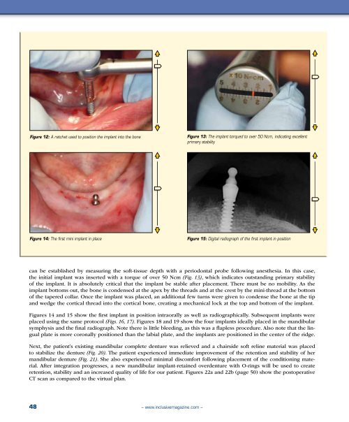
Figure 12: A ratchet used to position the implant into the bone<br />
Figure 13: The implant torqued to over 50 Ncm, indicating excellent<br />
primary stability<br />
Figure 14: The first mini implant in place<br />
Figure 15: Digital radiograph of the first implant in position<br />
can be established by measuring the soft-tissue depth with a periodontal probe following anesthesia. In this case,<br />
the initial implant was inserted with a torque of over 50 Ncm (Fig. 13), which indicates outstanding primary stability<br />
of the implant. It is absolutely critical that the implant be stable after placement. There must be no mobility. As the<br />
implant bottoms out, the bone is condensed at the apex by the threads and at the crest by the mini-thread at the bottom<br />
of the tapered collar. Once the implant was placed, an additional few turns were given to condense the bone at the tip<br />
and wedge the cortical thread into the cortical bone, creating a mechanical lock at the top and bottom of the implant.<br />
Figures 14 and 15 show the first implant in position intraorally as well as radiographically. Subsequent implants were<br />
placed using the same protocol (Figs. 16, 17). Figures 18 and 19 show the four implants ideally placed in the mandibular<br />
symphysis and the final radiograph. Note there is little bleeding, as this was a flapless procedure. Also note that the lingual<br />
plate is more coronally positioned than the labial plate, and the implants are positioned in the center of the ridge.<br />
Next, the patient’s existing mandibular complete denture was relieved and a chairside soft reline material was placed<br />
to stabilize the denture (Fig. 20). The patient experienced immediate improvement of the retention and stability of her<br />
mandibular denture (Fig. 21). She also experienced minimal discomfort following placement of the conditioning material.<br />
After integration progresses, a new mandibular implant-retained overdenture with O-rings will be used to create<br />
retention, stability and an increased quality of life for our patient. Figures 22a and 22b (page 50) show the postoperative<br />
CT scan as compared to the virtual plan.<br />
48<br />
– www.inclusivemagazine.com –


