

Inclusive - Glidewell Dental Labs
Inclusive - Glidewell Dental Labs
Inclusive - Glidewell Dental Labs

You also want an ePaper? Increase the reach of your titles
YUMPU automatically turns print PDFs into web optimized ePapers that Google loves.
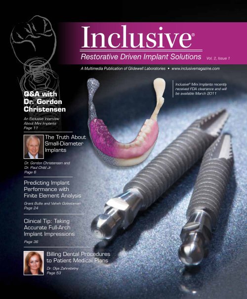
<strong>Inclusive</strong>®<br />
Restorative Driven Implant Solutions Vol. 2, Issue 1<br />
A Multimedia Publication of <strong>Glidewell</strong> Laboratories • www.inclusivemagazine.com<br />
Q&A with<br />
Dr. Gordon<br />
Christensen<br />
<strong>Inclusive</strong> ® Mini Implants recently<br />
received FDA clearance and will<br />
be available March 2011<br />
An Exclusive Interview<br />
About Mini Implants<br />
Page 11<br />
The Truth About<br />
Small-Diameter<br />
Implants<br />
Dr. Gordon Christensen and<br />
Dr. Paul Child Jr.<br />
Page 6<br />
Predicting Implant<br />
Performance with<br />
Finite Element Analysis<br />
Grant Bullis and Vaheh Golestanian<br />
Page 24<br />
Clinical Tip: Taking<br />
Accurate Full-Arch<br />
Implant Impressions<br />
Page 36<br />
Billing <strong>Dental</strong> Procedures<br />
to Patient Medical Plans<br />
Dr. Olya Zahrebelny<br />
Page 53
On the Web<br />
Log on to www.inclusivemagazine.com<br />
for bonus content and in-depth coverage.<br />
ONLINE Video Presentations<br />
Watch Drs. Alexandre-Amir Aalam and Mamaly<br />
Reshad present a systematic approach to advanced<br />
implant treatment planning in a Lecture-on-Demand<br />
from gIDE. Also, see how Dr. Timothy Kosinski uses<br />
digital treatment planning and guided surgery in<br />
combination with narrow-body implants for mandibular<br />
overdenture stabilization.<br />
ONLINE Photo Essays<br />
Gain further insight into Dr. Bradley Bockhorst’s<br />
technique for obtaining accurate full-arch implant impressions<br />
by viewing the photo slide show that accompanies<br />
his article. Plus, check out clinical photos<br />
from Dr. Raymond Choi’s presentation on surgically<br />
placing mini implants to support and retain a denture<br />
in an edentulous mandible.<br />
ONLINE CE credit<br />
Earn free CE credit for the material you’ve seen in<br />
this issue. You will earn two hours for each test you<br />
complete and pass. Take the CE tests on our website,<br />
www.inclusivemagazine.com.<br />
PLUS...<br />
New this issue, read <strong>Inclusive</strong> magazine on the go<br />
using your smartphone. You can now enjoy exclusive<br />
Web content anywhere you go, including video presentations,<br />
photo slide shows and featured articles.<br />
Check us out online to see what the implant<br />
industry is buzzing about.<br />
When you see these icons, it means we have even more information on<br />
that topic available at www.inclusivemagazine.com.<br />
– www.inclusivemagazine.com –
Contents<br />
Features<br />
6 The Truth About Small-Diameter<br />
Implants<br />
24 Predicting the Performance of<br />
Mini Implant-Retained Prostheses<br />
Using Finite Element Analysis<br />
36 Clinical Tip: A Technique for<br />
Obtaining Accurate Full-Arch<br />
Implant Impressions<br />
11<br />
18<br />
30<br />
43<br />
53<br />
Mini Implants: An Interview with<br />
Dr. Gordon Christensen<br />
Leading proponent of mini dental implants Dr. Gordon Christensen<br />
shares his experience on the subject in an exclusive Q&A interview<br />
with <strong>Inclusive</strong> magazine. Learn about Dr. Christensen’s placement<br />
approach, and find out what he considers to be the primary benefits<br />
of small-diameter implants and how to utilize them successfully.<br />
Mini <strong>Dental</strong> Implants for Every Dentist<br />
Dr. Raymond Choi delves into the world of mini dental implants<br />
with a discussion of their benefits and limitations, as well as their<br />
indications and contraindications. He also details a case in which<br />
“minis” are surgically placed to support and retain a denture in an<br />
edentulous mandible.<br />
Mandibular Denture Retention:<br />
The Mini Implant Solution<br />
According to Drs. Stephen Wagner and A. Burton Melton, edentulous<br />
patients in need of complete dentures were, until the standardization<br />
of dental implants, destined to wear prostheses that provided<br />
them with low levels of overall satisfaction. Today, mini implants<br />
are proving to be a viable alternative to standard-diameter implants<br />
for a variety of permanent applications, due to factors such as their<br />
lower cost and less-invasive surgical protocol.<br />
Utilizing Digital Treatment Planning and Guided<br />
Surgery in Conjunction with Narrow-Body Implants<br />
Dr. Timothy Kosinski presents a case study, utilizing CT diagnosis,<br />
digital treatment planning and guided surgery, for the immediate<br />
stabilization of a mandibular implant-retained overdenture. Included<br />
is a flapless surgical technique, immediate implant placement<br />
and resulting improved stability of the patient’s existing mandibular<br />
denture prior to delivery of the final prosthesis.<br />
Billing Patient Medical Plans:<br />
There’s Nothing Illegal About It!<br />
Seeking to dispel the misconception held among some dentists<br />
that they can only access benefits from dental plans, Dr. Olya<br />
Zahrebelny argues that numerous routine procedures performed in<br />
general and specialty dental practices are covered by patient medical<br />
benefit plans. Before clinicians bill procedures to these medical<br />
plans, however, she advises that they adhere to some basic rules.<br />
– Contents – 1
Letter from the Editor<br />
We saw many changes in implantology in 2010, including rapid growth in monolithic restorations<br />
such as BruxZir ® and IPS e.max ® , as well as the use of CAD/CAM technology in<br />
the design and fabrication of custom abutments, crowns, overdenture bars and frameworks.<br />
Digital treatment planning and guided surgery continued to grow with expanded access to<br />
CBCT scanners. Local reaction to the expansion of national chains brought about renewed<br />
interest in screw-retained dentures. And these were just a few of the changes.<br />
This issue focuses on small-diameter implants (SDIs). While controversial, mini implants are<br />
gaining popularity, so we asked several clinicians who have significant experience using SDIs<br />
to share their observations with us. Included is a featured reprint by Drs. Gordon Christensen<br />
and Paul Child, followed by an exclusive interview with Dr. Christensen, one of the most<br />
outspoken proponents of SDIs. Reading these articles should help you decide if minis have a<br />
place in your implant practice.<br />
Another subject garnering discussion is insurance coverage of implant cases. While we are<br />
all familiar with the ins and outs of dental insurance, Dr. Olya Zahrebelny discusses how you<br />
may also be able to access patient medical benefits by following some basic rules.<br />
The featured Clinical Tip outlines what we have found to be a highly predictable procedure<br />
for obtaining accurate full-arch implant impressions. Although the accuracy of CAD/CAM<br />
designed and fabricated titanium frameworks has greatly improved and simplified restoring<br />
full-arch cases, marking an end to the days of cutting and soldering conventional cast<br />
frameworks to achieve a passive fit, an extremely accurate impression and master cast are<br />
still critical requirements.<br />
Our goal with <strong>Inclusive</strong> in 2011 is to keep you up to date on the latest products, procedures<br />
and dental materials in implantology. We will also demonstrate our commitment to continuing<br />
education with the opening of our 2,800-square-foot training and education facility.<br />
(Watch for updates on upcoming courses and programs.) For expanded magazine content,<br />
please visit www.inclusivemagazine.com.<br />
Thank you for your continued support. We wish you and your practice a prosperous new year!<br />
Regards,<br />
Dr. Bradley C. Bockhorst<br />
Editor-in-Chief, Clinical Editor<br />
inclusivemagazine@glidewelldental.com<br />
– Letter from the Editor – 3
Publisher<br />
Jim <strong>Glidewell</strong>, CDT<br />
Editor-in-Chief<br />
Bradley C. Bockhorst, DMD<br />
Managing Editors<br />
Jim Shuck; Mike Cash, CDT<br />
Creative Director<br />
Rachel Pacillas<br />
Clinical Editor<br />
Bradley C. Bockhorst, DMD<br />
Contributing editors<br />
Dzevad Ceranic, CDT; Greg Minzenmayer<br />
senior Copy Editor<br />
Jennifer Holstein<br />
copy editors<br />
Melissa Manna, Eldon Thompson<br />
Graphic Designers/Web Designers<br />
Jamie Austin, Deb Evans, Joel Guerra, Lindsey Lauria,<br />
Phil Nguyen, Kelley Pelton, Ty Tran<br />
Photographers/Clinical Videographers<br />
Sharon Dowd, James Kwasniewski, Sterling Wright<br />
Illustrators<br />
Phil Nguyen<br />
coordinatorS/AD Representatives<br />
Teri Arthur, Vivian Tsang<br />
If you have questions, comments or suggestions, e-mail us at<br />
inclusivemagazine@glidewelldental.com. Your comments may be<br />
featured in an upcoming issue or on our website.<br />
© 2011 <strong>Glidewell</strong> Laboratories<br />
Neither <strong>Inclusive</strong> magazine nor any employees involved in its publication<br />
(“publisher”) makes any warranty, express or implied, or assumes<br />
any liability or responsibility for the accuracy, completeness, or usefulness<br />
of any information, apparatus, product, or process disclosed, or<br />
represents that its use would not infringe proprietary rights. Reference<br />
herein to any specific commercial products, process, or services by<br />
trade name, trademark, manufacturer or otherwise does not necessarily<br />
constitute or imply its endorsement, recommendation, or favoring<br />
by the publisher. The views and opinions of authors expressed<br />
herein do not necessarily state or reflect those of the publisher and<br />
shall not be used for advertising or product endorsement purposes.<br />
CAUTION: When viewing the techniques, procedures, theories and materials<br />
that are presented, you must make your own decisions about<br />
specific treatment for patients and exercise personal professional judgment<br />
regarding the need for further clinical testing or education and<br />
your own clinical expertise before trying to implement new procedures.<br />
<strong>Inclusive</strong> is a registered trademark of <strong>Glidewell</strong> Laboratories.<br />
Contributors<br />
■ Bradley C. Bockhorst, DMD<br />
After receiving his dental degree from<br />
Washington University School of <strong>Dental</strong><br />
Medicine, Dr. Bradley Bockhorst served<br />
as a Navy <strong>Dental</strong> Officer. Dr. Bockhorst is<br />
Director of Clinical Technologies at <strong>Glidewell</strong><br />
Laboratories, where he oversees <strong>Inclusive</strong><br />
® Digital Implant Treatment Planning<br />
Services and is editor-in-chief and clinical editor of <strong>Inclusive</strong><br />
magazine. A member of the CDA, ADA, AO, ICOI and<br />
AAID, Dr. Bockhorst lectures internationally on an array<br />
of dental implant topics. He maintains a private practice<br />
focused on implant prosthetics in Mission Viejo, Calif. Contact<br />
Dr. Bockhorst at 800-521-0576 or inclusivemagazine@<br />
glidewelldental.com.<br />
■ GRANT BULLIS<br />
Grant Bullis, director of implant R&D and<br />
digital manufacturing at <strong>Glidewell</strong> Laboratories,<br />
began his career in the dental<br />
industry at Steri-Oss in 1997. After Nobel<br />
Biocare acquired Steri-Oss, Grant worked<br />
in the R&D department. Since joining the<br />
lab in March 2007, Grant has been integral<br />
in obtaining FDA 510(k) clearances for the company’s<br />
<strong>Inclusive</strong> Implant Abutments. In 2010, he was promoted to<br />
director and now oversees all aspects of CAD/CAM, implant<br />
product development and manufacturing at <strong>Glidewell</strong>.<br />
Grant has a degree in mechanical CAD/CAM from Irvine<br />
Valley College in Orange County, Calif., and an MBA from<br />
Keller Graduate School of Management. Contact him at<br />
inclusivemagazine@glidewelldental.com.<br />
■ VAHEH GOLESTANIAN<br />
Vaheh Golestanian received a master’s<br />
degree in biomedical engineering at Iran<br />
University of Science and Technology in<br />
Tehran. In 2008, he joined <strong>Glidewell</strong> Laboratories’<br />
Implant R&D and Digital Manufacturing<br />
department as a manufacturing<br />
engineer. Vaheh has eight years’ experience<br />
as a mechanical engineer focused on finite element<br />
analysis and CNC programming. A member of the Society<br />
of Manufacturing Engineers, he recently co-authored a<br />
technical paper on using finite element methods to design<br />
zirconia abutments. Contact him at inclusivemagazine@<br />
glidewelldental.com.<br />
4<br />
– www.inclusivemagazine.com –
■ Paul L. Child Jr., DMD, CDT<br />
Dr. Paul Child graduated from Case Western<br />
Reserve University School of <strong>Dental</strong><br />
Medicine, completed a prosthodontic residency<br />
at Louisiana State University School<br />
of Dentistry and is a CDT. He also maintains<br />
a private practice at the CR <strong>Dental</strong><br />
Health Clinic in Provo, Utah. As CEO of<br />
CR Foundation ® , Dr. Child conducts extensive research<br />
in all areas of dentistry and directs the publication of the<br />
Gordon J. Christensen CLINICIANS REPORT ® and other<br />
publications. He lectures nationally and is a member of<br />
many professional associations and academies. Contact<br />
him at 801-226-2121 or toni@cliniciansreport.org.<br />
■ Raymond Choi, DDS<br />
Dr. Raymond Choi graduated from USC<br />
School of Dentistry, where he was an assistant<br />
clinical professor in the TMJ and<br />
Facial Pain Clinic for 10 years, and completed<br />
a TMJ/Facial Pain Preceptorship at<br />
UCLA. He is also a graduate of the Misch<br />
International Implant Institute, Fellow of<br />
the ICOI and Associate Fellow of the AAID. Dr. Choi was one<br />
of the first dentists on the West Coast to start using IMTEC<br />
Sendax Mini <strong>Dental</strong> Implants for lower denture stabilization.<br />
Founder and president of the Global Mini Implant<br />
Institute (GMI), Dr. Choi maintains a general private practice<br />
in Tustin, Calif. He has been conducting mini implant<br />
seminars and surgical workshops for dentists since 2002<br />
and lectures extensively in the U.S. and internationally.<br />
Contact him at www.miniimplanteducation.com.<br />
■ Gordon J. Christensen, DDS, MSD, Ph.D<br />
Dr. Gordon Christensen is a practicing<br />
prosthodontist in Provo, Utah. His degrees<br />
include DDS, University of Southern<br />
California; MSD, University of Washington;<br />
and Ph.D, University of Denver. He<br />
is a Diplomate of the American Board of<br />
Prosthodontics; Fellow and Diplomate<br />
of the ICOI; Fellow of the AO, ACD, ICD, ACP and Royal<br />
College of Surgeons of England; Honorary Fellow of the<br />
AGD; and Associate Fellow of the AAID. Drs. Gordon and<br />
Rella Christensen are cofounders of the nonprofit Gordon<br />
J. Christensen CLINICIANS REPORT. Contact him at<br />
801-226-6569 or info@pccdental.com.<br />
■ TIMOTHY F. KOSINSKI, DDS, MAGD<br />
Dr. Timothy Kosinski graduated from University<br />
of Detroit Mercy School of Dentistry<br />
and received a master’s degree in biochemistry<br />
from Wayne State University School of<br />
Medicine. An adjunct assistant professor<br />
at Mercy School of Dentistry, he serves on<br />
the editorial boards of numerous dental<br />
journals and is a Diplomate of the ABOI/ID, ICOI and AO;<br />
Fellow of the AAID; and Master of the AGD. Contact him at<br />
248-646-8651, drkosin@aol.com or www.smilecreator.net.<br />
■ A. Burton Melton, DDS<br />
Dr. Burton Melton received his undergraduate<br />
degree from BYU, his DDS from<br />
Baylor College of Dentistry and his Diploma<br />
in Prosthodontics from the University<br />
of Missouri School of Dentistry. He has<br />
practiced prosthodontics in Albuquerque<br />
and Santa Fe, N.M., since 1972. Dr. Melton<br />
has presented programs to audiences in the U.S., Japan,<br />
Korea, Mexico, Taiwan and England, and has appeared<br />
as a guest lecturer at dental schools across the U.S. Contact<br />
him at 505-883-7744 or abmeltonnm@aol.com.<br />
■ Stephen A. Wagner, DDS<br />
Dr. Stephen Wagner is in his 32nd year of<br />
private practice in Albuquerque, N.M. He<br />
received his dental degree from USC School<br />
of Dentistry and his prosthodontic training<br />
from MD Anderson Cancer Center in<br />
Houston, Texas. Dr. Wagner serves on the<br />
review board of the International Journal<br />
of Oral and Maxillofacial Implants and is a Diplomate<br />
of the American Board of Prosthodontics. Contact him at<br />
bigjawbone@mac.com.<br />
■ Olya Zahrebelny, DDS<br />
Dr. Olya Zahrebelny graduated from the<br />
University of Toronto Faculty of Dentistry<br />
and completed a general practice residency.<br />
She has practiced in hospital and private-practice<br />
settings. A former insurance<br />
plan consultant, she has taught at three<br />
dental schools and was an attending physician<br />
at the University of Illinois Medical Center. Repeatedly<br />
named a leader in continuing education and dental<br />
consulting by Dentistry Today, she has lectured nationally<br />
and internationally. Contact her at drz@thezgroupllc.com.<br />
– Contributors – 5
The Truth About<br />
Small-Diameter Implants<br />
Go online for<br />
in-depth content<br />
by Gordon J. Christensen, DDS, MSD, Ph.D and Paul L. Child Jr., DMD, CDT<br />
If we listened to and believed some of the comments about small-diameter implants<br />
(SDIs or “mini” implants) that we hear coming from some areas of surgical dentistry,<br />
we would be led to think that these devices simply do not work. However, the truth is diametrically<br />
opposed to what some are saying, and it has been our observation that some of the most severely negative<br />
comments come from dentists who have never placed SDIs.<br />
This article includes: the definition of mini implants or SDIs; a discussion of the evolution of SDIs, including<br />
their clearance by the U.S. Food and Drug Administration (FDA) and research support; reasons for SDI<br />
use instead of conventional-diameter implants; the indications for SDI use; and suggestions on how to use<br />
them successfully.<br />
THE EVOLUTION OF THE<br />
SMALL-DIAMETER IMPLANT CONCEPT<br />
There is no question among dentists that root-form implants,<br />
3.0 mm in diameter and more, are one of the most<br />
successful and important additions to clinical dentistry<br />
in the entire history of dentistry. The FDA cleared these<br />
conventional-diameter root-form implants for clinical<br />
use in 1976. Millions of conventional-diameter implants<br />
have been placed for more than four decades, and their<br />
cumulative success rate of around 95 percent is impressive.<br />
In many situations, it has been our experience that<br />
the conventional prosthodontic portion of implant treatment<br />
fails faster than the properly integrated root-form<br />
implants themselves.<br />
In the early 1990s, some innovative practitioners started<br />
using SDIs (up to 2.9 mm in diameter) for long-term use in<br />
situations with insufficient bone. At that time, SDIs were<br />
considered to be for transitional use only. Also, orthodontists<br />
began using SDIs, also known as temporary anchorage<br />
devices, for anchorage for difficult tooth movement<br />
situations. It soon became obvious to those practitioners<br />
that properly placed SDIs were working adequately.<br />
As a result of their obvious clinical success, SDIs were<br />
cleared by the FDA for “long-term intrabony applications,”<br />
with the help of IMTEC (a 3M ESPE company), in 1997.<br />
Subsequently, numerous other SDI brands have received<br />
similar FDA clearance. Thousands of these SDIs are now<br />
in successful restorative use, with a reported 91 percent<br />
to 97 percent survival rate. Numerous surveys, testimonials,<br />
research projects, and satisfied dentists and patients<br />
attest to that fact. 1–6 Many more positive references are<br />
available in the restorative, prosthodontic and orthodontic<br />
literature.<br />
WHY USE SMALL-DIAMETER<br />
IMPLANTS IF CONVENTIONAL-DIAMETER<br />
IMPLANTS ARE SO SUCCESSFUL?<br />
The authors of this article are prosthodontists who place<br />
conventional- and small-diameter root-form implants. The<br />
following are their observations on the desirability of SDI<br />
use compared to conventional-diameter implant use.<br />
Inadequate Bone Quantity<br />
Conventional-diameter implants, averaging about 3.5 mm<br />
in diameter, require minimally about 6 mm of bone in<br />
a facial-lingual dimension, and about 10 mm of bone in<br />
a crestal-apical dimension, for uncomplicated placement<br />
without grafting. Some patients accept the overall implant<br />
concept, but they have inadequate bone quantity and do<br />
not want to undergo significant bone grafting.<br />
6<br />
– www.inclusivemagazine.com –
If SDIs were used only for patients with edentulous<br />
mandibles, roughly 35 to 40 million edentulous patients<br />
in the U.S. alone would have better fitting and retained<br />
complete dentures.<br />
SDIs can be placed in as little as 3 mm of<br />
bone in a facial-lingual dimension and 10 mm<br />
of bone in a crestal-apical dimension. In fact,<br />
often bone 3 mm or 4 mm in a facial-lingual<br />
dimension is ideal, because the cortical bone<br />
plate on the facial has nearly approximated the<br />
lingual cortical plate, and this dense bone holds<br />
the SDI securely. Some experienced implant<br />
surgeons may question this, until they consider<br />
the fact that the SDI is usually a “screw,”<br />
expanding bone instead of cutting it away.<br />
Inadequate Financial Resources<br />
Some patients have inadequate bone, accept<br />
the implant concept, accept the need for extensive<br />
bone grafting, and they are ready to<br />
accept the treatment with the following exceptions:<br />
the cost of the grafting is too high, the<br />
expense of the restorative treatment is high<br />
and conventional-diameter implant treatment<br />
is denied. SDIs frequently solve this challenge,<br />
as stated in the previous point.<br />
Compromised Physical Condition<br />
Many physically debilitated patients do not<br />
have the ability to tolerate conventional-diameter<br />
implant placement, but they can tolerate<br />
Table 1: Use of SDIs in Approximate Order<br />
of Decreasing Frequency of Use<br />
Edentulous mandible<br />
Removable partial denture<br />
Edentulous maxilla (this use has a higher<br />
failure rate than edentulous mandibles)<br />
Augmentation of fixed prosthesis<br />
Sole support of fixed prosthesis<br />
the simple, few-minute placement of SDIs<br />
without a flap. Recent research has shown<br />
that flapless implant placement may accelerate<br />
osseointegration and produce quicker<br />
healing. Debilitated persons can benefit<br />
from these simple procedures.<br />
MAJOR USES OF<br />
SMALL-DIAMETER IMPLANTS<br />
SDIs are listed in Table 1 in approximate<br />
order of decreasing frequency of use, as<br />
noted in the previously referenced articles,<br />
our own use patterns and our observation<br />
of other practitioners.<br />
If SDIs were used only for patients with<br />
edentulous mandibles, roughly 35 to 40<br />
million edentulous patients in the U.S.<br />
alone would have better fitting and retained<br />
complete dentures. These implants<br />
are so easy to use in most edentulous<br />
mandibles that it is upsetting to us they<br />
are not used more in the profession for<br />
the indications.<br />
WHY DO SOME DENTISTS<br />
STATE THAT SMALL-DIAMETER<br />
IMPLANTS ARE NOT<br />
SUCCESSFUL?<br />
Many of the same surgical dentists now<br />
condemning SDIs stated 40 years ago that<br />
conventional root-form implants would not<br />
work. It appears that their opinion is that<br />
anything new is automatically bad! SDIs<br />
are new, but they are proving themselves.<br />
This section is probably the most important<br />
part of this article. Some SDIs fail. We<br />
have experienced a few failures ourselves<br />
over the past nine years of use. These failures<br />
were almost always related to one or<br />
more of the following errors:<br />
Salvage of previously made prosthesis<br />
– The Truth About Small-Diameter Implants – 7
a<br />
b<br />
Figures 1a, 1b: A typical conventional-diameter implant has a blunt end necessitating<br />
cutting a hole in the bone for placement. A typical SDI has a screw<br />
configuration that expands minimal bone on placement. This difference is one<br />
that allows SDIs to be placed in bone as thin as 3 mm to 4 mm in the faciallingual<br />
dimension.<br />
Figure 2: SDIs placed in a mandible model. Small spheres on the implants and<br />
rubber washers in housings in the denture support and retain the denture.<br />
■ Too much thickness of the soft tissue. If the ratio of<br />
the coronal portion of the SDI to the portion placed in<br />
the bone is excessive, a long lever arm is present. This<br />
situation stresses the SDI and may lead to failure of<br />
the implant. If the soft tissue, through which the SDI<br />
is to be placed, is thicker than about 2 mm, it should<br />
be reduced by removing a wedge of tissue from the<br />
coronal portion of the ridge. This can be done before<br />
the implants are placed, allowing for adequate healing,<br />
or at the time of implant placement. This surgery may<br />
be done with a scalpel, or some lasers may be used<br />
around implants to accomplish this task without causing<br />
damage to the implant osseointegration potential.<br />
■ Improper parallelism of implants. SDIs should be as<br />
parallel as possible. If these implants are much more<br />
than 15 degrees from parallelism, technical difficulty at<br />
placement of the prosthesis and subsequent potential<br />
clinical failure can be anticipated.<br />
■ Inadequate preoperative radiographs. Poor bone<br />
is commonly present in some areas of edentulous patients.<br />
We discourage using only two-dimensional conventional<br />
panoramic radiographs because you cannot<br />
determine the quality or quantity of the bone in a facial-lingual<br />
dimension. Coarsely trabeculated bone is<br />
not appropriate for SDIs. The more dense the bone,<br />
the better. To determine the density of the bone, faciallingual<br />
oriented radiographs are strongly suggested.<br />
These include tomograph or cone-beam radiographs.<br />
Most communities now have accessibility to some form<br />
of the suggested facial-lingual orientation radiographs<br />
at moderate cost.<br />
■ Poor bone density in the posterior maxillary<br />
tuberosity areas. Usually, the dense Type I bone<br />
of the resorbed anterior mandible is excellent for<br />
SDIs. The worst bone, contraindicated for SDI<br />
placement by most experienced practitioners, is<br />
the posterior maxillary tuberosity, with its porous<br />
type IV bone. A careful analysis of the density of<br />
the bone in any other part of the oral cavity is<br />
suggested, as they too may have poor bone density<br />
contraindicating SDIs.<br />
To determine the density<br />
of the bone, facial-lingual<br />
oriented radiographs are<br />
strongly suggested. These<br />
include tomograph or<br />
cone-beam radiographs.<br />
8<br />
– www.inclusivemagazine.com –
Figure 3: Atlas ® (Dentatus) implants placed in a mandible model. Small spheres<br />
on the implants and soft denture reline material in the denture support and<br />
retain the dentures.<br />
Figure 4: Zimmer (Sterngold) implants placed in a mandible model with ERA ®<br />
attachments on the implants. Reciprocal ERAs in the denture base support and<br />
retain the denture.<br />
■ Too few SDIs are often placed. It has been suggested<br />
in both empirical and research reports that the minimal<br />
number of SDIs for edentulous mandibles is four, evenly<br />
spaced from the left canine area to the right canine<br />
area. This is double the minimal number of implants<br />
suggested for conventional-diameter implants. The ratio<br />
appears to be two SDIs where one conventional-diameter<br />
implant would usually be used. Some companies<br />
are suggesting six SDIs instead of four for edentulous<br />
maxillas, evenly spaced from the canine area to the opposite<br />
canine areas. However, the more dense the bone,<br />
the fewer SDIs that are needed.<br />
■ SDIs are too short. The most popular average length<br />
for SDIs is 13 mm. It appears from clinical observation<br />
and research that this is a predictable and successful<br />
length. The implants must be used in adequate bone,<br />
according to the literature of reported successful use of<br />
thousands of SDIs and to the discussions with manufacturers<br />
about clinician reports to them.<br />
■ Poorly adjusted occlusion, or loading the implants<br />
too soon. Most SDIs are loaded immediately<br />
on placement. Occlusion needs to be adjusted<br />
perfectly on placement of the prosthesis. Allowing<br />
heavy occlusion to traumatize these small implants<br />
is asking for early failure. If questionable bone quality<br />
or quantity is present, soft denture reline material<br />
may be placed in the denture around the area of the<br />
implants for several weeks to ensure that they have optimum<br />
time for initiation of osseointegration.<br />
SUMMARY AND CONCLUSION<br />
SDIs that are treatment planned correctly, placed and<br />
loaded properly, and are within a well-adjusted occlusion,<br />
are working in an excellent manner for the patients described<br />
in this article. It is time for those practitioners<br />
unfamiliar with SDIs and their uses to discontinue their<br />
discouragement of this technique. SDIs are easily placed,<br />
minimally invasive and a true service to those patients<br />
described. They do not replace conventional-diameter<br />
implants; however, they are a significant and important<br />
augmentation to the original root-form implant concept.<br />
There is obvious evidence of the growing acceptance<br />
of small-diameter implants by both general practitioners<br />
and specialists.<br />
References<br />
1. Bulard RA, Vance JB. Multi-clinic evaluation using mini-dental implants for<br />
long-term denture stabilization: a preliminary biometric evaluation. Compend<br />
Contin Educ Dent. 2005;26:892–97.<br />
2. Clinical Research Associates. Small-diameter “mini” implants — user status<br />
report. CRA Newsletter. 2007;31:1–2.<br />
3. Griffitts TM, Collins CP, Collins PC. Mini dental implants: an adjunct for retention,<br />
stability and comfort for the edentulous patient. Oral Surg Oral Med Oral<br />
Pathol Oral Radiol Endod. 2005;100:e81–e84.<br />
4. Morneburg TR, Pröschel PA. Success rates of micro-implants in edentulous<br />
patients with residual ridge resorption. Int J Oral Maxillofac Implants. 2008;<br />
23:270–76.<br />
5. Shatkin TE, Shatkin S, Oppenheimer BD, et al. Mini dental implants for longterm<br />
fixed and removable prosthetics: a retrospective analysis of 2,514 implants<br />
placed over a five-year period. Compend Contin Educ Dent. 2007;28:92–99.<br />
6. Vigolo P, Givani A. Clinical evaluation of single-tooth mini-implant restorations:<br />
a five-year retrospective study. J Prosthet Dent. 2000;84:50–54.<br />
Disclosure: Dr. Christensen and Dr. Choi report no conflicts of interest.<br />
Reprinted by permission of Dentistry Today, ©2010 Dentistry Today.<br />
– The Truth About Small-Diameter Implants – 9
Mini Implants:<br />
An Interview with<br />
Dr. Gordon Christensen<br />
Interview of Gordon J. Christensen, DDS, MSD, Ph.D<br />
by Bradley C. Bockhorst, DMD<br />
Dr. Gordon Christensen is a leading proponent of mini implants. For<br />
this issue of <strong>Inclusive</strong> magazine, he was kind enough to discuss his<br />
views on the subject in an exclusive phone interview.<br />
Dr. Bradley Bockhorst: I enjoyed your presentation at the American Academy of Implant<br />
Dentistry’s 2010 Annual Meeting in Boston, Mass., last October. You are one of the most outspoken<br />
proponents on mini implants and, as such, I appreciate you sharing your experience with us.<br />
Dr. Gordon Christensen: Thank you. By the way, I’m very pleased to take part in this interview.<br />
There are so many questions regarding small-diameter implants, and I’m happy to answer any<br />
questions related to them.<br />
BB: To start things off, how many years have you been placing small-diameter implants? And how<br />
many have you placed to date?<br />
GC: Probably the best answer to that question is going back to our CRA ® Newsletter report (now<br />
the Gordon J. Christensen CLINICIANS REPORT ® ), which was published in November 2007. At that<br />
time, I had been placing mini implants since 1997 — only about three or four years. We had 200<br />
people in that report who stated what they had observed during their use of mini implants. I had<br />
done only a few hundred at that particular point. The 200 respondents were all CRA Newsletter<br />
subscribers. The respondents were from five to 65 years out of dental school, with a mean of 27.<br />
They were in 34 states, Canada and elsewhere. Ninety-five percent were general practitioners,<br />
4 percent were prosthodontists and 1 percent were periodontists. They had been in implant dentistry<br />
an average of 13 years. Approximately 74 percent of them did surgery and prosthodontics.<br />
The rest performed either surgery or prosthodontics. As for in-house education, depending on the<br />
brand, most of them had taken a short course on mini implants. So that group represented basically<br />
thousands of mini implants among the 200 people who responded.<br />
– Mini Implants: An Interview with Dr. Gordon Christensen – 11
BB: And within your practice, what percentage of implants placed<br />
would you say are small-diameter or mini implants?<br />
GC: I’ve been doing surgery and placing implants for 25 years.<br />
Each indication, of course, would have different percentages. Right<br />
now, in edentulous mandibles, I would say at least 50 percent<br />
of what I’m doing is small-diameter implants versus conventionaldiameter<br />
implants.<br />
The primary benefit<br />
of mini implants is<br />
for the person who<br />
is too debilitated<br />
to undergo the<br />
surgery necessary<br />
for conventional<br />
implant placement;<br />
the person who does<br />
not have the money<br />
for a complex case;<br />
or the person who<br />
will not accept, or<br />
cannot have for<br />
health reasons, a<br />
major bone graft.<br />
BB: For the sake of some of our readers who may not be as aware<br />
of mini implants as others, what would you say are the primary<br />
benefits for this type of implant?<br />
GC: The primary benefit of mini implants is for the person who is<br />
too debilitated to undergo the surgery necessary for conventional<br />
implant placement; the person who does not have the money for<br />
a complex case, which very often might be better; or the person<br />
who will not accept, or cannot have for health reasons, a major<br />
bone graft.<br />
BB: What would you say are the primary benefits, from the clinician’s<br />
standpoint?<br />
GC: Simplicity. Going back to that November 2007 CRA Newsletter<br />
report, when asked about difficulty of implant placement, respondents<br />
reported placement without a flap as “simple” and placement<br />
with a flap as “slightly more difficult.” That’s about what we saw<br />
for the major advantage to the clinician. I delivered a program at<br />
the World Congress of Minimally Invasive Dentistry, and it was on<br />
about 20 different minimally invasive techniques. And this was one<br />
of the major benefits for smaller-diameter implants.<br />
Another significant advantage is that they can be immediately loaded<br />
in bone that is adequate. With Type I bone, there’s no question;<br />
I’ve loaded hundreds of them immediately.<br />
BB: Initially, mini implants were primarily marketed as temporary<br />
or provisional implants. What would you say has changed to make<br />
them a viable long-term option?<br />
GC: Initially, I was using them as transitional implants when I had<br />
placed conventional implants and just wanted something to hold<br />
the denture or the fixed bridge in place while the conventionaldiameter<br />
implants integrated. I found, after three or four months<br />
of waiting for the conventional-diameter implants to integrate, that<br />
I seldom could take the mini implants out easily. In fact, I had a<br />
couple that I practically had to cut out. That was my turning event.<br />
When the initial transitional implants were introduced, they were<br />
pure titanium. They were so weak that you could bend them with<br />
your finger. They were not adequate. However, Dr. Victor Sendax<br />
(a periodontist based in New York) got together with the IMTEC<br />
Corporation and alloyed them, and they became stronger. The combination<br />
of strength and ease of placement, and the fact that they<br />
could be loaded immediately, made me change my mind about<br />
using transitional implants.<br />
12<br />
– www.inclusivemagazine.com –
BB: As with conventional-diameter implants, have manufacturers<br />
added different types of surfaces to increase bone-to-implant contact?<br />
GC: Yes, they have. There are numerous companies now involved<br />
with making small-diameter implants. By far the most well known<br />
is IMTEC. Intra-Lock is making some major introductions as well.<br />
We’re seeing about 10 companies that are involved with smalldiameter<br />
implants at this point, and at least three of them are<br />
major companies.<br />
BB: One of the challenges in gaining widespread acceptance of<br />
smaller diameters is the perception of a lack of long-term published<br />
studies. I know you just referred us to the CRA study. Are there any<br />
other studies out there for people to reference for relieving that fear?<br />
GC: These are from various years, but Shatkin 1 was one of the major<br />
ones, from Evolution Lab out of Amherst, N.Y., that found 94.2 percent<br />
retention. Other studies found 91 percent retention, 97.4 percent<br />
retention, 94.2 percent retention and 95.5 percent retention. 2–5 So<br />
there are some good studies out there and surely more to come.<br />
BB: As far as implant diameters, IMTEC started out with the 1.8<br />
and has added larger diameters. Other companies, such as OCO<br />
Biomedical, started with the 3.0 and are adding narrower diameters.<br />
So several companies now have this range. What diameter do<br />
you typically prefer?<br />
GC: It depends on the bone. For Type I bone, I still very strongly<br />
prefer the 1.8 mm diameter. As you know, a small-diameter implant<br />
would go, in the current marketplace, from 1.8 mm to 2.9 mm. That<br />
directly relates to the FDA clearance of these types of implants back<br />
in 1997 — 14 years ago. Standard-diameter implants — 3 mm and<br />
larger — were cleared by the FDA a long time ago, in 1976. So the<br />
differentiation of small diameter versus conventional diameter would<br />
be at the 3.0 mm diameter level. The sizes of mini-diameter implants<br />
typically start at 1.8 mm and go to 2.4 mm, 2.5 mm, 2.9 mm.<br />
The combination of<br />
strength and ease<br />
of placement, and<br />
the fact that they<br />
could be loaded<br />
immediately, made<br />
me change my<br />
mind about using<br />
transitional implants.<br />
In good Type III bone, which is obviously softer but usually more<br />
homogeneous, I would use a 2.4 mm diameter. And in the lower<br />
anterior, I tend to prefer the 1.8 mm because, when going to a<br />
larger diameter, I have broken them from the torque required to<br />
insert them. I’ve only broken one screwing it in, but I have broken<br />
one. And I had one break, as I mentioned at the AAID meeting in<br />
October, in service in Type I bone. So it varies with the bone density.<br />
The more dense the bone, the smaller the diameter. The more<br />
porous the bone, the larger the diameter.<br />
BB: Along that same line, most of the clinicians who are placing<br />
implants are putting them in with a winged driver, not with a torque<br />
wrench. Is there a maximum torque where you say, OK, stop, let’s<br />
run the drill down the osteotomy again?<br />
GC: Yes, you need about 30 Ncm to feel like you’re at an appropriate<br />
level of primary stability. If at 30 Ncm you’re not making<br />
any progress, then that makes me quite nervous. So I would screw<br />
– Mini Implants: An Interview with Dr. Gordon Christensen – 13
I strongly suggest a facial-lingual radiograph for any<br />
treatment plan — either a tomograph or a CBCT scan.<br />
the thing out and make a little deeper cut or use a widerdiameter<br />
implant. As a dentist becomes more familiar with<br />
these, they will soon sense when threading it into place<br />
whether the torque is approaching 30 Ncm. Let me put it<br />
this way: Dentists need to have a torque wrench.<br />
BB: One of the other presentations at the AAID meeting<br />
was by Dr. Sendax. His takeaway message was bicortical<br />
stabilization. That’s doable in the symphysis region. How<br />
do you handle that in other regions, such as the posterior<br />
mandible? Are you a proponent of bicortical stabilization,<br />
or do you have a different approach as far as placement?<br />
GC: Type II bone typically found in the posterior<br />
mandible is not a particularly good indication for smalldiameter<br />
implants, in my opinion, because the bone density<br />
of the cortical plate may be 1000 on the Hounsfield<br />
unit (HU) scale. However, the cancellous bone may be as<br />
low as 40 or 50 HU. The bottom line is, I’m usually reaching<br />
for a large-diameter implant, like a 6 mm, to better<br />
engage the cortical bone. With the exception of the small<br />
triangle of bone that’s usually directly distal to the mental<br />
foramen, I’m wary of small-diameter implants in Type II<br />
bone in the posterior mandible.<br />
BB: What’s your approach in the maxilla?<br />
GC: About the same. As you approach the sinus, which<br />
would be the first and second premolar, that Type III<br />
bone is going to be relatively dense. Anything distal to<br />
that — wow! — is not for mini implants, in my opinion.<br />
BB: That’s great feedback. Besides the quality of the bone,<br />
what are other caveats when considering mini implants?<br />
GC: There are many reasons for mini-implant failure.<br />
Over the 10 years I’ve done this, I have lost only 10. Now<br />
that sounds like a pretty egotistical statement, but when<br />
they have failed, and when I’ve seen them fail as they’ve<br />
come into our lab — and we do not solicit people sending<br />
their cases to our lab — is when we see these things:<br />
Improper radiography and lack of thorough treatment<br />
planning. I strongly suggest a facial-lingual radiograph for<br />
any treatment plan — either a tomograph or a CBCT scan.<br />
The quality and quantity of bone, as well as the ideal location<br />
of the implant, can be evaluated pre-surgically.<br />
Too much soft-tissue thickness on the ridge is another<br />
issue. If the thickness of the soft tissue is over 2 mm,<br />
the clinician should take a V-wedge out and allow the<br />
soft tissue to heal before he or she even considers making<br />
any kind of an impression. About 2 mm should<br />
be the maximum on the crest. I’ve seen clinicians stick<br />
the implant through 4 mm or 5 mm of soft tissue. That’s<br />
like sticking a 6-foot beanpole in a foot of mud — just<br />
absolute stupidity.<br />
Too few implants placed can also be a major problem.<br />
For Type I bone, four mini implants in the anterior region<br />
of an edentulous mandible is more than enough. I<br />
usually say two small-diameter implants would equal one<br />
conventional-diameter implant. Having done hundreds<br />
of cases with conventional-diameter implants in the<br />
two canine areas, we’re now putting four in the anterior<br />
edentulous mandible, spread either equally across from<br />
what was canine to what was canine, or emphasizing the<br />
canine areas with two in that area spread 4 mm or 5 mm<br />
apart, and the same in the other canine area. And then<br />
on the upper, IMTEC and others say six implants. I have,<br />
in relatively dense Type III bone, gotten along with four<br />
implants very nicely on the maxillary arch, although it’s<br />
safer to place six. I’ve seen clinicians try to put two minis<br />
in an edentulous case. You need four to six in an edentulous<br />
case — four in the mandible, six in the maxilla.<br />
Besides diameter, the length of the implant must be<br />
considered. Ten mm is very borderline. If you’re going<br />
through 2 mm of soft tissue, you really don’t have enough<br />
bone-to-implant contact. Thirteen is the standard length<br />
used by the profession.<br />
14<br />
– www.inclusivemagazine.com –
Regarding lining up the implants — and this is totally<br />
empirical — I do not like anything greater than 15 degrees<br />
from parallel. Usually, the housings will compensate for<br />
that quite nicely. If the divergence is too great, the O-rings<br />
will wear more quickly. It has also been reported in the<br />
literature that the metal ball of the implant may wear if<br />
the housing is sliding off and on at too odd an angle.<br />
And poorly adjusted occlusion is a total killer. If clinicians<br />
attempt to put minis in the mouth of a bruxer, they’re kidding<br />
themselves. I don’t even like conventional-diameter<br />
implants in that situation. So, poorly adjusted occlusion<br />
or not respecting the fact that he or she is dealing with<br />
aggressive occlusion is problematic.<br />
BB: As far as four or six implants in an edentulous maxilla,<br />
do you typically go with the palate, or is a palate-less<br />
overdenture an option?<br />
GC: That’s a question that comes up routinely. I try to<br />
keep a person with a palate if I possibly can. Usually<br />
they’ve had a palate before and, with the exception of the<br />
few people who would complain about the palate and<br />
opt not to have it, I try to get them into a palate because<br />
then we have some hard bone for support, and the lever<br />
going distal from the implant is reduced. I prefer to have<br />
a palate present.<br />
But if I don’t have a palate present, the minis have to be<br />
placed more distally. And you know the problem there. In<br />
the maxillary arch, you might get one mini distal to where<br />
the canine was, and you’ve still got a Class I lever going<br />
distally. I would strongly prefer to have the palate, even if<br />
six implants are planned.<br />
BB: What is the minimum vertical space needed to make<br />
sure there is enough room for the O-ring housing?<br />
GC: The minimum — and this would relate not only to<br />
small-diameter implants but also conventional-diameter<br />
implants — is about 4 mm, and that’s very borderline. I<br />
would like 5 mm of acrylic resin around the housing.<br />
BB: OK. From a lab perspective, if we’re going to process<br />
a new denture, we would recommend some kind of<br />
a casting.<br />
GC: Exactly. Especially if the occlusion is intense. A very<br />
thin chrome-cobalt framework processed into the denture<br />
with a finger extending over the top of the housing adds<br />
strength and will prevent the attachment from breaking<br />
through the denture.<br />
BB: If you’re going to have a new denture made, do you<br />
typically want the lab to process the housings in, or do you<br />
prefer to pick them up chairside?<br />
GC: Now, I know who I’m talking to, and I know you<br />
guys do excellent work; however, if the dentist is using<br />
a lab that has not done this, picking them up is far better.<br />
I have to say only about 5 percent of mine have been<br />
picked up. I strongly prefer to have the lab do it, but the<br />
lab has to know what it’s doing.<br />
BB: If you’re going to pick them up chairside, and say<br />
you’re dealing with four implants, how many housings<br />
could you pick up at once, versus trying to pick up all<br />
four at the same time?<br />
Making minis succeed means adequate numbers of<br />
implants and adequate planning. It means parallelism.<br />
It also means not having too much soft tissue coronal to<br />
the bone. It means adjusting occlusion impeccably well<br />
after the prosthesis is delivered.<br />
– Mini Implants: An Interview with Dr. Gordon Christensen – 15
GC: You know as well as I, if there is too much material placed into the well that they have cut in the denture, the<br />
denture will lift right off the base. I would recommend a small vent hole be drilled somewhere in the palate, or in<br />
the lingual if it’s a mandibular overdenture, so that as they chew on that material for several minutes, the denture can<br />
completely seat. I don’t like to do more than two at a time. Then I want to make sure the occlusion is correct and that<br />
there is material oozing out of the vent hole. To just stuff a large amount of resin in the denture and have the patient<br />
bite down is, again, stupid. The prosthesis will not be completely seated.<br />
BB: Is there a particular material you prefer to use when you’re picking up the housings?<br />
GC: There are many out there, as you know, but I prefer Sterngold pick-up material.<br />
BB: OK, very good. To wrap things up, do you have any tips or techniques you would recommend to our audience as far<br />
as minis, either from the surgical or the prosthetic side, that you would like to share at this time?<br />
GC: The list I gave you addressed the main reasons for mini failure. There are people placing them in Type IV bone.<br />
There are people placing them in Type II bone, with nothing on the internal portion of the cortical plate. We know what<br />
makes them fail. We know what makes them succeed. Let’s just kind of cap it off with that.<br />
Making minis succeed means adequate numbers of implants and adequate planning. It means parallelism. It also means<br />
not having too much soft tissue coronal to the bone. It means adjusting occlusion impeccably well after the prosthesis<br />
is delivered. And — something I haven’t mentioned yet — in the event that the bone is of questionable quality, it means<br />
waiting, with soft denture reline, for several months before loading. It may be three to four months until they are actually<br />
loaded. However, most of the small-diameter implants I have placed were loaded immediately.<br />
But there is a cautionary note, and that is recognizing what makes them fail. If clinicians respect the several points<br />
I’ve mentioned, the minis will work. If they don’t respect those points, the minis will indeed fail, much faster than<br />
conventional-diameter implants.<br />
BB: That brings us to the final question. How can clinicians obtain more information on small-diameter implants and<br />
get adequate training?<br />
GC: There are numerous courses given by manufacturers, and they’re fine. But as with any implant, I usually suggest<br />
to a person who wants information about large-diameter implants or mini-diameter implants that they take an eclectic,<br />
broadly based course first.<br />
We have our own course that we have given now since the advent of minis. It’s a two-day course presented in Provo,<br />
Utah, and it covers several brands of small-diameter implants. The course has been highly popular, and I cannot give<br />
enough of them — every time I open one up, it’s filled. The website is www.pccdental.com.<br />
If clinicians do that and still feel uncomfortable, there are courses given by the specific manufacturer once they have<br />
decided which implant system they would prefer to use. Some laboratories are giving courses as well.<br />
BB: As always, it was a pleasure speaking with you. I appreciate you taking the time to share your insights with me and<br />
the <strong>Inclusive</strong> audience.<br />
References<br />
1. Shatkin TE, Shatkin S, Oppenheimer BD, et al. Mini dental implants for long-term fixed and removable prosthetics: a retrospective analysis of 2,514 implants placed<br />
over a five-year period. Compend Contin Educ Dent. 2007;28:92–99.<br />
2. Bulard RA, Vance JB. Multi-clinic evaluation using mini-dental implants for long-term denture stabilization: a preliminary biometric evaluation. Compend Contin Educ<br />
Dent. 2005;26:892–97.<br />
3. Griffitts TM, Collins CP, Collins PC. Mini dental implants: an adjunct for retention, stability and comfort for the edentulous patient. Oral Surg Oral Med Oral Pathol Oral<br />
Radiol Endod. 2005;100:e81–e84.<br />
4. Vigolo P, Givani A. Clinical evaluation of single-tooth mini-implant restorations: a five-year retrospective study. J Prosthet Dent. 2000;84:50–54.<br />
5. Morneburg TR, Pröschel PA. Success rates of micro-implants in edentulous patients with residual ridge resorption. Int J Oral Maxillofac Implants. 2008;23:270–76.<br />
16<br />
– www.inclusivemagazine.com –
Mini <strong>Dental</strong> Implants for Every Dentist<br />
by Raymond Choi, DDS<br />
Go online for<br />
in-depth content<br />
In this article, I will discuss the exciting world<br />
of mini dental implants, from their benefits<br />
and limitations to their indications and contraindications.<br />
I will also present a case study<br />
that covers the surgical placement of “minis”<br />
in the edentulous mandible, as well as prosthetic<br />
protocols for the patient.<br />
18
Introduction<br />
Since the 1980s, conventional implants have drastically<br />
changed the way we practice dentistry. Mini implants are<br />
just as their name implies — a smaller version of conventionally<br />
sized implants. They are made of the same material,<br />
and they have the same design and surface treatment.<br />
Everything about them is virtually identical, except for<br />
their size. In the coming years, I am very excited to see<br />
how much more we will be able to do with mini implants.<br />
Benefits of Mini Implants<br />
A primary benefit of mini implants is their minimally<br />
invasive surgical protocol. As you will see in the case<br />
study that starts on page 20, you only need to take a small<br />
pilot drill and go through the soft tissue until you touch<br />
the bone. Then you start drilling in order to get through<br />
the superior cortical bone of the ridge. As soon as you tap<br />
through that ridge, stop drilling.<br />
That’s all there is to the surgical protocol for mini dental<br />
implants. It truly is minimally invasive, and there is hardly<br />
any postoperative discomfort and soft-tissue healing<br />
due to very minimal bone drilling. Even patients who are<br />
medically compromised can tolerate this process, whereas<br />
they could not tolerate the vigors or the invasiveness<br />
of conventional implant surgery.<br />
A second benefit of mini implants is that they can be<br />
immediately loaded. This means that on the day the<br />
implants are placed, they can be activated and the case<br />
can be loaded. In most lower edentulous mandible cases,<br />
implants can be placed and loaded in a single treatment<br />
session, which shortens the overall treatment duration<br />
and minimizes patient discomfort.<br />
A third, and perhaps the most important, benefit of mini<br />
implants is their affordability. Very often, the treatment<br />
cost of mini implants is a fraction of that of conventional<br />
implants. Many patients who would not be able to afford<br />
A primary benefit of<br />
mini implants is their<br />
minimally invasive<br />
surgical protocol.<br />
Lower edentulous cases —<br />
patients who are wearing<br />
loose lower dentures — are<br />
the number one indication<br />
for mini implants.<br />
conventional implant treatment, especially in this tough<br />
economy, will be able to benefit from mini implants<br />
because of their affordable cost.<br />
Limitations of Mini Implants<br />
Because FDA approval of mini implants only came about<br />
11 years ago, no long-term studies are available. The<br />
long-term outlook of mini implants currently is not well<br />
known. However, there are many short-term — five- to<br />
six-year — studies available, which state the success rate<br />
of mini implants is quite comparable to that of conventional<br />
implants. In time, I believe there will be some good<br />
long-term studies to validate the clinical efficacy of mini<br />
dental implants.<br />
Indications of Mini Implants<br />
Lower edentulous cases — patients who are wearing loose<br />
lower dentures — are the number one indication for mini<br />
implants. University of the Pacific School of Dentistry<br />
published a study in 2008 in which more than 600 smalldiameter<br />
implants were placed. Over the study’s six-year<br />
period, a 92.6 percent success rate was achieved. With a<br />
university study like this, I think it is safe to say that lower<br />
edentulous cases can be routinely helped when treated<br />
with mini dental implants.<br />
Of course, any removable prosthesis can be stabilized<br />
using mini implants. This includes upper dentures —<br />
although protocols are slightly different — and partial<br />
dentures, as well as stayplates.<br />
Another indication of mini implants is the fixed application.<br />
Yes, you read that right: fixed crown & bridge applications.<br />
Many dentists have been doing crown & bridge<br />
applications on minis and have had success. Yes, they<br />
– Mini <strong>Dental</strong> Implants for Every Dentist – 19
are smaller, and we have to be a little more cautious in<br />
using these implants in areas where there are a lot of<br />
heavy functional and parafunctional forces. But this is<br />
one area you will see more from. For now, they should be<br />
performed in areas where there are limited mesial-distal<br />
spaces and buccal-lingual bone width.<br />
Contraindications of<br />
Mini Implants<br />
Because mini implants are simply a smaller version of conventional<br />
implants, all the contraindications we know of<br />
for conventional implants apply to minis as well. Although<br />
the minimally invasive surgical protocol of minis allows<br />
us to help a lot of people who could not be helped with<br />
conventional implants, we should be cautious, as I stated<br />
above, about using minis in areas under heavy functional<br />
and parafunctional forces.<br />
We should be cautious …<br />
about using minis in areas<br />
under heavy functional and<br />
parafunctional forces.<br />
Case Study:<br />
Surgical Mini Implant Placement for Edentulous Mandible<br />
Figure 1: Edentulous mandible with resorbed alveolar ridge<br />
Figure 2: Implant sites marked with dye<br />
Figure 3: Pilot hole drilled through the cortical plate<br />
Figure 4: Implant delivered to prepared site<br />
20<br />
– www.inclusivemagazine.com –
Figure 5: Implant advanced with finger driver<br />
Figure 6: Implant advanced with winged thumb wrench<br />
Figure 7: Final seating with torque wrench<br />
Figure 8: Implants in situ<br />
The patient in our case study has been edentulous for many years. There is hardly any mandibular ridge left (Fig. 1).<br />
First, locate the mental foramen through manual palpation. Once you locate the mental foramen, mark the ridge with<br />
an indelible pencil. Measure 7 to 8 mm anterior from that location, and that will be the most distal implant position. As<br />
you can see, I have planned to place four minis between two mental foramina (Fig. 2).<br />
Then, take a very thin pilot drill and go through the soft tissue until you feel the bone (Fig. 3). Once you feel the bone,<br />
step on your rheostat and the drill will go through the superior cortical layer. This can take a few seconds, but when<br />
it goes through you will feel a change in density because the drill drops more easily into the cancellous bone. At that<br />
point, stop drilling. In most cases, that is the extent of drilling I recommend.<br />
Next, carry the implant into that site (Fig. 4). You will start the insertion process using a series of hand instruments.<br />
Establish the direction of the mini implant with the first driver and slowly insert the implant into place (Fig. 5). As you<br />
go farther, you will encounter more resistance because of the dense bone. This is a good time to change out to other<br />
hand instruments that can deliver a higher torque (Figs. 6, 7).<br />
Once the implants are in place (Fig. 8), measure the resistance torque of the implants with a torque driver and determine<br />
if the implants can be loaded immediately. As far as the surgical placement of the implants, this is pretty much it.<br />
Afterward, there is hardly any bleeding because we really haven’t made any incisions or raised a flap. It is truly a simple<br />
and minimally invasive procedure.<br />
– Mini <strong>Dental</strong> Implants for Every Dentist – 21
Figure 9: Bite registration using existing prosthesis<br />
Figure 10: PVS material applied to tissue surface of denture to identify<br />
implant positions<br />
Figure 11: Relieving existing denture<br />
Figure 12: Receptor sites to accommodate metal housings<br />
Figure 13: Block-out shims covering the head of each implant<br />
Figure 14: Metal housings placed over the implant heads<br />
Prosthetic Mini Implant Protocol for Edentulous Mandible<br />
Now, let’s discuss prosthetic protocol for a loose lower denture using mini implants. Before you begin surgical placement,<br />
you will take a bite registration of the upper and lower dentures intraorally (Fig. 9). This will become very useful<br />
after you place the mini implants.<br />
Then, find any really fast-setting chairside material. This could be a bite registration material, PVS, chairside relining<br />
material, soft relining material, Fit Checker — anything that sets fast. You’ll load that material onto the tissue side of<br />
the denture (Fig. 10), and then seat it in the patient’s mouth. Take it out after a couple minutes and you will see holes<br />
corresponding to the locations of the O-balls of the mini implants you’ve just placed.<br />
Next, score into your plastic — the tissue side of the denture — with some kind of a drill (Fig. 11). These holes will then<br />
be enlarged at the laboratory, until they are big enough to be seated passively over the mini implants and metal housings<br />
22<br />
– www.inclusivemagazine.com –
Figure 15: Pick-up material placed in relieved areas<br />
Figure 16: Block-out shims removed after pick-up of metal housings<br />
Figure 17: Metal housings with retentive O-rings<br />
Figure 18: Proper occlusion verified<br />
in the patient’s mouth (Fig. 12).<br />
Before you finalize the process, put a plastic shim over the shoulder of each mini implant to prevent locking of the<br />
denture when you do the chairside pickup (Fig. 13). After doing that, put on the metal housings of your choice (Fig. 14).<br />
Make sure the denture fits completely passively over the metal housing-implant complex. The vertical dimension and<br />
occlusion should also be confirmed.<br />
Once that is done, take a dimensionally stable chairside hard reline material and load it into the holes you just created<br />
on the tissue side of the denture (Fig. 15). Take it to the patient’s mouth and have the patient bite at the centric using<br />
the bite registration that you took at the beginning of the procedure.<br />
After seven to 10 minutes of setting time, remove the prosthesis from the patient’s mouth. Make sure you remove the<br />
shims from the patient’s mouth as well (Fig. 16). After the prosthesis is cleaned up and polished at the laboratory, it is<br />
ready to deliver to the patient (Fig. 17).<br />
The patient should be given postoperative instructions. Generally speaking, patients should be told to keep the denture<br />
in place for the first 24 to 48 hours (Fig. 18). A follow-up appointment should be scheduled for the patient to come back<br />
in a week or two-week period.<br />
Conclusion<br />
As you just read, the placement of mini implants is a simple process that is affordable for the patient and relatively<br />
stress free for the clinician. Every clinician should take proper implant training courses before he or she starts placing<br />
mini dental implants. Once you start doing them, I think you will really enjoy placing mini implants, and your career<br />
and practice will never be the same.<br />
– Mini <strong>Dental</strong> Implants for Every Dentist – 23
Predicting the Performance of<br />
Mini Implant-Retained Prostheses<br />
Using Finite Element Analysis<br />
by Grant Bullis and Vaheh Golestanian<br />
24<br />
– www.inclusivemagazine.com –
n Discussion n n n n n n<br />
Patients with resorbed residual ridges may not have adequate bone volume for standard implants. If site augmentation<br />
isn’t an acceptable option for the patient, then small-diameter “mini” implants can be used to retain a prosthesis<br />
when there is sufficient bone volume and density for primary stability.<br />
Mini implants can provide anchorage points for orthodontic treatment and retention for removable prostheses, such<br />
as dentures. They are indicated for several situations where conventional implants are not suitable 1 :<br />
For patients with inadequate bone width<br />
For older or medically compromised patients (flapless insertion of a mini implant preserves continuous blood<br />
flow to the area)<br />
For financially challenged patients who cannot afford conventional implant treatment<br />
For patients who are unwilling to undergo extensive bone augmentation<br />
For patients who are unwilling to wait the several months of healing frequently associated with conventional<br />
implant treatment<br />
When mini implants are used to retain a denture, a sufficient quantity of implants must be placed to adequately<br />
distribute loads generated during mastication. Using multiple mini implants to retain removable prostheses reduces<br />
the forces experienced by individual implants. If too few implants are used, cyclic occlusal loading may fatigue the<br />
small-diameter implant neck to the point of fracture. 2 The quantity of mini implants used can vary, but four implants<br />
in the anterior mandible is a common configuration.<br />
Patients with implant-retained mandibular overdentures opposing complete maxillary dentures can generate significantly<br />
higher maximum bite forces than those with conventional complete dentures. 3 Care must be taken with<br />
prosthesis design and implant placement to minimize off-axis loading. The prosthesis may be relieved in centric<br />
occlusion to reduce cyclic loading and occlusal force impact. 2<br />
One method available to analyze occlusal force transfer from the prosthesis to the implants and the supporting bone<br />
is finite element analysis (FEA). The history of FEA dates back to 1943, when Richard Courant used triangular mesh<br />
elements to study torsion of a cylinder. 4 There are several FEA software programs currently available. They are used<br />
extensively by the automotive and aerospace industries to simulate, visualize and optimize structures for strength<br />
and stiffness.<br />
Human tissue and bone are challenging to model accurately. Bone is not a homogeneous material, and its quality<br />
and quantity can vary considerably between individuals. When applying FEA to implant biomechanics, some simplifications<br />
and assumptions are necessary in order to build a model that can be solved. These assumptions can have<br />
a significant effect on the accuracy of the results. They include 5 :<br />
The detailed geometry of the bone and the implant to be modeled<br />
Material properties<br />
Boundary conditions<br />
The interface between the bone and the implant<br />
FEA gives engineers and product designers a powerful tool to evaluate the efficacy of their designs under simulateduse<br />
conditions. It allows them to apply boundary conditions and forces to their designs and evaluate their expected<br />
performance. At <strong>Glidewell</strong> Laboratories, we use this technology to assess product designs before, and in conjunction<br />
with, physical prototypes. 6<br />
– Predicting the Performance of Mini Implant-Retained Prostheses Using Finite Element Analysis – 25
n Simulation Techniques<br />
For our simulation, we used a stereolithographic<br />
(STL) model of a mandible created<br />
from an optical scan. The STL model was<br />
imported into a CAD program. Four 2 mm<br />
mini implants were placed anterior to the<br />
mental foramina. Cylindrical features with<br />
diameters and depths corresponding to<br />
the surgical site preparation were subtracted<br />
from the implant sites.<br />
Figure 1: Four mini implants placed in the STL model<br />
The model was printed using a rapid prototyping<br />
machine. The printed model had<br />
four holes at the locations of the implants.<br />
Mini implants were placed in these locations<br />
(Fig. 1). Soft tissue was then added<br />
over the implant sites (Fig. 2).<br />
Figure 2: The STL model with implants and soft tissue<br />
26<br />
– www.inclusivemagazine.com –
A denture was then fabricated to fit the<br />
model (Fig. 3). O-ring housings typical of<br />
those used to retain dentures were incorporated<br />
into the denture at each implant<br />
site.<br />
Figure 3: The model and the denture<br />
Next, an STL model of the denture was<br />
created from an optical scan. The model<br />
was then imported into a CAD program.<br />
In the CAD program, all the parts (jaw,<br />
implants, O-rings, implant housings and<br />
denture) were assembled (Fig. 4).<br />
Figure 4: CAD assembly of jaw with implants, O-rings and implant housings<br />
– Predicting the Performance of Mini Implant-Retained Prostheses Using Finite Element Analysis – 27
n Boundary Conditions<br />
and Meshing<br />
The mandible was restrained at the temporomandibular<br />
joint. A force of 200 N<br />
was applied approximately at the locations<br />
shown by the arrows (Fig. 5). The<br />
assembly was meshed and refined. A default<br />
element size of 2.5 mm was defined.<br />
Figure 5: Forces and constraints applied to the assembly<br />
n Analysis and Results<br />
After establishing the boundary conditions<br />
and meshing on the assembly, FEA was<br />
conducted on the assembly. The analysis<br />
took approximately 1.5 hours to complete<br />
due to the complex shape of the model<br />
and the small size of the elements. The results<br />
of the FEA indicated that bite forces<br />
are distributed relatively evenly across the<br />
implants, except when force is applied at<br />
the middle of the denture, and that stresses<br />
are higher in the outer implants compared<br />
to the middle implants (Fig. 6).<br />
Figure 6: Simulation results<br />
28<br />
– www.inclusivemagazine.com –
n Conclusion n n n n n n<br />
The results of the simulation indicate a safety factor of 3x relative to the strength of the implants when<br />
a 200 N force is applied across a mandibular overdenture retained by four mini implants in the symphysis<br />
region. The number of implants, bone quality and anterior-posterior spread of the implants are<br />
significant factors in how effectively occlusal loads are distributed. It must also be considered that occlusal<br />
loads that are significantly divergent to the axes of the implants introduce stress gradients at the<br />
implant sites that are not apparent in this simulation.<br />
The forces that act on removable implant-retained prostheses have complex spatial and temporal distributions,<br />
which currently are not practical to define and simulate. The prosthesis incorporates compliant<br />
attachments to the implants that undergo semi-restrained axial loading. Defining the bone-biomaterial<br />
interface also presents significant challenges. The displacements at the interface are small. In addition,<br />
bone is not a homogeneous material and its structural characteristics can vary greatly between patients.<br />
To simplify the model enough to perform an analysis, assumptions have to be made that affect the accuracy<br />
of the results.<br />
More research on the composition and simulation of the interaction of anterior excursive forces on<br />
implant-retained overdentures is necessary to better approximate actual-use conditions. Finite element<br />
analysis is an approximate method of predicting how implant and prosthetic designs will perform. Testing<br />
is ultimately necessary to confirm that the results of the analysis are predictively valid.<br />
n References n n n n n n<br />
1. Ahn MR, An KM, Choi JH, Sohn DS. Immediate loading with mini dental implants in the fully edentulous mandible. Implant Dent.<br />
2004;13(4):367–72.<br />
2. Flanagan D. Implant-supported fixed prosthetic treatment using very small-diameter implants: a case report. J Oral Implantol.<br />
2006;32(1):34–37.<br />
3. Rismanchian M, Bajoghli F, Mostajeran Z, Fazel A, Eshkevari P. Effect of implants on maximum bite force in edentulous patients.<br />
J Oral Implantol. 2009;35(4):196–200.<br />
4. Courant R. Variational methods for the solutions of problems of equilibrium and vibrations. Bull Amer Math Soc. 1943;49:1–23.<br />
5. Geng J, Yan W, Xu W, eds. Application of the Finite Element Method in Implant Dentistry (Advanced Topics in Science and Technology<br />
in China). Springer. Jointly published with Zhejiang University Press; 2008.<br />
6. Golestanian V, Leeson D, Bullis G, Zhang W. Design of zirconia abutments for dental implants by finite element analysis approach.<br />
SME Technical Paper. 2010. 10 p. Report No: TP10PUB113.<br />
– Predicting the Performance of Mini Implant-Retained Prostheses Using Finite Element Analysis – 29
Mandibular Denture Retention:<br />
The Mini Implant Solution<br />
by Stephen A. Wagner, DDS and A. Burton Melton, DDS<br />
This is a historic moment for dentistry,<br />
especially for dentists who provide removable<br />
complete dentures in their practice. More patients than ever<br />
before are edentulous and in need of complete dentures — and<br />
their number is rising. Until recently, these patients were destined to<br />
wear prostheses that, in many cases, were unstable or even unwearable.<br />
Their ability to chew was limited and, even with clinicians’ best<br />
efforts, their overall level of satisfaction was disappointing.<br />
That all changed in 1984. We now have dental implants in our<br />
armamentarium and proven techniques that allow the average dentist<br />
to provide stable, retentive dentures that offer patients a great deal<br />
of satisfaction. Previously, the biggest impediment to providing this<br />
service was cost: the cost of placement, the cost of the implants themselves<br />
and the cost of the final denture. Mini implants have changed<br />
all of that, making the implant-retained denture and the satisfaction<br />
that comes with it an attainable reality for virtually all patients. 1<br />
Figure 1: Case at 21-year follow-up appointment<br />
The case in Figure 1 shows three mini implants that were placed<br />
21 years ago. The mandibular overdenture was remade after 10 years<br />
of service. The O-ring housings can be seen in the intaglio surface of<br />
the denture (Fig. 2).<br />
◊ What Are Mini Implants?<br />
The FDA granted clearance to standard-diameter implants (implants<br />
with a diameter of 3 mm or larger) in 1976. Since then, dental implants<br />
have become the foundation of an industry and have been<br />
used with great success and increasing sophistication. But even today,<br />
implant placement is limited by a lack of bone structure to support<br />
the implant body, a lack of attached gingiva in the desired implant<br />
area and the cost of the implants themselves.<br />
Enter mini implants. Most mini implants are less than 3 mm in diameter<br />
and feature a one-piece construction. They offer either an O-ring<br />
retention system or, more recently, proprietary retentive elements<br />
such as the ERA ® (Sterngold; Attleboro, Mass.) and implant-based<br />
Figure 2: Intaglio surface of mandibular overdenture<br />
30<br />
– www.inclusivemagazine.com –
More patients than ever before are<br />
edentulous and in need of complete<br />
dentures — and their number is rising.<br />
Locator ® Abutment (Zest Anchors; Escondido, Calif.). The cost per implant is low, ranging in price from<br />
about $70 to $150, and many of them come with the retentive fixture included in the cost of the implant. 2<br />
Mini implants are indicated where bone is limited, especially in the labial-lingual dimension, and when<br />
patient cost is an issue. These factors come into play particularly in the severely resorbed anterior edentulous<br />
mandible, where use of a traditional implant may require extensive bone modification, such as bone<br />
grafting. They must be used in areas with attached mucosa and placed as close to parallel as possible.<br />
Many companies currently produce mini implants, including Dentatus, <strong>Dental</strong> Implant Technologies, Implant<br />
Direct, OCO Biomedical, IMTEC, Intra-Lock International and Sterngold. And legions of companies are introducing<br />
their own mini implant systems every year.<br />
◊ Indications of Mini Implants<br />
In these authors’ opinion, mini implants are perfect for retaining mandibular dentures. The implants should<br />
be placed mesial to the mental foramina to avoid damage to the mandibular nerve. All implants should be<br />
placed through attached gingiva and where parallelism of the implants is possible.<br />
In the maxilla, the use of mini implants is limited primarily by the surgeon’s inability to place the implants<br />
in a parallel fashion. The anatomy of the edentulous maxilla often requires the implants to be placed with<br />
the heads of the implants buccal to the implant body. This compromises the parallel placement of the implants<br />
and, in turn, decreases the ability of the restorative dentist to successfully place the O-ring abutments<br />
on the retentive element of the implant body. If mini implants with integrated Locator Abutments (OCO<br />
Biomedical) are used, the implants can be placed with some degree of non-parallelism, but parallelism of<br />
all implants is preferred.<br />
◊ Parallelism Is Key<br />
Mini implants with O-ring abutments must be placed as parallel as possible to obtain effective retention with<br />
the denture and to prevent wear of the O-rings over time. This requires a skilled surgeon with impeccable<br />
technique, a surgical guide that angles the surgeon’s drill in a predetermined direction, or any number of<br />
commercial devices designed to obtain parallelism at the time of surgery.<br />
In general, the remaining bone found in the anterior edentulous mandible is conducive to parallel implant<br />
placement, but the bone of the residual edentulous maxilla is not. The pattern of bone loss in the maxilla requires<br />
that the implants be placed with the heads of the implants labial or buccal to the implant body, resulting<br />
in divergent implant placement. In many cases, the retentive O-ring in the denture cannot effectively engage<br />
the undercut of the implant, preventing it from becoming an effective retentive feature of the final denture.<br />
◊ Does Diameter Matter?<br />
Narrow-diameter implants were designed for use in residual ridges that were too narrow for regular implants.<br />
They were first considered transitional implants that were to be used for temporary stabilization prior<br />
to engaging standard abutments. Narrow-diameter implants are also indicated when the bone in the implant<br />
site is limited in a buccal-lingual dimension and bone grafting is not possible or permitted.<br />
One theoretical disadvantage of a narrow-diameter implant is the reduction of resistance to occlusal loading.<br />
However, in animal studies, the retention of an implant was directly connected to the length of the implant<br />
– Mandibular Denture Retention: The Mini Implant Solution – 31
and not to the diameter, suggesting that a narrow-diameter implant of<br />
significant length is acceptable in most situations. This is an area that<br />
will require further study before a definitive conclusion can be made.<br />
◊ Optimal Number of Implants<br />
Most clinicians feel that four implants placed in the area of tooth #22,<br />
#24, #25 and #27 are optimal for retention and stability of a mandibular<br />
denture when using mini implants with O-ring retention elements.<br />
Two implants do not afford enough retention in most cases. Placing<br />
five to six implants in the symphysis often creates a situation where<br />
the implants are overcrowded, which prevents effective use of the<br />
implants (Fig. 3). Increasing the number of implants often results in a<br />
straight-line configuration, thereby creating a fulcrum on which the<br />
denture will rotate (Fig. 4). 3<br />
The most effective placement of implants is 5 mm mesial to the mental<br />
foramina with two implants placed as anteriorly as possible, without<br />
overcrowding. The goal is to place the implants with a maximum<br />
anterior-posterior dimension, thereby creating a stable four-implant<br />
“table leg” position and providing maximum stability for the implantsupported<br />
denture. 4<br />
Figure 3: Too many implants<br />
◊ Surgical Technique<br />
When placing mini implants in the anterior, the clinician must decide<br />
whether to place the implants directly through the mucosa using<br />
a “flapless” technique, or to expose the bone of the anterior mandible<br />
with a releasing incision and identify the position of the mental<br />
foramina and placement of the implants with the basal bone exposed.<br />
The flapless technique has the advantage of speed because<br />
the time required to lay a flap is eliminated. Most implant companies<br />
promote this technique because it relieves any fear associated with<br />
the flapping open of the mucosa. It is believed that most general<br />
dentists with limited surgical experience will feel more comfortable<br />
with this procedure.<br />
Figure 4: Implants with no anterior-posterior spread<br />
The main disadvantage of a flapless technique is that the clinician<br />
essentially is performing the procedure blind, which greatly limits his or her ability to evaluate the shape and<br />
quality of the existing bone, as well as the final result. Critics often assume that implants placed using a blind<br />
technique are not secured in bone at all, but rather placed in bone and in the soft tissue of the lingual floor of<br />
the mouth. This would be caused by the implant drill being guided to the lingual by the sloping ridge shape<br />
of a residual mandibular alveolus. Regardless, there is no way for the surgeon to evaluate the postoperative<br />
placement unless a cone-beam CT (CBCT) scan is taken. A panorex radiograph will not show the labiallingual<br />
positioning of the implants, only their position in the mesiodistal plane.<br />
These authors believe that narrow-diameter implants should be<br />
placed using a technique that exposes the body of<br />
the anterior mandible, which allows the<br />
surgeon to adequately evaluate implant<br />
placement.<br />
32
Mini implants are indicated where bone is limited,<br />
especially in the labial-lingual dimension,<br />
and when patient cost is an issue.<br />
Using a flapless technique should be limited to treating those few patients with an obvious, rounded alveolar<br />
ridge with adequate bone or those cases where digital treatment planning and guided surgery are utilized.<br />
◊ Surgical Planning<br />
Surgical planning for mini implants should include the use of a completed denture or trial denture constructed<br />
with the proper vertical dimension of occlusion (VDO). An existing denture that does not demonstrate<br />
the proper VDO or tooth placement is not sufficient. A radiograph that demonstrates the quality and quantity<br />
of bone in the proposed surgical site is required. Depending on the situation, this can be a panographic<br />
radiograph or a CBCT scan. It must be understood that the distance between the inferior bony margin of the<br />
mandible and the alveolar ridge is increased and cannot be relied upon to give an accurate measurement<br />
with most panoramic radiographs. A panorex also does not give any indication of the three-dimensional<br />
shape of the mandible. A CBCT typically produces a panoramic radiograph that can be used to accurately<br />
measure the inferior and superior length of the anterior mandible and produce cross sections of the anterior<br />
mandible, which will demonstrate the lingual concavity of the mandible, if present.<br />
◊ Using a Surgical Stent<br />
A surgical stent can be used to aid the surgeon in determining the position of the mental foramina in cases<br />
where a flapless technique will be used. First, an impression of the mandibular alveolar ridge is made.<br />
Next, the surgeon attempts to identify the mental foramina by palpation, then transfers that position to the<br />
resultant cast. A resin stent is made and two 5 mm ball bearings are inserted into the base to indicate the<br />
positions of the mental foramina. A panorex radiograph is then made with the base in place to determine<br />
the accuracy of the positioning of the mental foramina. Any discrepancy between the placement of the ball<br />
bearings in relation to the radiographic presentation of the mental foramina should be noted. This will<br />
enable the surgeon to place the distal implants in the maximum anterior-posterior placement. Utilizing<br />
a surgical guide based on a CBCT scan provides another approach to precisely place the implants in a flapless<br />
procedure.<br />
◊ Positioning the Implants<br />
The ideal position of a four-implant overdenture is two implants placed as distally as possible and<br />
two implants placed as anteriorly as the arch of the mandible permits. Research has shown that the distal<br />
implants must be positioned at least 5 mm mesial to the mental foramina. Invading the 5 mm boundary can<br />
temporarily injure the mental nerve or cause permanent nerve damage in the area. The anterior<br />
implants should be placed in the area of tooth #24 and #25 in such a way that does not position<br />
them too close together.<br />
It is not uncommon to find two implants positioned so close together that one implant prevents<br />
the attachment of one of the retentive elements. However, the goal is to create the greatest anteriorposterior<br />
spread possible for a stable table-leg effect. If the implants are too close to each other in an<br />
anterior-posterior position, the denture will rock around a fulcrum created by the implants, which will<br />
make the denture feel unstable when seated in the patient’s mouth.<br />
◊ Mandible Versus Maxilla<br />
Mini implants, when used to support complete dentures, are more appropriate for the mandible than the<br />
maxilla. More specifically, implants have the highest success rate when placed in the alveolar and basal bone<br />
– Mandibular Denture Retention: The Mini Implant Solution – 33
of the anterior mandible as determined by the right and left mental<br />
foramina. In most cases, the bone is dense and the implants can be<br />
placed parallel to one another and still be positioned in solid bone.<br />
This bone differs from the bone of the maxilla. In many cases, the<br />
bone of the maxilla is poorly trabeculated, and the structure of the<br />
bone dictates that the implants be placed divergent to one another.<br />
This divergence can decrease the implants’ retentive ability and, in<br />
some cases, can prevent the O-ring retainer from attaching to the<br />
undercut of the implant body.<br />
◊ Immediate or Delayed Loading?<br />
Many of the implant companies recommend that you immediately<br />
load a mini implant at the time of surgery. They reason that an implant<br />
placed in solid bone will withstand the retentive stresses created by<br />
the prosthesis and will give the patient the immediate satisfaction of<br />
a solid denture.<br />
Figure 5: Implants placed in the mucosa<br />
However, these authors recommend delaying the definitive placement<br />
of the O-ring retainers and instead to use a soft denture reline material, such as COE-SOFT (GC America;<br />
Alsip, Ill.), to provisionally retain the denture during the healing phase following implant placement. The<br />
O-ring retainers can be placed at a later date following complete healing of the bone and soft tissue, and the<br />
patient still will experience the sense of security that comes with an implant-retained denture.<br />
◊ Role of Attached Gingiva<br />
Every implant must be placed through keratinized attached epithelium. Unfortunately, many implants are<br />
placed through unattached mucosa (Fig. 5). When this occurs, the tissues will not heal properly and will<br />
continuously be irritated by the movement of the labial or buccal vestibules. Thus, it is very important that<br />
adequate attached gingiva be identified before mini implants are prescribed. 5<br />
◊ Conclusion<br />
Traditionally reserved for provisional applications during the osseointegration phase of standard-diameter<br />
implants, small-diameter or mini implants are quickly proving to be a viable alternative for a variety of permanent<br />
applications. Among the most common is the retention of a full denture in the edentulous mandible.<br />
With their low cost compared to standard-diameter implants, relatively noninvasive surgical protocol and<br />
ability to be prescribed in instances of severely resorbed alveolar ridges, mini implants have resulted in the<br />
increased satisfaction of a growing number of denture-wearing patients, and provide fresh hope to those<br />
who may confront this challenge in the future.<br />
References<br />
1. Christensen GJ. The ‘mini’-implant has arrived. J Am Dent Assoc. 2006;137(3):387–90.<br />
2. Bryant SR, MacDonald-Jankowski D, Kim K. Does the type of implant prosthesis affect outcomes for the completely edentulous arch? Int J Oral<br />
Maxillofac Implants. 2007;22 Suppl:117–39.<br />
3. Bulard RA, Vance JB. Multi-clinic evaluation using mini-dental implants for long-term denture stabilization: a preliminary biometric evaluation. Compend<br />
Contin Educ Dent. 2005;26(12):892–97.<br />
4. Rodriguez AM, Orenstein IH, Morris HF, Ochi S. Survival of various implant-supported prosthesis designs following 36 months of clinical function.<br />
Ann Periodontol. 2000;5(1):101–08.<br />
5. Nedir R, Bischof M, Szmukler-Moncler S, Belser UC, Samson J. Prosthetic complications with dental implants: from an up-to-eight-year experience in<br />
private practice. Int J Oral Maxillofac Implants. 2006;21(6):919–28.<br />
34<br />
– www.inclusivemagazine.com –
Clinical Tip:<br />
A Technique for Obtaining Accurate<br />
Full-Arch Implant Impressions<br />
Go online for<br />
in-depth content<br />
One of the most critical requirements for<br />
implant-borne restorations is a passive fit of<br />
the prosthesis. If this is not obtained, the resulting stress<br />
could result in loosening of the abutment screws or, in<br />
the worst-case scenario, damage to the implants or surrounding<br />
bone. 1–7 In the past, casting large frameworks<br />
resulted in inherent errors. Frequently, the restorative<br />
dentist would cut and solder the framework until a passive,<br />
stress-free fit was achieved. 8–10 With the evolution of<br />
CAD/CAM technology in dentistry, frameworks can now<br />
be virtually designed and milled to an extreme level of<br />
accuracy (Figs. 1, 2). 11 Therefore, it is imperative that clinical<br />
and laboratory procedures, including taking the final<br />
impression and fabricating the master cast, be executed<br />
in such a way as to optimize the fit of screw-retained<br />
prostheses. 1,4,8,9 by Bradley C. Bockhorst, DMD<br />
Figure 1: CAD workstation with virtual framework design<br />
One of the most<br />
critical requirements<br />
for implant-borne<br />
restorations is a<br />
passive fit of<br />
the prosthesis.<br />
Figure 2: The framework is milled from a solid block of titanium.<br />
36<br />
– www.inclusivemagazine.com –
With the evolution of CAD/CAM technology in<br />
dentistry, frameworks can now be virtually designed<br />
and milled to an extreme level of accuracy.<br />
Numerous impression techniques have evolved to improve<br />
accuracy. 12–16 One technique involves tying opentray<br />
impression copings together with floss, then splinting<br />
them together with self-curing resin. Another technique<br />
entails the fabrication of an implant verification jig (IVJ).<br />
In this procedure, a conventional implant-level impression<br />
is made and the master cast is poured. Temporary<br />
cylinders are splinted together on the model with resin or<br />
acrylic to fabricate the IVJ. The splinted assembly is then<br />
tried-in clinically. If the IVJ seats passively, the accuracy<br />
of the master cast is verified. If the IVJ does not fit passively,<br />
it is sectioned and reluted together in the mouth.<br />
The IVJ is then removed, implant analogs are attached to<br />
the copings, and the bases of the copings are seated in a<br />
stone base. Then, the IVJ is used to reposition the offending<br />
implant analog in the master cast. In both of these<br />
techniques, the copings are rigidly splinted together to<br />
prevent movement.<br />
Figure 3: Sectioned IVJ on soft tissue model<br />
Following is a clinical technique we have found to be<br />
simple but accurate. It involves luting a sectioned IVJ<br />
together intraorally, then picking up the assembly with an<br />
open-tray impression technique.<br />
In this case, six implants were placed in the patient’s mandible<br />
to support a screw-retained denture. Following a<br />
four-month healing period, a preliminary implant-level<br />
impression was made and a soft tissue model was fabricated.<br />
Non-engaging temporary cylinders were luted<br />
together with Triad ® Provisional Material (DENTSPLY;<br />
York, Pa.). A disc was used to cut thin slices between the<br />
cylinders, and the sections were numbered for easy identification<br />
(Fig. 3). A custom tray was then fabricated with<br />
access openings for the guide pins (Fig. 4).<br />
Figure 4: Custom open-face impression tray<br />
– Clinical Tip: A Technique for Obtaining Accurate Full-Arch Implant Impressions – 37
To verify a passive fit<br />
of the IVJ, tighten one<br />
guide pin at a time.<br />
Figure 5: Healing abutments are removed.<br />
Clinical Procedure<br />
(Clinical dentistry by Drs. Hugh Murray and Al Manesh)<br />
The healing abutments were removed from the implants<br />
(Fig. 5). The sections of the IVJ were seated as they<br />
were on the model and the guide pins were tightened<br />
(Fig. 6). If necessary, periapical radiographs can be taken<br />
to verify complete seating of the temporary cylinders on<br />
the implants. The custom tray was tried in (Fig. 7). The<br />
heads of the guide pins should extend through the access<br />
openings. The tray should be adjusted if it comes into<br />
contact with the guide pins or the IVJ.<br />
Figure 6: IVJ sections are seated and the guide pins are tightened.<br />
NOTE: If an overdenture is the planned prosthesis,<br />
the custom tray can be border-molded following standard<br />
procedures.<br />
The IVJ sections were then luted together (Fig. 8). Once<br />
set or cured (Fig. 9), the IVJ was removed and inspected.<br />
Additional material can be added, if necessary, to strengthen<br />
the joints.<br />
Figure 7: Custom tray is tried in.<br />
NOTE: In this case, Triad DuaLine was used. The curing<br />
light provides an initial set. The assembly should<br />
be placed in the Triad Curing Unit for a complete cure.<br />
The assembly was reseated on the implants. To verify<br />
a passive fit of the IVJ, one guide pin was tightened at<br />
a time. No lifting of the IVJ should occur (Fig. 10). The<br />
adjacent guide pin was tightened. The previous screw<br />
was removed and the fit verified. This procedure was<br />
repeated for each implant (Fig. 11).<br />
Figure 8: IVJ sections are luted together.<br />
Rope wax can be placed over the access openings to control<br />
excess impression material (Fig. 12). The intaglio surface<br />
of the tray was painted with adhesive. Impression<br />
material was injected under the IVJ (Fig. 13).<br />
NOTE: This is important: The area under the IVJ<br />
should be completely filled with impression material<br />
to capture the soft tissue contours.<br />
Figure 9: The resin is light-cured.<br />
38<br />
– www.inclusivemagazine.com –
The combination of an<br />
extremely accurate<br />
impression and<br />
CAD/CAM fabrication<br />
of the framework<br />
has brought implant<br />
prosthetics to<br />
a new level.<br />
Figure 10: Fit of IVJ is verified.<br />
Figure 11: The procedure is repeated for each implant.<br />
The filled tray was completely seated, ensuring that<br />
the guide pins extended through the access openings<br />
(Fig. 14). Once the impression material had set and the<br />
guide pins were completely loosened (Fig. 15, next page),<br />
the impression was carefully removed and examined for<br />
accuracy (Fig. 16, next page). The IVJ was picked up in<br />
the impression.<br />
Figure 12: Access openings are covered with soft wax.<br />
NOTE: The non-engaging cylinders allow the impression<br />
to be pulled even if the implants are divergent.<br />
Master Cast Fabrication<br />
Implant analogs were seated on the temporary cylinders<br />
and the guide pins were tightened. A soft tissue model<br />
was poured (Fig. 17, next page).<br />
Figure 13: Impression material is injected under the IVJ.<br />
Precautions<br />
While this technique is relatively straightforward, there<br />
are situations that could complicate the procedure. The<br />
patient must have adequate vertical opening to accommodate<br />
the guide pins. The more posterior the implants,<br />
the more challenging it can become to seat the tall guide<br />
pins. Shorter screws may be an option. If there are trajectory<br />
issues, angle-shouldered/multi-unit abutments may<br />
be required. In these cases, the final impression should<br />
Figure 14: Custom tray is seated.<br />
– Clinical Tip: A Technique for Obtaining Accurate Full-Arch Implant Impressions – 39
e made at the abutment level, rather than at the implant<br />
level. Whether angled abutments are necessary can be<br />
determined by working the case up through a trial denture<br />
set-up. The trajectory of the implant can then be<br />
evaluated in relation to the positions of the teeth to be<br />
replaced on the model. Once the appropriate abutments<br />
are selected, the sectioned IVJ and custom tray can be<br />
fabricated and the final impression made.<br />
Conclusion<br />
Splinting temporary cylinders together intraorally and<br />
then picking the assembly up in an open-tray procedure<br />
provides an accurate impression technique. The combination<br />
of an extremely accurate impression and CAD/<br />
CAM fabrication of the framework has brought implant<br />
prosthetics to a new level. Taking one’s time and paying<br />
attention to detail during the impression procedure will<br />
simplify the rest of the restorative process and result in a<br />
superior prosthesis.<br />
Figure 15: Once the impression material has set, the guide pins are<br />
loosened.<br />
References<br />
1| Taylor TD. Prosthodontic problems and limitations associated with osseointegration.<br />
J Prosthet Dent. 1998;79:74–78.<br />
2| Goodacre CJ, Kan JY, Rungcharassaeng K. Clinical complications of osseointegrated<br />
implants. J Prosthet Dent. 1999;81:537–52.<br />
3| Jemt T, Book K. Prosthesis misfit and marginal bone loss in edentulous<br />
patients. Int J Oral Maxillofac Implants. 1996;11:620–25.<br />
4| Kan JY, Rungcharassaeng K, Bohsali K, Goodacre CJ, Lang BR. Clinical<br />
methods for evaluating implant framework fit. J Prosthet Dent. 1999;<br />
81:7–13.<br />
5| Skalak R. Biomechanical considerations in osseointegrated prostheses.<br />
J Prosthet Dent. 1983;49:843–48.<br />
6| Branemark PI. Osseointegration and its experimental background.<br />
J Prosthet Dent. 1983;50:399–410.<br />
7| Zarb GA, Schmitt A. The longitudinal clinical effectiveness of osseointegrated<br />
dental implants: the Toronto study. Part III: problems and complications<br />
encountered. J Prosthet Dent. 1990;64:185–94.<br />
8| Cobb GW, Metcalf AM, Parsell D, Reeves GW. An alternate treatment<br />
method for a fixed-detachable hybrid prosthesis: a clinical report.<br />
J Prosthet Dent. 2003;89:239–43.<br />
9| Misch CE. Contemporary Implant Dentistry. 2nd ed. St Louis: Mosby.<br />
1999;69:549–93.<br />
10| Watzek G. Endosseous Implants: Scientific and Clinical Aspects. Chicago:<br />
Quintessence. 1996:342–54.<br />
11| Ortorp A, Jemt T, Back T, Jalevik T. Comparisons of precision of fit between<br />
cast and CNC-milled titanium implant frameworks for the edentulous<br />
mandible. Int J Prosthodont. 2003;16:194–200.<br />
12| Lee H, So JS, Hochstedler JL, Ercoli C. The accuracy of implant impressions:<br />
a systematic review. J Prosthet Dent. 2008;100:285–91.<br />
13| Chee W, Jivaj S. Impression techniques for implant dentistry. Br Dent J.<br />
2006;201(7):429–32.<br />
14| Cabral LM, Guedes CG. Comparative analysis of four impression techniques<br />
for implants. Implant Dent. 2007;16(2).<br />
15| Assif D, Marshak B, Schmidt A. Accuracy of implant impression techniques.<br />
Int J Oral Maxillofac Implants. 1996;11:216–22.<br />
16| Carr AB. Comparison of impression techniques for a five-implant mandibular<br />
model. Int J Oral Maxillofac Implants. 1991;6:448–55.<br />
Figure 16: The impression is pulled.<br />
Figure 17: The master cast is poured.<br />
40<br />
– www.inclusivemagazine.com –
Utilizing Digital Treatment Planning<br />
and Guided Surgery in Conjunction<br />
with Narrow-Body Implants<br />
by Timothy F. Kosinski, DDS, MAGD<br />
Go online for<br />
in-depth content<br />
I<br />
mplant dentistry is undergoing some amazing transformations. With the help<br />
of CT diagnosis and digital treatment planning, general dentists can predictably<br />
surgically place and restore many different designs of dental implants<br />
to improve quality of life for their patients. Innovations in design and materials<br />
have made our job as clinicians less complicated and more successful. Implant<br />
dentistry has entered into the mainstream mindset as well, with many patients<br />
asking about the benefits of dental implants. However, we sometimes are presented<br />
with challenges relating to bone quality or quantity, anatomic restraints<br />
and esthetic complications. The therapeutic goal of implant dentistry is oral<br />
rehabilitation and improved form and function. When we include dental implants<br />
in our diagnostic armamentarium, we provide patients with hope for a long-term<br />
positive result. Modern technology and design enable us to surgically place<br />
implants in compromised areas.<br />
– Utilizing Digital Treatment Planning and Guided Surgery in Conjunction with Narrow-Body Implants – 43
The patient in this case, a 64-year-old white female, had<br />
been edentulous for more than 25 years. Although her<br />
maxillary conventional complete denture was retentive<br />
and provided lip support for esthetics, her mandibular<br />
conventional complete denture was never completely<br />
stable and dramatically diminished her perceived quality<br />
of life. Although well made, the mandibular conventional<br />
denture provided her with decreased chewing<br />
function. As the years progressed, the denture became<br />
less tolerable due to the instability of the appliance, and<br />
the patient’s interest in dental implant reconstruction became<br />
more apparent. There were no apparent medical<br />
complications for this procedure. Oral and radiographic<br />
examination revealed good vertical height of the mandibular<br />
ridge.<br />
CT diagnosis allowed us to visualize the patient’s available<br />
bone in three dimensions and to virtually place the<br />
implants prior to any surgical intervention. CT scanning<br />
has fast become an important tool in the diagnosis and<br />
treatment of dental implant position and placement. Especially<br />
in areas of the mouth where bone contours are difficult<br />
to determine with conventional radiography and oral<br />
palpation alone, CT scanning software allows for determination<br />
of bone quantity and quality. 1 For the less experienced<br />
clinician, this tool also helps determine potential<br />
risks involved in surgical placement. Correct position in<br />
bone and angulation are determined prior to any surgical<br />
intervention. CT planning software also helps eliminate<br />
potential manual placement errors and can match the surgical<br />
planning to the eventual prosthetic reconstruction.<br />
Finally, the software allows for surgery to be less invasive<br />
and more predictable because there is no longer a need<br />
for full-thickness flap procedures. Instead, the implants<br />
are placed using a flapless approach, which is more<br />
comfortable for the patient and allows for improved postoperative<br />
healing.<br />
When considering dental implant placement in the mandibular<br />
arch, compromised conditions can result in poor<br />
positioning or angulation, which will make fabrication<br />
CT diagnosis allowed us to<br />
visualize the patient’s available<br />
bone in three dimensions and to<br />
virtually place the implants prior<br />
to any surgical intervention.<br />
of the final prosthesis difficult. The main objective in<br />
dental implant placement is improving stability of the<br />
denture and providing retention, thus increasing chewing<br />
efficiency. Over time, the implants will minimize further<br />
bone loss.<br />
A patient’s understanding of the benefits of dental implant<br />
therapy is certainly a motivator to patient acceptance.<br />
Wearing a removable appliance can be difficult to tolerate<br />
physically and emotionally. Creating an appliance with<br />
improved retention and stability helps alleviate functional<br />
and psychological concerns.<br />
The 3.0 mm I-Mini ® IOT/O-ball dental implant (OCO<br />
Biomedical; Albuquerque, N.M.) is proving to be an outstanding<br />
small-diameter implant for immediate overdenture<br />
stabilization. It is a one-piece implant designed with<br />
a ball that serves as the male component to the long-used<br />
O-ring system. The design is intended for immediateload<br />
and immediate-function capability. The mini cortical<br />
thread pattern at the top of the implant locks into the<br />
cortical bone, and a bull-nose “auger” design at the apex<br />
condenses bone around the tip and the threads. Dual stabilization<br />
locks the implant into place, providing a true<br />
mechanical lock. The minimally invasive system offers an<br />
outstanding choice in areas of narrow bone. An advantage<br />
of this system is that a transitional prosthesis can<br />
be immediately placed. In this case, the patient’s existing<br />
mandibular complete denture was relieved and relined to<br />
immediately stabilize her prosthesis. 2<br />
The surgical technique used to place the 3.0 mm I-Mini<br />
IOT/O-ball implants was simple and precise. Chairtime<br />
was reduced dramatically following diagnosis using<br />
CT scanning software and virtual placement of the dental<br />
implants using Blue Sky Plan ® treatment planning software<br />
(Blue Sky Bio; Grayslake, Ill.).<br />
The technology behind digital treatment planning and<br />
guided surgery is based on long-used planning algorithms.<br />
CT scans and 3-D software improve predictability<br />
and safety for the clinician, especially in areas of anatomic<br />
concern, such as a thin mandible, as demonstrated in this<br />
case. The surgery is driven by the demands of the final<br />
esthetic and functional prosthetics. Final tooth position<br />
is established prior to any surgical intervention to ensure<br />
compatibility between the surgical placement and the clinician’s<br />
and the patient’s expectations of the final result.<br />
CT planning and placement systems provide an increased<br />
level of comfort and safety for patients by reducing surgical<br />
and restorative chairtime. This is accomplished by utilizing<br />
an accurate three-dimensional plan prior to surgical<br />
placement of the implants. 3 There are several advantages<br />
to doing this. The case is more easily presented to the<br />
44<br />
– www.inclusivemagazine.com –
patient. There is reduced surgical chairtime and reduced<br />
prosthetic chairtime because the implants are properly<br />
positioned. And in compromised situations, there is reduced<br />
stress to the clinician because accurate surgical<br />
placement is predetermined, which results in fewer clinical<br />
surprises that could cause the clinician to abort the case.<br />
Prior to the CT scan, the dental lab fabricated a scan appliance,<br />
which aids in visualizing the optimal prosthetic<br />
outcome. The teeth are then properly positioned in wax,<br />
or the patient’s existing denture can be duplicated if it is<br />
well fitting. Either way, a hard appliance is processed to<br />
illustrate what the case will look like when finished,<br />
before it is even started.<br />
In this particular case, the patient’s existing conventional<br />
complete denture was duplicated. All appropriate dental<br />
anatomy was included. The scan appliance was placed<br />
into the mouth during the CT scan. 4 Doing this enables<br />
the clinician to see the ideal position of the teeth in a<br />
3-D model. The entire 3-D image is analyzed, and the<br />
implant planning and simulation of implant placement<br />
completed using the computer. The surgical placement of<br />
the implants can be done in a conventional manner using<br />
the newly created surgical guide to help direct the drills<br />
in the ideal direction. Often this can become a flapless<br />
procedure. The implants are then simply placed in the<br />
desired depth and direction using the computer software<br />
and the surgical guide. 5<br />
In this case, a surgical guide based on the virtual plan<br />
and fabricated by <strong>Glidewell</strong> Laboratories was utilized.<br />
It consisted of 1.8 mm sleeves, which allowed for directional<br />
determination of the dental implants to be placed.<br />
This surgical guide was fabricated using the information<br />
Final tooth position is established<br />
prior to any surgical intervention<br />
to ensure compatibility between<br />
the surgical placement and<br />
the clinician’s and the patient’s<br />
expectations of the final result.<br />
created by the CT scanning software. The guide was<br />
used to determine correct directional placement of the<br />
implants in the patient’s symphysis area. Because bone<br />
and soft tissue were compensated for in the fabrication of<br />
the surgical guide, no reflection or flap was needed in the<br />
placement of the implants.<br />
Although placing mini implants is marketed as a simple<br />
procedure, the clinician must understand the patient’s<br />
anatomy, including the quantity and quality of the bone,<br />
as well as the location of the inferior alveolar nerve.<br />
This can be accomplished by reflecting a soft-tissue flap.<br />
Or, if you select a flapless approach, as in this case, a<br />
CBCT scan can be taken and the implants planned in a<br />
virtual environment.<br />
Figure 1 shows the preoperative bone morphology in a<br />
two-dimensional radiograph. Clinically, there appears to<br />
be a thin mandibular ridge (Fig. 2). However, the 3-D view<br />
Figure 1: Preoperative radiograph of the edentulous ridge<br />
Figure 2: Occlusal view of the thin mandibular ridge<br />
– Utilizing Digital Treatment Planning and Guided Surgery in Conjunction with Narrow-Body Implants – 45
Figure 3: CT scan with virtually placed implants<br />
Figure 4: Surgical guide based on the virtual plan<br />
Figure 5: Surgical guide in situ<br />
Figure 6a: Small-diameter drill aligned with the implant site<br />
Figure 6b: Small-diameter drill going through sleeve of surgical guide<br />
Figure 7: Preparation through soft tissue in a flapless procedure<br />
46<br />
– www.inclusivemagazine.com –
Figure 8: A 1.8 mm pilot drill used to depth<br />
Figure 9: Digital radiograph of pilot drill at proper angulation and depth<br />
Figure 10: Final 2.4 mm drill for 3.0 mm implant<br />
Figure 11: Implant hand-tightened into the osteotomy site<br />
reveals a relatively short, wide ridge with a thick cortical plate (Fig. 3). Utilizing the patient’s existing denture as a scan<br />
appliance, we were able to plan the case from a surgical and a prosthetic perspective. Four 3.0 mm I-Mini IOT/O-ball<br />
implants were planned in the symphysis. The implants were angled slightly to the labial to maximize the length, thus<br />
establishing bicortical stabilization and locating the O-ball head of the implants under the anterior teeth of the denture.<br />
The surgical guide with directional sleeves for the pilot drill was fabricated based on the CT scan of the patient’s existing,<br />
well-fitting conventional complete denture (Figs. 4, 5). A small-diameter pilot drill was used through the sleeves of<br />
the surgical guide to create proper angulation and position of the four small-diameter implants (Figs. 6a, 6b). Perforations<br />
were made through the soft tissue and ridge crest, allowing for directional placement of the implants (Fig. 7).<br />
The directional guide was then removed and a 1.8 mm drill used to depth as determined by analysis of the CT planning<br />
software (Fig. 8). The directional openings were used to correctly angle the drills to the predetermined depths —<br />
14 mm for the #23 and #25 implants, and 12 mm for the #22 and #27 implants. A radiograph was taken to ensure proper<br />
depth (Fig. 9). A 2.4 mm drill was then used to the predetermined depth to create the osteotomy site for the 3.0 mm<br />
mini implant (Fig. 10). Because we were using a self-tapping implant, there was no need to use a thread former. The<br />
final drill used to form the osteotomy was side-cutting only. The pilot drill alone determined the depth of the implant<br />
placement. There were no intermediate-sized drills used in this technique.<br />
The implant was then hand-tightened to establish initial stability, and a torque ratchet was used to thread the implant<br />
to the desired depth (Figs. 11, 12). Compensation of soft tissue can be determined by using the CT scan, or it<br />
– Utilizing Digital Treatment Planning and Guided Surgery in Conjunction with Narrow-Body Implants – 47
Figure 12: A ratchet used to position the implant into the bone<br />
Figure 13: The implant torqued to over 50 Ncm, indicating excellent<br />
primary stability<br />
Figure 14: The first mini implant in place<br />
Figure 15: Digital radiograph of the first implant in position<br />
can be established by measuring the soft-tissue depth with a periodontal probe following anesthesia. In this case,<br />
the initial implant was inserted with a torque of over 50 Ncm (Fig. 13), which indicates outstanding primary stability<br />
of the implant. It is absolutely critical that the implant be stable after placement. There must be no mobility. As the<br />
implant bottoms out, the bone is condensed at the apex by the threads and at the crest by the mini-thread at the bottom<br />
of the tapered collar. Once the implant was placed, an additional few turns were given to condense the bone at the tip<br />
and wedge the cortical thread into the cortical bone, creating a mechanical lock at the top and bottom of the implant.<br />
Figures 14 and 15 show the first implant in position intraorally as well as radiographically. Subsequent implants were<br />
placed using the same protocol (Figs. 16, 17). Figures 18 and 19 show the four implants ideally placed in the mandibular<br />
symphysis and the final radiograph. Note there is little bleeding, as this was a flapless procedure. Also note that the lingual<br />
plate is more coronally positioned than the labial plate, and the implants are positioned in the center of the ridge.<br />
Next, the patient’s existing mandibular complete denture was relieved and a chairside soft reline material was placed<br />
to stabilize the denture (Fig. 20). The patient experienced immediate improvement of the retention and stability of her<br />
mandibular denture (Fig. 21). She also experienced minimal discomfort following placement of the conditioning material.<br />
After integration progresses, a new mandibular implant-retained overdenture with O-rings will be used to create<br />
retention, stability and an increased quality of life for our patient. Figures 22a and 22b (page 50) show the postoperative<br />
CT scan as compared to the virtual plan.<br />
48<br />
– www.inclusivemagazine.com –
Figure 16: Preparation of the subsequent implant sites<br />
Figure 17: Placement of the remaining implants<br />
Figure 18: Four small-diameter implants ideally placed in the symphysis<br />
area<br />
Figure 19: Postoperative radiograph of implants in position<br />
Figure 20: Soft reline of patient’s existing denture over the implant<br />
heads<br />
Figure 21: Existing dentures in place. New denture will be fabricated<br />
with O-ring retention housings following osseointegration.<br />
– Utilizing Digital Treatment Planning and Guided Surgery in Conjunction with Narrow-Body Implants – 49
Figure 22a: Postoperative CT scan compared to preoperative plan<br />
Figure 22b: Digital plan based on the CBCT scan<br />
The OCO Biomedical 3.0 mm I-Mini one-piece overdenture implant system allowed us to strategically place predictable<br />
implants to improve the patient’s functional ability. This was a difficult case because the patient had lost significant horizontal<br />
and vertical bone. As cost is a factor, especially in today’s economy, the small-diameter implant was an excellent<br />
option for this patient to improve stability and retention of her mandibular denture. The use of CT diagnosis improved<br />
my confidence in proper placement of the dental implants using a flapless procedure, which is much less invasive than<br />
cutting a long, full-thickness flap. The patient experienced little to no discomfort following the procedure and appreciated<br />
the immediate improvement in the fit of her mandibular denture.<br />
This case demonstrates just one of the innovative techniques available to clinicians and our patients. CT scans and<br />
digital planning software make the surgical placement of dental implants simple and effective. Anatomical anomalies<br />
are virtually determined prior to any surgical intervention. Using this system, implants can be ideally placed and maintenance<br />
is routine.<br />
REFERENCES<br />
1. Williams PA, ed. Overdenture construction of implants directionally placed using CT scanning techniques. <strong>Dental</strong> Implantation and Technology. Nova Science Publishers.<br />
2010;197–208.<br />
2. Fortin T, Champleboux G, Bianchi S, Buatois H, Coudert, JL. Precision of transfer of pre-operative planning for oral implants based on cone-beam CT-scan images<br />
through a robotic drilling machine: an in vitro study. Clinical Oral Implants Res. 2002;13:651-56.<br />
3. Fortin T, Bosson JL, Isidori M, Blanchet E. Effect of flapless surgery on pain experienced in implant placement using an image-guided system. Int J Oral Maxillofac<br />
Implants. 2006;21(2):298–304.<br />
4. Kosinski T, Kline R. Case presentation: overdenture alternative with narrow-body implants. Implant News and Views. 2006;8(5):1.<br />
5. Kosinski T. CT planning for implants: don’t let a panoramic fool you. <strong>Inclusive</strong>. 2010;1(4):43–50.<br />
50<br />
– www.inclusivemagazine.com –
Billing Patient Medical Plans:<br />
There’s Nothing Illegal About It!<br />
Go online for<br />
in-depth content<br />
by Olya Zahrebelny, DDS<br />
Dentists have always wondered whether they could legally<br />
bill their services to medical plans. Traditionally, this has<br />
been an area of great concern. It is a commonly held misconception<br />
that only physicians can obtain medical benefits for services provided,<br />
whereas dentists, with perhaps the exception of oral surgeons, can only<br />
access benefits from dental plans. Nothing could be further from the<br />
truth. A large number of routine procedures performed in general and<br />
specialty dental practices are billable to and, more importantly, are covered<br />
by patient medical benefit plans. In addition, many procedures that<br />
are typically billed only to dental plans can also be billed simultaneously<br />
to the medical carrier, with no fear of legal ramifications. However, some<br />
basic premises and rules must be followed.<br />
Medically Billable Procedures<br />
It is a commonly<br />
held misconception<br />
that only physicians<br />
can obtain medical<br />
benefits for<br />
services provided.<br />
Procedures can be divided into “oral” and “dental”<br />
procedures. “Oral” procedures are those performed<br />
on the bone and soft tissue. “<strong>Dental</strong>” procedures are<br />
those performed on or in the tooth itself. Procedures<br />
that are covered under medical contracts fall into these<br />
four categories:<br />
1. Inflammation and infections — problems not<br />
treatable by entry through the tooth<br />
2. Pathology — hard and soft tissue<br />
3. Dysfunction — skeletal dysplasias, sleep apnea,<br />
oral dysfunction<br />
4. Trauma — anything and everything related<br />
to traumatic injury<br />
– Billing Patient Medical Plans: There’s Nothing Illegal About It! – 53
Let’s take a look at individual treatment categories and the procedures<br />
that are medically billable in each.<br />
Don’t be afraid<br />
to bill procedures<br />
to medical plans.<br />
Learn the codes<br />
and the language,<br />
the documentation<br />
and claim-filing<br />
requirements, and<br />
the billing protocol.<br />
Diagnostic: This includes examinations, consultations, radiographs<br />
(orthopantographs, cephalometric X-rays, occlusal films, lateral jaw<br />
projections) and other diagnostic procedures such as photos, models and<br />
diagnostic/surgical/healing stents that are required for surgery and/or fall<br />
into any of the aforementioned four categories. This also includes impacted<br />
teeth (including wisdom teeth, supernumeraries, congenitally missing<br />
teeth). Injections that are needed to determine the cause and origin of<br />
pain, as well as bacterial testing, are also covered under this category.<br />
Medical (i.e., Non-Surgical) Treatment: All non-trauma-related<br />
emergency procedures, such as incision and drainage (I&D), curettage<br />
of periodontal abscesses and irrigation of tissue overlying wisdom teeth,<br />
are typically covered under the medical contracts. Also included in this<br />
category are nightguards, TMD orthotics and sleep apnea appliances.<br />
Fluoride trays for at-home use are also billable when indicated for patients<br />
undergoing radiation/chemotherapy, and for those with psychological/GI<br />
disorders such as anorexia and bulimia.<br />
Surgical Treatment: The removal of teeth in non-traumatic and nonemergency<br />
situations, including any type of impactions, as well as teeth<br />
requiring removal prior to radiation therapy, transplantation of organs or<br />
other similar circumstances on the recommendation of the physician, are<br />
also covered benefits. In addition, the biopsy or excision of any hard- or<br />
soft-tissue lesions is billable to medical plans. The surgical placement of<br />
implants and periodontal hard- and soft-tissue (non-cosmetic) procedures,<br />
such as osseous contouring, gingivectomy, alveolectomy, removal of tori/<br />
exostoses and frenectomy, are included in this category.<br />
Treatment for Traumatic Injury: Any and all treatment of the teeth, supporting<br />
structures and surrounding tissues that are damaged during a traumatic<br />
incident is covered under the comprehensive portion of the medical<br />
plan. This includes, but is not limited to, restorative, endodontic, surgical,<br />
implant replacement, removable and fixed prosthodontic treatment, as well<br />
Procedures Billable<br />
by All <strong>Dental</strong> Providers<br />
• Exams<br />
• Consults<br />
• Orthopantograms, CT scans, jaw X-rays<br />
• Appliances — TMD/sleep apnea/habit breaking<br />
• Cancer screenings and biopsies<br />
54<br />
– www.inclusivemagazine.com –
Procedures Performed Mainly<br />
by Specialists<br />
General dentists and specialists can bill the previously<br />
mentioned procedures. There is also a wide array of<br />
procedures performed by specialists that are medically<br />
billable and should be routinely submitted to a patient’s<br />
medical plan for reimbursement. These include, but are<br />
not limited to, the following:<br />
1. Periodontal<br />
• Osseous surgery to correct defects<br />
• Soft- and hard-tissue grafting (non-cosmetic)<br />
• Soft- and hard-tissue augmentation (non-cosmetic)<br />
• Implants<br />
2. OMFS<br />
• Sinus elevation<br />
• LeFort procedures<br />
• Implants and related procedures<br />
• Treatment of anomalies affecting function<br />
• TMD surgery<br />
• LAUP<br />
• Laser procedures<br />
• Dermatologic/plastic surgery procedures<br />
It is prudent … to<br />
bill dental plans only<br />
for tooth-related<br />
procedures and<br />
then to bill all other<br />
procedures to the<br />
medical plans.<br />
3. Endodontic<br />
• Apicoectomies<br />
• Hemi-sections<br />
• Endodontic therapy related to traumatic injury<br />
4. Orthodontic<br />
• Nightguards<br />
• Palatal expanders for skeletal anomalies<br />
• Habit-breaking appliances (including those for<br />
tongue thrusting and thumb sucking)<br />
5. Pedodontic<br />
• Emergency procedures<br />
• Analgesia/anesthesia for the control of behavior<br />
problems in the office<br />
– Billing Patient Medical Plans: There’s Nothing Illegal About It! – 55
as orthotics, stents and the like. None of these procedures are restricted to<br />
a specific specialty, and may be performed by any dentist that is licensed<br />
and properly trained to perform the procedure in question.<br />
When a large<br />
comprehensive<br />
treatment is<br />
planned, the<br />
diagnostics can<br />
be billed to both<br />
plans in order to<br />
satisfy the medical<br />
deductible.<br />
NOW WHAT?<br />
The various aforementioned procedures are billable, for the most<br />
part, to both medical and dental plans and can be billed simultaneously.<br />
It is prudent, however, to bill dental plans only for tooth-related<br />
procedures (procedures on or in the tooth, not involving traumatic injury)<br />
and then to bill all other procedures to the medical plans. Not<br />
only will this leave the paltry annual dental benefits for strictly “dental”<br />
services, but will also allow the patient the luxury of having more necessary<br />
services performed with a decreased out-of-pocket expense. When a<br />
large comprehensive treatment is planned, the diagnostics can be billed<br />
to both plans in order to satisfy the medical deductible. This, in turn,<br />
allows for maximum coverage for the surgical portion of treatment, such<br />
as periodontal and implant services.<br />
CONCLUSION<br />
Don’t be afraid to bill procedures to medical plans. Learn the codes and<br />
the language, the documentation and claim-filing requirements, and the<br />
billing protocol — and then go for it! Not only will the patient be able<br />
to undergo all the necessary treatment in a shorter time frame, but the<br />
out-of-pocket costs will also be much more manageable, resulting in a<br />
happy and satisfied patient and a dentist thrilled with the ability to complete<br />
the total treatment in a shorter time period.<br />
v v v v v<br />
Dr. Zahrebelny’s 2011 medical billing manual, “Accessing Medical Benefits in the Comprehensive and<br />
Surgical <strong>Dental</strong> Practice” (aka the “Z Book”), and further information can be obtained by e-mail request<br />
at drz@thezgroupllc.com.<br />
56<br />
– www.inclusivemagazine.com –


