

PDF Download - Glidewell Dental Labs
PDF Download - Glidewell Dental Labs
PDF Download - Glidewell Dental Labs

Create successful ePaper yourself
Turn your PDF publications into a flip-book with our unique Google optimized e-Paper software.
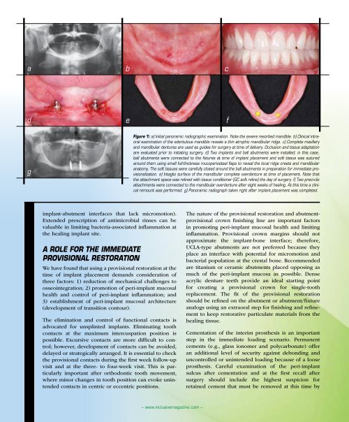
a<br />
b<br />
c<br />
d<br />
e<br />
f<br />
g<br />
Figure 1: a) Initial panoramic radiographic examination. Note the severe resorbed mandible. b) Clinical intraoral<br />
examination of the edentulous mandible reveals a thin atrophic mandibular ridge. c) Complete maxillary<br />
and mandibular dentures are used as guides for surgery at time of delivery. Occlusion and tissue adaptation<br />
are evaluated prior to initiating surgery. d) Two implants and ball abutments were installed; in this case,<br />
ball abutments were connected to the fixtures at time of implant placement and soft tissue was sutured<br />
around them using small full-thickness mucoperiosteal flaps to reveal the local ridge crests and mandibular<br />
anatomy. The soft tissues were carefully closed around the ball abutments in preparation for immediate provisionalization.<br />
e) Intaglio surface of the mandibular complete overdenture at time of placement. Note that<br />
the attachment space was relined with tissue conditioner (GC soft reline) the day of surgery. f) Two preci-clix<br />
attachments were connected to the mandibular overdenture after eight weeks of healing. At this time a clinical<br />
remount was performed. g) Panoramic radiograph taken right after implant placement was completed.<br />
implant-abutment interfaces that lack micromotion).<br />
Extended prescription of antimicrobial rinses can be<br />
valuable in limiting bacteria-associated inflammation at<br />
the healing implant site.<br />
A Role for the Immediate<br />
Provisional Restoration<br />
We have found that using a provisional restoration at the<br />
time of implant placement demands consideration of<br />
three factors: 1) reduction of mechanical challenges to<br />
osseointegration; 2) promotion of peri-implant mucosal<br />
health and control of peri-implant inflammation; and<br />
3) establishment of peri-implant mucosal architecture<br />
(development of transition contour).<br />
The elimination and control of functional contacts is<br />
advocated for unsplinted implants. Eliminating tooth<br />
contacts at the maximum intercuspation position is<br />
possible. Excursive contacts are more difficult to control;<br />
however, development of contacts can be avoided,<br />
delayed or strategically arranged. It is essential to check<br />
the provisional contacts during the first week follow-up<br />
visit and at the three- to four-week visit. This is particularly<br />
important after orthodontic tooth movement,<br />
where minor changes in tooth position can evoke unintended<br />
contacts in centric or eccentric positions.<br />
The nature of the provisional restoration and abutmentprovisional<br />
crown finishing line are important factors<br />
in promoting peri-implant mucosal health and limiting<br />
inflammation. Provisional crown margins should not<br />
approximate the implant-bone interface; therefore,<br />
UCLA-type abutments are not preferred because they<br />
place an interface with potential for micromotion and<br />
bacterial population at the crestal bone. Recommended<br />
are titanium or ceramic abutments placed opposing as<br />
much of the peri-implant mucosa as possible. Dense<br />
acrylic denture teeth provide an ideal starting point<br />
for creating a provisional crown for single-tooth<br />
replacement. The fit of the provisional restoration<br />
should be refined on the abutment or abutment/fixture<br />
analogs using an extraoral step for finishing and refinement<br />
to keep restorative particulate materials from the<br />
healing tissue.<br />
Cementation of the interim prosthesis is an important<br />
step in the immediate loading scenario. Permanent<br />
cements (e.g., glass ionomer and polycarbonate) offer<br />
an additional level of security against debonding and<br />
uncontrolled or unintended loading because of a loose<br />
prosthesis. Careful examination of the peri-implant<br />
sulcus after cementation and at the first recall after<br />
surgery should include the highest suspicion for<br />
retained cement that must be removed at this time by<br />
– www.inclusivemagazine.com –


