infertility treatments, genes and chromosomes
infertility treatments, genes and chromosomes
infertility treatments, genes and chromosomes

Create successful ePaper yourself
Turn your PDF publications into a flip-book with our unique Google optimized e-Paper software.
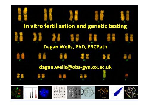
In vitro fertilisation <strong>and</strong> genetic testing<br />
Dagan Wells, PhD, FRCPath<br />
dagan.wells@obs‐gyn.ox.ac.ukgyn.ox.ac.uk
Infertility<br />
• Unprotected intercourse for 1 year without conception<br />
• Very common – 1 in 6 couples affected<br />
• In vitro fertilization first birth in 1978<br />
• More than 3,000,000 IVF babies worldwide<br />
• 1‐5% of all births in Western countries
Indications for IVF/ICSI<br />
Unexplained<br />
9%<br />
Other<br />
9%<br />
Uterine<br />
2%<br />
Male<br />
27%<br />
Anovulation<br />
12%<br />
Endometriosis<br />
15%<br />
Tubal<br />
26%
Ovarian stimulation<br />
• Ideally need 10‐1515 high quality eggs<br />
• 3 types of drugs<br />
• GnRH (gonadotropin<br />
releasing hormone agonist/antagonist)<br />
Suppresses luteinizing hormone (released from pituitary)<br />
LH surge would cause premature ovulation<br />
• Follicle stimulating hormone (FSH)<br />
Stimulates development of multiple follicles (structures that<br />
contain eggs)<br />
• Human chorionic gonadotropin (HCG)<br />
Causes final maturation of eggs in follicles
Egg collection procedure
Sperm preparation<br />
Semen analysis, wash <strong>and</strong> centrifugation
Fertilization<br />
Mixing of sperm <strong>and</strong> eggs
Fertilization using intracytoplasmic sperm injection<br />
(ICSI)<br />
• Method developed for men with sperm deficiencies<br />
Low concentration of sperm in ejaculate<br />
Sperm with motility problems<br />
Sperm unable to penetrate the egg<br />
• First reported in 1992. Now very widely applied
Embryo development<br />
2‐cell embryo<br />
2 PN<br />
8‐cell<br />
embryo<br />
Fallopian<br />
Tube<br />
morula<br />
Blastocyst<br />
Uterus<br />
Hatched<br />
blastocyst
Success rates of IVF –affect of maternal age<br />
50<br />
45<br />
40<br />
35<br />
30<br />
25<br />
20<br />
15<br />
10<br />
5<br />
0<br />
27 29 31 33 35 37 39 41 43 45 47<br />
Age<br />
Donor Eggs<br />
Own Eggs<br />
SART 1999
Genetic analysis of embryos
Testing <strong>chromosomes</strong> in embryos<br />
produced during IVF treatment:<br />
Why bother?
Aneuploidy <strong>and</strong> IVF failure<br />
Chromosome abnormality is extremely common in oocytes<br />
Problem increases with advancing maternal age<br />
As aneuploidy increases age, so implantation rate decreases<br />
50%<br />
40%<br />
Aneuploidy<br />
30%<br />
20%<br />
Implantation<br />
10%<br />
20‐34<br />
35‐39<br />
39 40‐45<br />
45 Maternal age
Preimplantation genetic screening (PGS)<br />
St<strong>and</strong>ard embryo evaluations do not reveal embryos with<br />
the wrong number of <strong>chromosomes</strong><br />
regular<br />
after chromosome<br />
screening<br />
or
Preimplantation genetic screening (PGS)<br />
Anticipated i db benefits for IVF patients<br />
Reduce aneuploid syndromes<br />
Reduce miscarriage<br />
Increase embryo implantation/pregnancy rate
PGS- the controversy<br />
• It is widely accepted that PGS reduces the risk of Down<br />
syndrome<br />
BUT…<br />
• Several r<strong>and</strong>omised trials have shown no improvement in<br />
pregnancy rates
Increase in implantation/pregnancy‐ controversy<br />
• Mastenbroek et al (2007), NEJM<br />
• Maternal age >35<br />
• 8 <strong>chromosomes</strong> assessed, r<strong>and</strong>omised<br />
• No significant improvement in implantation
BUT….<br />
Problems with negative PGS studies<br />
• Many patients with
Problems with negative PGS studies<br />
Critically<br />
damaged by<br />
biopsy<br />
Only 28% of<br />
aneuploidies<br />
detected<br />
No result<br />
Pool of embryos reduced while little selective advantage has been gained
Legitimate criticisms of traditional PGS methods<br />
• Methodologies are not robust, limiting application<br />
• Biopsy can have a serious impact if poorly performed<br />
• Mosaicism will lead to the exclusion of a small number of<br />
potentially viable embryos<br />
• No r<strong>and</strong>omized study has proven that PGS is beneficial
Microarray comparative genomic hybridization<br />
• Rapid –results in 24 hours<br />
• Allows the copy number of every chromosome to be determined<br />
d<br />
Embryo DNA<br />
Normal DNA<br />
Gutierrez‐Mateo et al., Fertility & Sterility 2010; Fragouli et al., Human Reproduction 2011
Microarray comparative genomic hybridization<br />
• Rapid –results in 24 hours<br />
• Allows the copy number of every chromosome to be determined<br />
d<br />
Embryo DNA<br />
Normal DNA<br />
Trisomy<br />
Monosomy<br />
Normal<br />
Gutierrez‐Mateo et al., 2010; Fragouli et al., 2010
46,XX‐10 +16<br />
Microarray‐CGH (array‐CGH or aCGH)
Clinical application of CGH<br />
Analysis of blastocyst stage<br />
Biopsy of several cells is possible<br />
Diagnosis robust <strong>and</strong> accurate<br />
Little or no impact of embryo biopsy<br />
Fragouli et al., 2010; Schoolcraft et al., 2010
Blastocyst CGH‐ clinical results<br />
Implantation rate<br />
Aneuploidy rate<br />
Cycles with all embryos<br />
aneuploid<br />
Wells <strong>and</strong> Fragouli, unpublished
Analysis of mutations in the DNA<br />
sequence<br />
(causing gsingle g gene disorders)
PGD of single gene disorders<br />
Alternative to prenatal diagnosis ‐ avoids pregnancy termination<br />
>200 different single gene disorders diagnosed using PGD<br />
PGD for any disease provided the causative mutation is known<br />
First disease diagnosed – cystic fibrosis, 1992
PGD of single gene disorders<br />
>100 diseases already approved by the HFEA<br />
Adrenoleukodystrophy (Adrenomyeloneuropathy)<br />
Agammaglobulinaemia<br />
Alpers Syndrome<br />
α thalassaemia/mental retardation syndrome<br />
Alport's Syndrome<br />
Alzheimers Disease ‐ early onset<br />
Anderson Fabry Disease<br />
Androgen Insensitivity Syndrome<br />
Aplastic anaemia ‐ severe<br />
Barth Syndrome<br />
Battens Disease (infantile)<br />
Beta Hydroxyisobuyryl CoA Hydrolase Deficiency<br />
(Methacryic<br />
Aciduria)<br />
Beta Thalassaemia<br />
Bilateral Frontoparietal Polymicrogyria<br />
BRCA 1 (increased susceptibility to breast cancer)*<br />
Bruton Agammaglobulinemia Tyrosine Kinase<br />
Cardiac Valvular Dysplasia<br />
Carney Complex<br />
Charcot Marie Tooth Disease<br />
Chondrodysplasia Punctata<br />
Choroideraemia<br />
Chronic Granulomatous Disease<br />
Coffin‐Lowry Syndrome<br />
Congenital Adrenal Hyperplasia<br />
Congenital Fibrosis of the Extraocular Muscles<br />
Congenital Stationary Night Blindness<br />
Crouzon Syndrome<br />
Cystic Fibrosis<br />
Cystinosis<br />
Diamond Blackfan Anaemia<br />
Dystonia 1 Torsion Autosomal Dominant (DYT1)<br />
Ectodermal dysplasia (Hypohidrotic<br />
Hypohidrotic)<br />
Epidermolysis Bullosa (Hallopeau<br />
Hallopeau‐Siemens & Herlitz<br />
junctional)<br />
Facioscapulohumeral Dystrophy<br />
Familial Adenomatous polyposis coli<br />
Fanconi's Anaemia A<br />
Fanconi's Anaemia C<br />
Fragile X Syndrome<br />
Gaucher's Disease (Type II)<br />
Gonadal mosaicism<br />
Greig's Cephalopolysyndactyly<br />
Haemophilia A
PGD of single gene disorders<br />
Diseases already approved by the HFEA<br />
Haemophilia B<br />
Hereditary diffuse gastric cancer*<br />
Hereditary motor <strong>and</strong> sensory neuropathies<br />
Homozygous Familial Hypercholesterolaemia<br />
Hunters Syndrome<br />
Huntington’s Disease<br />
Hydrocephalus<br />
Hydroxyisobuyryl CoA Hydrolase Deficiency<br />
Hyper IgM Syndrome ‐ Hypogammaglobulinaemia<br />
Hypospadias (severe)<br />
Ichthyosis<br />
Incontinentia Pigmenti<br />
Juvenile Retinoschisis<br />
Krabbe Disease<br />
Leber's hereditary optic neuropathy / Lebers Optic<br />
atrophy<br />
Leigh's (subacute<br />
necrotising encephalopathy of<br />
childhood)<br />
Lenz syndrome<br />
Lesch Nyhan Syndrome<br />
Leukocyte Adhesion Deficiency (Type I)<br />
Li‐Fraumeni<br />
Syndrome<br />
Lymphoproliferative Syndrome<br />
Lynch Syndrome (MLH 2)<br />
Lynch syndrome (MLH 1)<br />
Macular Dystrophy y(<br />
(childhood onset ‐ variant of<br />
Retinitis pigmentosa)<br />
Marfan Syndrome<br />
Medium‐chain<br />
acyl‐Co A dehydrogenase<br />
MELAS (Mitochondrial encephalomyopathy, p y<br />
lactic<br />
acidosis <strong>and</strong> stroke‐like episodes)<br />
Menkes Syndrome<br />
Myoclonic epilepsy <strong>and</strong> ragged red fibres (MERFF)<br />
Metachromatic Leukodystrophy<br />
y<br />
Multiple Endocrine Neoplasia (Type I)<br />
Multiple Exostoses<br />
Muscular Dystrophy (Beckers<br />
Beckers)<br />
Muscular Dystrophy (Duchenne<br />
Duchenne)<br />
Muscular dystrophy (Occulopharangeal)<br />
Myotonic Dystrophy<br />
Myotublar myopathy<br />
Neurogenic muscle weakness, ataxia, retinitis<br />
pigmentosa (NARP)<br />
Neurofibromatosis type I
PGD of single gene disorders<br />
Diseases already approved by the HFEA<br />
Neurofibromatosis type II<br />
Niemann Pick Disease Type C<br />
Ornithine carbamoyl transferase Deficiency (OTC)<br />
Ornithine transcarbamylase deficiency (OTD)<br />
Osteo<strong>genes</strong>is Imperfecta (Type II)<br />
Ostheopathia Striata with Cranial Sclerosis<br />
Otopalatodigital syndrome (Type 2)<br />
Partial Lipodystrophy, Familial (Type 2)<br />
Pelizaeus Merzbacher Disease<br />
Phenylketonuria (PKU)<br />
Plakophilin 1 (PKP1) associated ectodermal dysplasia<br />
syndrome<br />
Polycystic kidney disease<br />
Pompe Disease (early onset)<br />
Prader Willi Syndrome<br />
Pyrodoxine‐dependent dependent seizures<br />
Recurrent Digynic Triploidy<br />
Recurrent hydatitiform mole<br />
Retinitis Pigmentosa<br />
Retinoblastoma<br />
Retinoschisis (Juvenile)<br />
S<strong>and</strong>hoff Disease<br />
Sensorineural deafness ‐ autosomal recessive non‐<br />
syndromic<br />
Severe Combined Immune Deficiency (x‐linked)<br />
Sickle Cell Anaemia<br />
Spastic paraplegia<br />
Spinal Muscular Atrophy (SMA1)<br />
Tay Sachs Disease (infantile onset)<br />
Torsion Dystonia<br />
Treacher Collins Syndrome<br />
Tuberous Sclerosis (TSC2)*<br />
Turner's syndrome (Mosaic)<br />
Von Hippel Lindau Syndrome*<br />
Wiscott‐Aldrich Syndrome*<br />
Wolman's Disease (Acid Lipase Deficiency)<br />
Chromosome rearrangements<br />
HLA‐typing
Cleavage stage biopsy<br />
•Used by majority of PGD labs<br />
•Biopsy at 6‐10 cell stage (day 3)<br />
•Blastomeres<br />
totipotent<br />
•1‐22 cells for analysis
DNA amplification: the polymerase chain reaction<br />
• Enzymatic method for copying specific DNA sequences<br />
C T T A C C G T G G T A A A T C G<br />
G A A T G G C A C C A T T T A G C<br />
• Essential for the analysis of <strong>genes</strong> in single cells<br />
See Wells & Sherlock 1998 for review
Generalized Diagnostic Methodology<br />
Biopsy<br />
PCR<br />
Mutation<br />
detection<br />
Polymorphism<br />
analysis<br />
Diagnosis<br />
Linked<br />
polymorphisms<br />
DNA<br />
fingerprint<br />
Report<br />
Contamination<br />
detection
Ethical questions<br />
Application of PGD to late onset disorders<br />
HLA typing<br />
Complex (polygenic) disorders