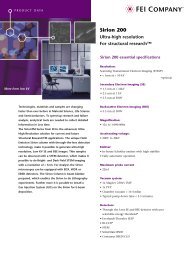
Tecnai™ G2 Spirit Product Brochure - FEI Company
Tecnai™ G2 Spirit Product Brochure - FEI Company
Tecnai™ G2 Spirit Product Brochure - FEI Company

Create successful ePaper yourself
Turn your PDF publications into a flip-book with our unique Google optimized e-Paper software.
Tecnai G 2 <strong>Spirit</strong> Highlights<br />
• High contrast, high resolution for 20 –120 kV operation<br />
• Optimized for 2D & 3D imaging of cells, cell organelles and soft matter<br />
• High level of automation: Auto-Gun and automatic tuning<br />
• Smart Tracking Position System for sample navigation<br />
• Customized protocols for diverse applications<br />
• Sample observation at liquid nitrogen temperature<br />
• Technology for sharp imaging of thicker samples<br />
• Ergonomic design for operational comfort<br />
<strong>FEI</strong> <strong>Company</strong><br />
World Headquarters and<br />
North American Sales<br />
5350 NE Dawson Creek Drive<br />
Hillsboro, OR 97124-5793 USA<br />
Tel: +1 503 726 7500<br />
Fax: +1 503 726 7509<br />
European Sales<br />
Tel: +31 40 27 66 768<br />
Fax: +31 40 27 66 786<br />
Asia-Pacific Sales<br />
Tel: +65 351 7671<br />
Fax: +65 354 0644<br />
e-mail: sales@feico.com<br />
www.feicompany.com<br />
www.tecnai.com<br />
032-PB00811 05/04<br />
©2004. We are constantly improving the performance of our products<br />
so all specifications are subject to change without notice. The <strong>FEI</strong> logo,<br />
Xplore3D, Tecnai, Tools for Nanotech and DualBeam are trademarks of<br />
<strong>FEI</strong> <strong>Company</strong>. Windows is a trademark of Microsoft Corporation.<br />
Tecnai G 2 <strong>Spirit</strong><br />
Next Generation Microscopy<br />
for Life Sciences<br />
The <strong>Spirit</strong> of Automation: High Quality 2D and 3D Imaging
2<br />
Meeting the needs of the scientific community<br />
Fundamental research within the scope of cell biology, structural biology, soft matter and nano-<br />
technology is increasingly focusing on understanding interactive biological processes, pathways and<br />
structures at the macromolecular level. Transmission electron microcopy (TEM) is of paramount<br />
importance to obtain high-magnification and high-resolution 2D and 3D information of cells and<br />
organelles or even smaller cell constituents. A pre-requisite for optimal 2D and 3D-tomographic<br />
imaging is a high level of microscope automation and intelligence. The new Tecnai G 2 <strong>Spirit</strong> – built<br />
on more than 50 years’ experience in Life Science – combines modern day technology to satisfy the<br />
stringent demands of both today’s and tomorrow’s scientific community.<br />
The Tecnai G 2 <strong>Spirit</strong> ensures high-quality images, is easy to use and is designed to facilitate the work<br />
of the microscope user. It has a high level of automation, yet doesn't limit full control for experienced<br />
expert users. The microscope is ready for novel applications like 3D-imaging using tomography<br />
and/or cryo-electron microscopy. The <strong>Spirit</strong>’s design philosophy ensures application flexibility for<br />
today and tomorrow, it holds a ticket to the future: functionality can be added or upgraded by<br />
uploading current and new application software. In the world of Life Sciences, developments occur<br />
at a high pace. <strong>FEI</strong> <strong>Company</strong> has ongoing collaborations with the relevant scientific communities<br />
to better understand their needs and co-develop dedicated tools.<br />
The Tecnai G 2 <strong>Spirit</strong> is building a further and more detailed understanding in the world of decreasing<br />
dimensions: today and in the future.<br />
3
4<br />
Figure 1:<br />
Diagnostic Research<br />
Lung biopsy from a patient<br />
suffering from SARS.<br />
Image courtesy of: Queen<br />
Elisabeth II Hospital<br />
Hong Kong.<br />
Figure 2:<br />
Cell Biology Research<br />
Immunogold labeling of<br />
plasmodesmata in genetically<br />
modified plants<br />
Diagnostic research<br />
As a consequence of the growing and<br />
aging world population, the number<br />
of patients needing advanced medical<br />
care and treatment inevitably increases.<br />
This stresses the urgency for fast,<br />
reliable screening of disease patterns.<br />
As a mainstream diagnostic modality<br />
in medical centers, the Tecnai <strong>G2</strong> <strong>Spirit</strong><br />
is a ready-to-use system that ensures a<br />
high level of automation and a high<br />
sample throughput to cope with the<br />
need for increasing the speed of diagnostics.<br />
Furthermore, the full digital<br />
Tecnai <strong>G2</strong> <strong>Spirit</strong> allows for on-line<br />
consultation with colleagues and<br />
experts in the field to enable a fast<br />
and correct diagnosis.<br />
Cell Biology research<br />
Achieving breakthroughs that can<br />
improve human health or increase<br />
the quality or yield of agricultural<br />
products requires research to the<br />
underlying fundamental processes.<br />
For studying the functionality of<br />
ongoing physiological and pathological<br />
pathways, the Tecnai <strong>G2</strong> <strong>Spirit</strong> is<br />
perfectly suited. For retrieval of cell<br />
biological data, the <strong>Spirit</strong> is set for<br />
fast and (semi-)automated 2D and 3D<br />
high-quality image acquisition and<br />
analysis. Its ease-of-use makes the<br />
instrument very attractive for multiuser<br />
scientific environments like<br />
universities and institutes.<br />
Pharmaceutics and Chemistry<br />
The Tecnai <strong>G2</strong> <strong>Spirit</strong> is an effective<br />
and efficient tool to gain insight into<br />
release mechanisms of drugs in the<br />
body. In addition, the effects of medicines<br />
on the morphology and functioning<br />
of tissues and organs need to<br />
be investigated. The <strong>Spirit</strong>, optimized<br />
for 2D and 3D imaging of cells, cell<br />
organelles and soft matter combined<br />
with a high level of automation can<br />
play an essential role in this and is of<br />
significant importance for continued<br />
advances in healthcare and chemistry.<br />
Figure 3:<br />
Pharmaceutics and Chemistry<br />
Cryo TEM image of cis-Pt particles packaged<br />
in PS liposomes. The liposomes are used as<br />
novel drug targeting entities in anti-cancer<br />
research.<br />
Image courtesy of Dr. P. Frederik, University of<br />
Maastricht, The Netherlands.<br />
High Image quality – throughout<br />
the entire magnification range<br />
All biological TEM specimens, ranging<br />
from ultra-thin sections to negative<br />
stained samples, have in common<br />
that for high-quality visualization<br />
maximized contrast and resolution<br />
are required. In general, the imaging<br />
magnifications are in the intermediate<br />
range, roughly from about 500 to<br />
100 000 times covering 95% of the<br />
used applications.<br />
The optics of the Tecnai <strong>G2</strong> <strong>Spirit</strong> –<br />
with the well proven and patented<br />
BioTWIN and TWIN concept – provides<br />
sharp, high-contrast and highresolution<br />
images at all magnifications.<br />
The electron source in combination<br />
with the excellent vacuum<br />
ensures a high brightness and a long<br />
life-time of both the tungsten and<br />
LaB6 filaments. To facilitate the practical<br />
work, one can store and re-load<br />
optical parameters like magnification<br />
and illumination settings, in conjunction<br />
with stage coordinates to<br />
re-allocate interesting areas of the<br />
specimen, at will. Moreover, alignments<br />
or operations parameters can<br />
be stored easily and retrieved at the<br />
push of a button. Each user has his<br />
or her own secure and customized<br />
microscope environment, and works<br />
with own or supervised settings and<br />
alignments. Microscope alignment<br />
efforts are reduced to a minimum so<br />
one can concentrate on getting the<br />
best out of the specimen.<br />
In addition, the user is given the flexibility<br />
to choose different imaging<br />
modes, e.g. rotation free magnification<br />
or isotropic chromatic aberration<br />
corrected magnification. In other<br />
words, the optical system can be<br />
adapted to the needs of biological<br />
research: easy zoom in or maximized<br />
sharpness, even for thicker samples.<br />
Figure 4<br />
Left-hand shows a typical image at a low magnification on the Tecnai G 2 <strong>Spirit</strong>: it demonstrates<br />
the value of the patented high contrast performance. The right-hand image shows<br />
the high-resolution performance. For reasons of reference a graphite sample was used.<br />
5
6<br />
Diffracted<br />
energy-loss<br />
electrons<br />
Electron beam<br />
Transmitted electrons<br />
Specimen<br />
Objective aperture<br />
short-focal-length lens<br />
Objective aperture<br />
long-focal-length lens<br />
Figure 5.<br />
In a long focal length lens system, the objective aperture stops<br />
many more diffracted electrons and energy loss electrons – and<br />
thereby provides higher contrast – than in a short focal length<br />
lens system. The Tecnai G 2 <strong>Spirit</strong> BioTWIN has the longest focal<br />
length worldwide.<br />
A B<br />
C D<br />
Figure 7.<br />
Displayed images are from an ultra-thin section of a kidney<br />
glomerulus post stained with lead citrate and uranyl acetate. The<br />
electron optics magnification of the images shown are taken at<br />
610x, 1250x, 2550x and 6000x (A,B,C,D). There is no limitation of<br />
the field of view in these images, not even at the lowest magnification<br />
(610x) and with the smallest objective aperture.<br />
Chromatic<br />
Blurring<br />
No Corrected System<br />
No Chromatic<br />
Blurring<br />
Corrected System<br />
1st Intermediate Image<br />
Slower<br />
Electron<br />
Faster<br />
Electron<br />
2nd Intermediate Image<br />
Figure 6.<br />
The chromatic error correction system of the Tecnai G 2 <strong>Spirit</strong> re-unites<br />
electrons of different energies (shown in red and blue), thereby<br />
giving sharp, high-quality images even for thick specimens. On<br />
microscopes without chromatic error correction, these electrons<br />
separate and cause blurring of the image.<br />
High Image quality – at various<br />
section thicknesses<br />
Typically, energy loss of transmitted<br />
electrons due to thicker samples will<br />
lead to unsharp, blurred images. The<br />
projection system of the Tecnai <strong>G2</strong> <strong>Spirit</strong> limits this effect since the<br />
objective aperture – due to the long<br />
focal length of the objective lens –<br />
blocks the majority of the electrons<br />
with multiple energy levels.<br />
Furthermore, the projection system<br />
can be used in such a way that both<br />
energy loss and zero-loss electrons<br />
are focused on the same point as the<br />
zero-loss electrons. This approach<br />
ensures sharp, high contrasted<br />
images even for thick specimens.<br />
This is in particular valuable for<br />
3D tomography applications where<br />
tilting can lead to tripling of thickness<br />
at high tilt. Image brightness<br />
remains high due to the Tecnai <strong>G2</strong> <strong>Spirit</strong> high coherence, high brightness<br />
electron gun.<br />
Operational comfort<br />
The Tecnai <strong>G2</strong> <strong>Spirit</strong> is a fully digital<br />
microscope allowing a high level of<br />
automation to facilitate its use for<br />
the various experience levels in the<br />
user community. Flexibility is paramount.<br />
The user can control the<br />
microscope manually or automatically<br />
at will. Besides via the software<br />
user interface – displayed on the LCD<br />
screen(s) – the microscope operator is<br />
also communicating with the system<br />
via the newly designed control panels.<br />
In addition to those buttons representing<br />
the standard microscope<br />
functions (i.e. magnification, focus,<br />
intensity), each user has the flexibility<br />
to assign various application dedicated<br />
functions to the programmable<br />
buttons. Moreover, the optical trackballs<br />
guarantee a smooth control of<br />
sample and beam shift. Needless to<br />
say that the trackballs can also be<br />
assigned to almost any user-defined<br />
microscope function.<br />
Automation – of essential microscope<br />
functions<br />
The Tecnai <strong>G2</strong> <strong>Spirit</strong> stands for an<br />
unprecedented automation of essential<br />
and most frequently applied<br />
microscope functions. With the Auto-<br />
Gun function, retrieving an optimal<br />
screen brightness is literally one<br />
mouse click away. Its intelligence<br />
thinks for you and corrects all gun<br />
alignments and conditions and saturates<br />
the filament fully automatically.<br />
Moreover, the image focus and objective<br />
astigmatism can be routinely corrected<br />
using the Auto Tune function,<br />
always giving the best possible result<br />
with the lowest effort. Needless to<br />
say that experienced users can always<br />
take full manual control of all micro-<br />
Figure 8.<br />
Hand Panel Controls<br />
scope functions. In line with the<br />
Auto-Gun and Auto-Tune automation,<br />
the Tecnai <strong>G2</strong> <strong>Spirit</strong> also introduces<br />
the Smart Tracking Position<br />
System (STPS). STPS keeps track of<br />
grid areas that have been screened<br />
and investigated. One click on the<br />
tracking line and the stage is automatically<br />
translocated to the preferred<br />
position on the grid.<br />
Additionally, positions can be stored<br />
and relocated based on comments<br />
rather than coordinates. This textual<br />
approach is a next step in the development<br />
of microscope intelligence,<br />
just for you!<br />
Figure 9a:<br />
Smart Tracking Position System<br />
module User Interface.<br />
Figure 9b:<br />
Auto Gun User Interface.<br />
7
8<br />
Figure 10.<br />
Illustrates an automatically stitched<br />
3k x 3k image composed out of<br />
9 individual 1k x 1k images.<br />
The result is a high-quality lowmagnification<br />
image.<br />
Figure 11.<br />
Shows a 3D-image of magnetosome<br />
chains in magnetotactic bacteria.<br />
Data automatically acquired on a<br />
200 nm section on a slow scan CCD<br />
camera and processed using the<br />
<strong>FEI</strong> Xplore3D software.<br />
Courtesy: Dr. Kobayashi, National<br />
Institute of Advanced Industrial<br />
Science and Technology, Osaka, Japan<br />
Automation – for optimal image<br />
acquisition and quality<br />
The functional combination of hardware,<br />
optics, stage and digital detectors<br />
like CCD cameras in an embedded<br />
software environment has<br />
opened the door for advanced, (semi-)<br />
automated microscope applications.<br />
One of the most frequently performed<br />
applications is image<br />
resolution enhancement by stitching<br />
multiple digital images together.<br />
In the Tecnai <strong>G2</strong> <strong>Spirit</strong>, stitching<br />
occurs fully automatically. Upon<br />
selection of the final resolution<br />
goal, all images are recorded and<br />
are stitched into one image within<br />
a few seconds time.<br />
Even though microscopy has entered<br />
the digital era, the microscope still<br />
features the option for conventional<br />
image recording on film plates. In<br />
line with this, software is available<br />
to allow photomontage in a guided<br />
manner.<br />
3D-imaging – “Confocal TEM”<br />
For the last 50 years, the typical<br />
result of a transmission electron<br />
microscopy session was a 2D-image<br />
of a 3D-structure. It takes a lot of<br />
experience to understand and interpret<br />
the information in the correct<br />
way. Confocal Laser Scanning<br />
Microscopy (CLSM) was the trigger to<br />
master the third dimension. A lot of<br />
cellular processes are easier to understand<br />
when 3D information of cells<br />
and cell structures can be obtained.<br />
The automation of the Tecnai <strong>G2</strong> <strong>Spirit</strong> microscope in combination<br />
with <strong>FEI</strong>’s proprietary intelligent<br />
tomography solution enables the user<br />
to achieve 3D TEM-results fully automatically<br />
and in a limited time, thus<br />
revealing high-resolution confocal<br />
TEM images ~ in analogy with<br />
confocal light microscopy (down to<br />
nanometer resolution scale).<br />
The Tecnai <strong>G2</strong> <strong>Spirit</strong> can completely<br />
and automatically acquire a tomographic<br />
data set. The <strong>Spirit</strong> features a<br />
comprehensive tomography data<br />
acquisition and processing software<br />
package. To acquire a series of images,<br />
samples are tilted in the electron<br />
microscope from + 70 to –70 degrees<br />
at user defined intervals to ensure the<br />
highest possible resolution and reconstruction<br />
quality.<br />
<strong>FEI</strong>’s Xplore3D automatically determines<br />
changes in image movement<br />
and focus after tilting. The software<br />
has a high degree of flexibility, allowing<br />
individual users to optimize the<br />
procedure for obtaining their best<br />
results. Image data are collected using<br />
a CCD camera and stored on hard<br />
disk or DVD.<br />
Cryo electron microcopy<br />
Cryo-fixation by rapid freezing has<br />
been accepted as a key-technology to<br />
study the natural state of a biological<br />
specimen. In this way, the integrity<br />
of the specimen is maintained during<br />
trasfer and data collection sessions<br />
in the microscope. The results<br />
obtained at high resolution contain<br />
the information to understand the<br />
processes and related structural<br />
changes during the many biological<br />
processes within a cell. The goal is<br />
ultimate specimen protection to<br />
achieve ultra-structural details down<br />
at the Ångstrom level.<br />
Many techniques have been designed<br />
to maintain specimen integrity dur-<br />
ing preparation, transfer and observation.<br />
For the vitrification process as<br />
such, the parameters to control are<br />
humidity and temperature. The quality<br />
of vitrification is reproducibly<br />
improved since the commercial<br />
availability of the Vitrobot.<br />
Once the vitrified sample is in the<br />
microscope, the clean vacuum<br />
ensures a contamination-free observation<br />
for long working sessions. The<br />
observation at liquid nitrogen temperature<br />
in combination with the low<br />
dose software gives the best protection<br />
for beam-sensitive specimens.<br />
Important other improvements are<br />
the optimization of the low-magnification<br />
high-resolution performance.<br />
The microscope is designed to obtain<br />
the ultimate resolution at low magnifications.<br />
Finally, in recent years a<br />
significant change has taken place<br />
in image recording. Highly sensitive<br />
CCD cameras are used to record<br />
cryo-results. The major advantage of<br />
this approach is the further automation<br />
of the cryo-observation process.<br />
Automation will inevitably lead to<br />
optimal, statistically validated results<br />
with minimal user effort – a prerequisite<br />
for single particle analysis.<br />
The system reliability ensures<br />
researchers have a high sample<br />
throughput.<br />
Figure 12.<br />
The picture illustrates a suspension of<br />
Cowpea Mosaic Virus (CPMV), vitrified and<br />
subjected to a single particle reconstruction<br />
procedure. The morphological relation<br />
between the outer protein shell (blue) and<br />
the inner viral genomic material (yellow) is<br />
clearly visible.<br />
9
10<br />
Embedded control of cameras<br />
and analytical technology<br />
Optimal information acquisition at<br />
maximum ease-of-use is a continuous<br />
challenge for further improvement of<br />
<strong>FEI</strong>’s instrumentation. A big step in<br />
realizing this is the embedded control<br />
of frequently used microscope functions<br />
(e.g. CCD image acquisition)<br />
and additional, analytical<br />
functions (e.g. STEM and EDX).<br />
Through the embedding concept,<br />
the Tecnai <strong>G2</strong> software interface<br />
basically has full control over these<br />
functions resulting in increased<br />
ease-of-use.<br />
Service support<br />
Customer surveys show that <strong>FEI</strong><br />
instruments have an excellent reputation<br />
for reliability and robustness,<br />
even after being in operation for<br />
more then a decade. Each microscope<br />
comes with detailed and complete<br />
digital service manuals as well as<br />
built-in self-diagnostics and on-line<br />
help manuals. Unsurpassed instrument<br />
up time is ensured by highquality,<br />
first-line service support,<br />
available throughout the world.<br />
Highly skilled service, development<br />
and factory engineers from the product<br />
divisions establish back up.<br />
Our philosophy of support<br />
Dedicated training is pivotal in <strong>FEI</strong>'s<br />
customer support philosophy.<br />
Training aims to share and transfer<br />
operational skills and to show the<br />
versatility and possibilities of various<br />
electron microscopy techniques.<br />
Above all, it strives to show you how<br />
to reach your best result for different<br />
applications. In other words: <strong>FEI</strong><br />
Academy – a mature competence<br />
center- helps you maximize the value<br />
of your installed equipment by optimizing<br />
the potential of the person<br />
controlling it. Please visit our website<br />
www.feiacademy.com to learn more<br />
about <strong>FEI</strong> Academy and the latest<br />
courses and workshops.<br />
For more information visit: www.feicompany.com<br />
or www.tecnai.com for more technical info.<br />
About <strong>FEI</strong>: Tools for Nanotech<br />
<strong>FEI</strong> <strong>Company</strong> provides advanced<br />
Tools for Nanotech to a range nanotechnology<br />
markets including materials<br />
science, life science, nanotechnology,<br />
semiconductors, and data storage.<br />
Its ion beam and electron beam products,<br />
market-leading DualBeam<br />
systems, and productivity-enhancing<br />
automation software packages enable<br />
researchers and manufacturers to fabricate<br />
and modify nanoscale structures<br />
and to view and characterize them in<br />
three dimensions down to the atomic<br />
level. The company has been paving<br />
the way for nanoscale exploration and<br />
discovery since 1946. <strong>FEI</strong> <strong>Company</strong><br />
currently has more than $360 million<br />
in revenue, enjoys consistent profitability,<br />
has manufacturing and development<br />
facilities in North America<br />
and Europe, and service operations in<br />
more than 40 countries worldwide.<br />
Tecnai G 2 <strong>Spirit</strong> Bio(TWIN) – Specifications<br />
Electron )ptics<br />
Line resolution 0.34 nm (0.20 nm)<br />
Information limit < 0.20 nm<br />
High tension (min step) 20 - 120 kV, [continuous- 10 V]<br />
HT stability ≤ 2 ppm/min<br />
Illumination modes Micro/nanoprobe<br />
Number of spot size 11<br />
Number of C1 apertures 1 fixed<br />
Number of C2 apertures 4 exchangeable<br />
Number of obj. apertures 4 exchangeable<br />
Focal length 6.1 mm (2.8) mm<br />
Cs 6.3 mm (2.2) mm<br />
Cc 5.0 mm (2.2) mm<br />
Minimum focal step 9.0 mm (3.0) nm<br />
Magnification 22 - 300 kx (18 - 650 kx)<br />
SA Camera Length 0.05 - 8.9 m (0.02 – 4.2 m)<br />
Operating System<br />
Controller PC [Windows 2000]<br />
User interface User defined<br />
Control Panels New ergonomics, with optical<br />
and removable trackball<br />
User levels Three<br />
Scripting facilities Via Tecnai Scripting [optional]<br />
Remote Operation Taro software [optional]<br />
Accessories (STEM, EDX, CCD) Embedded<br />
Vacuum<br />
Gun vacuum 1 x 10⁻⁶ Pa<br />
Column vacuum 1 x 10⁻⁵ Pa<br />
Filament lifetime ≥ 1 year<br />
Filament exchange time < 5 min<br />
Air lock pumping Oil-free<br />
Differential aperture 200 µm<br />
Pumping time for sample holder 10 –180 secs<br />
Pumping time for (film)chamber 5 min<br />
Cold trap Optional [for cryo applications]<br />
Specimen Manipulation<br />
spec. alfa tilt angle ± 80° (70°)<br />
BioTWIN (TWIN) BioTWIN (TWIN)<br />
x,y direction 2.0 / 2.0 mm<br />
z direction 0.75 mm<br />
Recalling accuracy < 0.1 µm<br />
Number of stored positions Infinite, including optics setting i.e.<br />
intensity, magnification, spotsizes<br />
Sample exchange time < 30 sec [without switching off<br />
High Tension and Filament]<br />
STPS Smart Tracking Position System Tracking<br />
interface for visualization of searching<br />
pathway incl. stored positions<br />
Intelligent Recall Comment mediated stage position recall<br />
Specimen drift ≤ 1.0 nm/min<br />
Accessories<br />
CCD cameras Embedded control for all supporting<br />
products: Gatan, SIS, Tietz<br />
Magnification calibration for<br />
double CCD cameras Automatic<br />
EDX detector resolution ≤ 135 eV<br />
EDX detector window SUTW<br />
EDX solid angle 0.13 str. (0.21)<br />
STEM resolution 1.0 nm<br />
STEM magnification 150 – 3.1 Mx<br />
Tomography<br />
Calibrations, tilt series acquisition,<br />
reconstruction and volume Fully automatic acquisition<br />
rendering Xplore3D intelligent solution<br />
Cryo TEM<br />
Transfer Standard Gatan holders [60˚/70˚]<br />
Sample analysis Low Dose supported<br />
Automation<br />
AutoGun module Enables automated gun conditioning,<br />
saturation, alignments. Including a one<br />
push button for direct light<br />
AutoTune module Enables automatic focus and astigmatism<br />
correction<br />
Tomography Automated calibrations and image<br />
series acquisition. Reconstruction and<br />
rendering through Xplore3D<br />
STPS Smart Tracking Position System for interactive<br />
and navigational stage control<br />
Applications support<br />
On-line help files Yes - English, Japanese, Chinese<br />
Application instructions Available<br />
Customer <strong>FEI</strong> Academy training Basic, Advance Materials,<br />
Advanced Life-sciences, Advanced Cryo<br />
and workshops in English<br />
11