How Do I Perform a Lumbar Puncture and Analyze the Results to ...
How Do I Perform a Lumbar Puncture and Analyze the Results to ...
How Do I Perform a Lumbar Puncture and Analyze the Results to ...

You also want an ePaper? Increase the reach of your titles
YUMPU automatically turns print PDFs into web optimized ePapers that Google loves.
DIAGNOSING BACTERIAL MENINGITIS BY LUMBAR PUNCTURE<br />
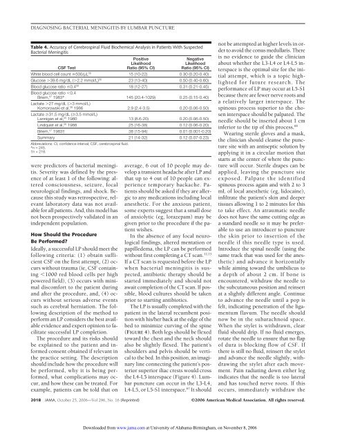
Table 4. Accuracy of Cerebrospinal Fluid Biochemical Analysis in Patients With Suspected<br />
Bacterial Meningitis<br />
CSF Test<br />
Positive<br />
Likelihood<br />
Ratio (95% CI)<br />
Negative<br />
Likelihood<br />
Ratio (95% CI)<br />
White blood cell count 500/µL 56 15 (10-22) 0.30 (0.20-0.40)<br />
Glucose 39.6 mg/dL (2.2 mmol/L) 56 23 (13-40) 0.50 (0.40-0.60)<br />
Blood glucose ratio 0.4 56 18 (12-27) 0.31 (0.21-0.45)<br />
Blood glucose ratio 0.4<br />
Briem, 57 1983* 145 (20.4-1029) 0.25 (0.15-0.40)<br />
Lactate 27 mg/dL (3 mmol/L)<br />
Komorowski et al, 58 1986 2.9 (2.4-3.5) 0.20 (0.06-0.50)<br />
Lactate 31.5 mg/dL (3.5 mmol/L)<br />
Lannigan et al, 55 1980 13 (8.6-20) 0.20 (0.06-0.50)<br />
Lindquist et al, 56 1988 25 (16-38) 0.12 (0.06-0.20)<br />
Briem, 57 1983† 38 (15-94) 0.01 (0.001-0.20)<br />
Summary 21 (14-32) 0.12 (0.07-0.23)<br />
Abbreviations: CI, confidence interval; CSF, cerebrospinal fluid.<br />
*n = 245.<br />
†n = 218.<br />
were predic<strong>to</strong>rs of bacterial meningitis.<br />
Severity was defined by <strong>the</strong> presence<br />
of at least 1 of <strong>the</strong> following: altered<br />
consciousness, seizure, focal<br />
neurological findings, <strong>and</strong> shock. Because<br />
this study was retrospective, relevant<br />
labora<strong>to</strong>ry data was not available<br />
for all patients. And, this model has<br />
not been prospectively validated in an<br />
independent population.<br />
<strong>How</strong> Should <strong>the</strong> Procedure<br />
Be <strong>Perform</strong>ed?<br />
Ideally, a successful LP should meet <strong>the</strong><br />
following criteria: (1) obtain sufficient<br />
CSF on <strong>the</strong> first attempt, (2) occurs<br />
without trauma (ie, CSF containing<br />
1000 red blood cells per high<br />
powered field), (3) occurs with minimal<br />
discomfort <strong>to</strong> <strong>the</strong> patient during<br />
<strong>and</strong> after <strong>the</strong> procedure, <strong>and</strong>, (4) occurs<br />
without serious adverse events<br />
such as cerebral herniation. The following<br />
description of <strong>the</strong> method <strong>to</strong><br />
perform an LP considers <strong>the</strong> best available<br />
evidence <strong>and</strong> expert opinion <strong>to</strong> facilitate<br />
successful LP completion.<br />
The procedure <strong>and</strong> its risks should<br />
be explained <strong>to</strong> <strong>the</strong> patient <strong>and</strong> informed<br />
consent obtained if relevant in<br />
<strong>the</strong> practice setting. The description<br />
should include how <strong>the</strong> procedure will<br />
be performed, why it is being performed,<br />
what complications may occur,<br />
<strong>and</strong> how <strong>the</strong>se can be treated. For<br />
example, patients can be <strong>to</strong>ld that on<br />
average, 6 out of 10 people may develop<br />
a transient headache after LP <strong>and</strong><br />
that up <strong>to</strong> 4 out of 10 people can experience<br />
temporary backache. Patients<br />
should be asked if <strong>the</strong>y are allergic<br />
<strong>to</strong> any medications including local<br />
anes<strong>the</strong>tic. For <strong>the</strong> anxious patient,<br />
some experts suggest that a small dose<br />
of anxiolytic (eg, lorazepam) may be<br />
given prior <strong>to</strong> <strong>the</strong> procedure if <strong>the</strong> patient<br />
wishes.<br />
In <strong>the</strong> absence of any focal neurological<br />
findings, altered mentation or<br />
papilledema, <strong>the</strong> LP can be performed<br />
without first completing a CT scan. 12,13<br />
If a CT scan is requested before <strong>the</strong> LP<br />
when bacterial meningitis is suspected,<br />
antibiotic <strong>the</strong>rapy should be<br />
started immediately <strong>and</strong> should not<br />
await completion of <strong>the</strong> CT scan. If possible,<br />
blood cultures should be taken<br />
prior <strong>to</strong> starting antibiotics.<br />
The LP is usually completed with <strong>the</strong><br />
patient in <strong>the</strong> lateral recumbent position<br />
with his/her back at <strong>the</strong> edge of <strong>the</strong><br />
bed <strong>to</strong> minimize curving of <strong>the</strong> spine<br />
(FIGURE 4). Both legs should be flexed<br />
<strong>to</strong>ward <strong>the</strong> chest <strong>and</strong> <strong>the</strong> neck should<br />
also be slightly flexed. The patient’s<br />
shoulders <strong>and</strong> pelvis should be vertical<br />
<strong>to</strong> <strong>the</strong> bed. In this position, an imaginary<br />
line connecting <strong>the</strong> patient’s posterior<br />
superior iliac crests would cross<br />
<strong>the</strong> L4-L5 interspace (Figure 4). <strong>Lumbar</strong><br />
puncture can occur in <strong>the</strong> L3-L4,<br />
L4-L5, or L5-S1 interspace. 67 It should<br />
not be attempted at higher levels in order<br />
<strong>to</strong> avoid <strong>the</strong> conus medullaris. There<br />
is no evidence <strong>to</strong> guide <strong>the</strong> clinician<br />
about whe<strong>the</strong>r <strong>the</strong> L3-L4 or L4-L5 interspace<br />
is <strong>the</strong> optimal site for <strong>the</strong> initial<br />
attempt, which is a <strong>to</strong>pic highlighted<br />
for future research. The<br />
performance of LP may occur at L5-S1<br />
because <strong>the</strong>re are fewer nerve roots <strong>and</strong><br />
a relatively larger interspace. The<br />
spinous process superior <strong>to</strong> <strong>the</strong> chosen<br />
interspace should be palpated. The<br />
needle should be inserted about 1 cm<br />
inferior <strong>to</strong> <strong>the</strong> tip of this process. 68<br />
Wearing sterile gloves <strong>and</strong> a mask,<br />
<strong>the</strong> clinician should cleanse <strong>the</strong> puncture<br />
site with an antiseptic solution by<br />
applying it in a circular motion that<br />
starts at <strong>the</strong> center of where <strong>the</strong> puncture<br />
will occur. Sterile drapes can be<br />
applied, leaving <strong>the</strong> puncture site<br />
exposed. Palpate <strong>the</strong> identified<br />
spinous process again <strong>and</strong> with 2 <strong>to</strong> 3<br />
mL of local anes<strong>the</strong>tic (eg, lidocaine),<br />
infiltrate <strong>the</strong> patient’s skin <strong>and</strong> deeper<br />
tissues allowing 1 <strong>to</strong> 2 minutes for this<br />
<strong>to</strong> take effect. An atraumatic needle<br />
does not have <strong>the</strong> same cutting edge as<br />
a st<strong>and</strong>ard needle so it may be preferable<br />
<strong>to</strong> use an introducer <strong>to</strong> puncture<br />
<strong>the</strong> skin prior <strong>to</strong> insertion of <strong>the</strong><br />
needle if this needle type is used.<br />
Introduce <strong>the</strong> spinal needle (using <strong>the</strong><br />
same track that was used for <strong>the</strong> anes<strong>the</strong>tic)<br />
<strong>and</strong> advance it horizontally<br />
while aiming <strong>to</strong>ward <strong>the</strong> umbilicus <strong>to</strong><br />
a depth of about 2 cm. If bone is<br />
encountered, withdraw <strong>the</strong> needle <strong>to</strong><br />
<strong>the</strong> subcutaneous position <strong>and</strong> reinsert<br />
at a slightly different angle. Continue<br />
<strong>to</strong> advance <strong>the</strong> needle until a pop is<br />
felt, indicating penetration of <strong>the</strong> ligamentum<br />
flavum. The needle should<br />
now be in <strong>the</strong> subarachnoid space.<br />
When <strong>the</strong> stylet is withdrawn, clear<br />
fluid should drip. If no fluid emerges,<br />
rotate <strong>the</strong> needle <strong>to</strong> ensure that no flap<br />
of dura is blocking flow of CSF. If<br />
<strong>the</strong>re is still no fluid, reinsert <strong>the</strong> stylet<br />
<strong>and</strong> advance <strong>the</strong> needle slightly, withdrawing<br />
<strong>the</strong> stylet after each movement.<br />
Pain radiating down ei<strong>the</strong>r leg<br />
indicates that <strong>the</strong> needle is <strong>to</strong>o lateral<br />
<strong>and</strong> has <strong>to</strong>uched nerve roots. If this<br />
occurs, immediately withdraw <strong>the</strong><br />
2018 JAMA, Oc<strong>to</strong>ber 25, 2006—Vol 296, No. 16 (Reprinted) ©2006 American Medical Association. All rights reserved.<br />
<strong>Do</strong>wnloaded from www.jama.com at University of Alabama-Birmingham, on November 8, 2006