2 Yeast Cell Architecture and Function
2 Yeast Cell Architecture and Function
2 Yeast Cell Architecture and Function

Create successful ePaper yourself
Turn your PDF publications into a flip-book with our unique Google optimized e-Paper software.
2 <strong>Yeast</strong> <strong>Cell</strong> <strong>Architecture</strong> <strong>and</strong> <strong>Function</strong><br />
2.1 General <strong>Cell</strong>ular Characteristics of <strong>Yeast</strong><br />
<strong>Yeast</strong> cells exhibit great diversity with respect to cell size, shape <strong>and</strong> colour. Even individual cells<br />
from a particular yeast strain of a single species can display morphological <strong>and</strong> colour heterogeneity.<br />
This is mainly due to alterations of physical <strong>and</strong> chemical conditions in the environment. Among<br />
different yeast species, cell size may vary widely. In the following we will concentrate on S. cerevisiae.<br />
More detailed information <strong>and</strong> references on yeast cytology can be found in [Walker, 1998].<br />
S. cerevisiae cells are generally ellipsoidal in shape ranging from 5 to 10 µm at the large diameter<br />
<strong>and</strong> 1 to 7 µm at the small diameter. Mean cell volumes are 29 or 55 µm 3 for a haploid or a diploid<br />
cell, respectively; cell size increases with age.<br />
Macromolecular constituents of yeast comprise proteins, glycoproteins, polysaccharides,<br />
polyphosphates, lipids, <strong>and</strong> nucleic acids (Table 2-1).<br />
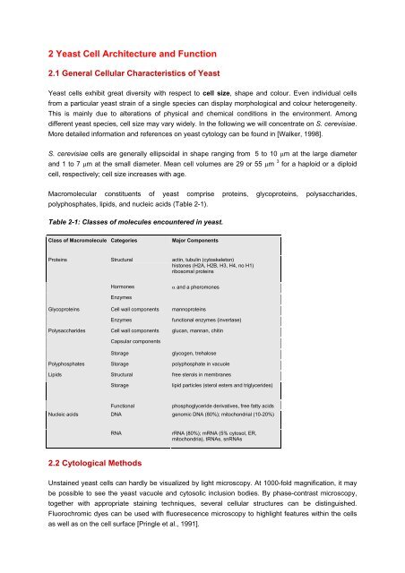
Table 2-1: Classes of molecules encountered in yeast.<br />
Class of Macromolecule Categories<br />
Major Components<br />
Proteins<br />
Structural<br />
actin, tubulin (cytoskeleton)<br />
histones (H2A, H2B, H3, H4, no H1)<br />
ribosomal proteins<br />
Glycoproteins<br />
Polysaccharides<br />
Hormones<br />
Enzymes<br />
<strong>Cell</strong> wall components<br />
Enzymes<br />
<strong>Cell</strong> wall components<br />
Capsular components<br />
Storage<br />
α <strong>and</strong> a pheromones<br />
mannoproteins<br />
functional enzymes (invertase)<br />
glucan, mannan, chitin<br />
glycogen, trehalose<br />
Polyphosphates Storage polyphosphate in vacuole<br />
Lipids<br />
Structural<br />
Storage<br />
free sterols in membranes<br />
lipid particles (sterol esters <strong>and</strong> triglycerides)<br />
Nucleic acids<br />
<strong>Function</strong>al<br />
phosphoglyceride derivatives, free fatty acids<br />
DNA genomic DNA (80%); mitochondrial (10-20%)<br />
RNA<br />
rRNA (80%); mRNA (5% cytosol, ER,<br />
mitochondria), tRNAs, snRNAs<br />
2.2 Cytological Methods<br />
Unstained yeast cells can hardly be visualized by light microscopy. At 1000-fold magnification, it may<br />
be possible to see the yeast vacuole <strong>and</strong> cytosolic inclusion bodies. By phase-contrast microscopy,<br />
together with appropriate staining techniques, several cellular structures can be distinguished.<br />
Fluorochromic dyes can be used with fluoresecence microscopy to highlight features within the cells<br />
as well as on the cell surface [Pringle et al., 1991].
A very convenient tool to localize <strong>and</strong> even to follow the movement of particular proteins within yeast<br />
cells is the use of the ‚green‘ fluorescent protein (GFP) from the jellyfish (Aequorea victoria) as a<br />
reporter molecule, <strong>and</strong> several derivatives of GFP with fluorescence spectra shifted to other<br />
wavelengths. Fusions of genes of interest with the GFP gene (N- or C-terminal) also allow to follow the<br />
expression <strong>and</strong> destiny of the fusion proteins followed by fluorescence microscopy [Niedenthal et al.,<br />
1996; see also chapter 4].<br />
The range of cellular features visualized is greatly increased, when monospecific antibodies raised<br />
against structural proteins are coupled to fluorescent dyes, such as fluorescein isothiocyanate (FITC)<br />
or Rhodamine B.<br />
Table 2-2: Structure-specific dyes for yeast cells.<br />
Dye Structures visualized Comments<br />
Methylene Blue Whole cells Non-viable cells stain blue<br />
Aminoacridine <strong>Cell</strong> walls Indicator of surface potential<br />
F-C ConA <strong>Cell</strong> walls Binds specifically to mannan<br />
Calcofluor white Bud scars Chitin in scar fluoresces<br />
DAPI (4,6-diamidino-2-phenylindole) Nuclei DNA fluoresces (Figure 2-1)<br />
Neutral red Vacuoles Vacuoles stain red-purple<br />
Iodine Glycogen deposits Glycogen stained red-brown<br />
DAPI Mitochondria Mitochondria fluoresce pink-white<br />
Rhodamine<br />
Mitochondria<br />
Figure 2-1: DAPI-staining of yeast cells.<br />
Flow cytometry has several applications in yeast studies. For example, fluorescence-activated cellsorting<br />
(FACS) can monitor yeast cell cycle progression, when cell walls are labelled with<br />
Concanavalin A conjugated to FITC <strong>and</strong> cell protein with tetramethylrhodamine isothiocyanate<br />
(TRITC). These tags enable to collect quantitative information on the growth properties of individual<br />
yeast cells as they progress through their cell cycle.
Organelle ultrastructure <strong>and</strong> macromolecular architecture can only be obtained with the aid of electron<br />
microscopy, which in scanning procedures is useful for studying cell topology, while ultrathin sections<br />
are essential in transmission electron microscopy to visualize intracellular fine structure. Atomic force<br />
microscopy can be applied to uncoated, unfixed cells of imaging the cell surfaces of different yeast<br />
strains or of cells under different growth conditions.<br />
2.3 <strong>Yeast</strong> <strong>Cell</strong> Organelles <strong>and</strong> Compartments<br />
Figure 2-2: Thin section of a yeast cell.<br />
In an idealized yeast cell (Figure 2-2), the following ultrastructural features can be observed (Figure<br />
2-3): cell wall; periplasm; plasma membrane; invagination; bud scar; cytosole; nucleus; mitochondrion;<br />
ER, endoplasmic reticulum; Golgi apparatus; secretory vesicles; vacuole; peroxisome. Obviously,<br />
yeast cells share most of the structural <strong>and</strong> functional features of higher eukaryotes, which has<br />
rendered yeast an ideal model for eukaryotic cell biology. In contrast to mammalian cells, peculiarities<br />
of yeast cells are that they are surrounded by a rigid cell wall <strong>and</strong> develope birth scars during cell<br />
division; the vacuole corresponds to lysomes in higher cells. Table 2-3 offers a list of marker enzymes<br />
that can be used to specifically identify these structures.<br />
Table 2-3: Organelles <strong>and</strong> compartments in a yeast cell.<br />
Organelle/Compartment<br />
<strong>Cell</strong> wall<br />
Plasma membrane<br />
Cytosole<br />
Nucleus<br />
Endoplasmic reticulum<br />
Vacuole<br />
Golgi apparatus<br />
periplasm<br />
secretory<br />
nucleoplasm<br />
nuclear envelope<br />
light microsomal<br />
fraction<br />
membrane<br />
sap<br />
Marker enzyme<br />
Invertase<br />
Acid phosphatase<br />
Vanadate-sensitive ATPase<br />
G-6-PDH<br />
RNA polymerase<br />
transmission EM<br />
NADPH:cytochrome c<br />
oxidoreductase<br />
α-Mannosidase<br />
Protease A <strong>and</strong> B<br />
ß-Glucan synthase;
Mitochondrion<br />
Peroxisome<br />
matrix<br />
intermembrane<br />
space<br />
inner membrane<br />
outer membrane<br />
mannosyltransferase<br />
Aconitase; fumarase<br />
Cytochrome c peroxidase<br />
Cytochrome c oxidase<br />
Kynurenine hydroxylase<br />
Catalase; isocitrate lyase; flavin<br />
oxidase<br />
Subcellular structures from yeast cells can be isolated from protoplasts or from intact cells by breaking<br />
the cell wall prior to differential centrifugation (see chapter 4: Techniques).<br />
Figure 2-3: Scheme of organelles <strong>and</strong> compartments in a yeast cell.<br />
2.3.1 <strong>Cell</strong> Envelope<br />
The yeast cell envelope is a protecting capsule, consisting of three major constituents (inside out):<br />
the plasma membrane, the periplasmic space, <strong>and</strong> the cell wall. In S. cerevisiae, the cell envelope<br />
takes ca. 15% of the total cell volume <strong>and</strong> has a major role in controlling the osmotic <strong>and</strong> permeabilty<br />
properties of the cell.<br />
The plasma membrane is about 7 nm thick, with some invaginations into the cytosole. Like other<br />
mebranes, it is a lipid bilayer with proteins inserted into this layer or traversing it as transmembrane<br />
proteins of various functions. The lipid composition comprises mainly phosphatidylcholine <strong>and</strong><br />
phosphatidylethanolamine, with minor proportions of phosphaditylinositol, phosphatidylserine or<br />
phosphadityl-glycerole, as well as sterols, mainly ergosterol <strong>and</strong> zymosterol. <strong>Yeast</strong> membrane<br />
proteins include the following catagories: (i) cytoskeleton anchors; (ii) enzymes for cell wall synthesis;<br />
(iii) proteins for transmembrane signal transduction; (iv) proteins for solute transport (permeases,<br />
channels, ATPases); (v) transport facilitators, such as the ABC (ATP binding cassette) proteins<br />
involved in multidrug transport.
Thus the primary functions of the yeast cell membrane is to provide selective permeability, i.e. to<br />
control what enters <strong>and</strong> what leaves the cytosole. Most important is the role of membrane proteins in<br />
regulating yeast nutrition, such as uptake of carbohydrates, nitrogenous compounds or ions, <strong>and</strong> the<br />
extrusion of molecules hazardous to the cell. Other important aspects include exo- <strong>and</strong> endocytosis of<br />
cargo molecules, stress responses, <strong>and</strong> sporulation.<br />
The yeast periplasm is a thin (35-45 A), cell wall associated region external to the plasma membrane<br />
<strong>and</strong> internal to the cell wall. It mainly contains secreted proteins (mannoproteins) that are unable to<br />
permeate the cell wall, but fulfill essential functions in hydrlolysing substrates that do not cross the<br />
plasma membrane: invertase converts sucrose into glucose <strong>and</strong> fructose; acid phosphatase catalyzes<br />
the liberation of free phosphate from organic compounds.<br />
2.3.2 <strong>Cell</strong> Wall<br />
The wall of a yeast cell is a remarkably thick (100 to 200 nm) envelope (Figures 2-4 <strong>and</strong> 2-5), which<br />
contains some 15 to 25% of the dry mass of the cell. Major structural constituents of the cell wall are<br />
polysaccharides (80-90%), mainly glucans <strong>and</strong> mannans, with a minor percentage of chitin. Glucans<br />
(both ß-2,6 <strong>and</strong> ß-1,3-linked glucans are represented) provide strength to the cell wall, forming a<br />
microfibrillar network. Mannans are present as an α -1,6-linked inner core with α -1,2- <strong>and</strong> α -1,3 side<br />
chains. Chitin is a polymer of N-acetylglucosamine representing only 2-4% of the cell wall <strong>and</strong> mainly<br />
located in bud scars. It may be interesting to note that some filamentous growing yeasts, such as<br />
C<strong>and</strong>ida albicans, have a higher content of chitin, while chitin is absent from many other yeast<br />
species. Other components of the cell wall are variable quantities of proteins, lipids, <strong>and</strong> inorganic<br />
phosphate. Preparation of cell walls are described in Zinser <strong>and</strong> Daum, 1995.<br />
Figure 2-4: <strong>Architecture</strong> of a yeast cell wall.
Table 2-4: Drugs to treat systemic mycoses.<br />
Compound<br />
Amphotericin<br />
Fluconazole<br />
5-Fluorocytosin<br />
Bacilysin<br />
Effects<br />
Complexes with membrane sterols (ergosterols) <strong>and</strong> leads to cell disruption<br />
Interferes with ergosterol biosynthesis (blocks P450-dependent demethylation<br />
step) <strong>and</strong> leads to accumulation of lanosterol<br />
Is deaminated to 5-Fluorouracil<br />
Incorporated in RNA, inhibits protein synthesis<br />
If converted to 5-Fluorodeoxyuridylate, is incorporated in DNA <strong>and</strong> inhibits<br />
thymidylate synthase<br />
Inhibitor of glucosamin-6-p synthesis from Fructose-6-p <strong>and</strong> glutamin<br />
Bud scars are specialized, ring-shaped convex protrusions at the cell surface which remain on the<br />
mother cells (of budding yeasts) after cell division <strong>and</strong> birth of daughter cells (Figure 2-6). The<br />
concave indentations remaining on the surface of the daughter cell after budding are called birth<br />
scars.<br />
Figure 2-5: Constituents of a yeast cell wall..<br />
Figure 2-6: The yeast bud.
If yeast cells are treated with lytic enzymes (e.g. Helicase from snail digestive juice; Zymolase or<br />
Lyticase from microbial sources) in the presence of osmotic stabilizers, the cell wall is removed giving<br />
rise to the formation of spheroblasts, which can be visualized as globular structures in the<br />
microscope. Spheroblast formation is often used prior to facilitate the isolation of subcellular<br />
components. Remarkably, spheroblasts keep the potential of regenerating the cell wall (Figure 2-7).<br />
Figure 2-7: Micrograph of a regenerating yeast protoplast.<br />
Flocculation (asexual cell aggregation) is a phenomenon of particular interest in brewing, because<br />
cells of brewer’s yeast strains tend to aggregate to a different extent. Bottom yeasts, normally used for<br />
larger fermentations, show a higher degree of flocculation (i.e. formation of macroscopic flocs by cell<br />
adherence) than top yeasts, which are used for making special kinds of beer. Most probably,<br />
flocculation is a consequence of the differential expression of flocculins, mannose-specific lectins of<br />
yeast cells, which interact with mannose receptors on the cell wall of neighbouring cells.<br />
2.3.3 Cytosol <strong>and</strong> Cytoskeleton<br />
The yeast cytoplasm is an acidic (pH 5.25) colloidal fluid, mainly containing ions <strong>and</strong> low or<br />
intermediate molecular weight organic compounds, <strong>and</strong> soluble macromolecules (e.g. enzyme<br />
proteins, factors, glycogen). The cytosolic enzymes of yeast include those of: (i) the glycolytic<br />
pathway, (ii) the fatty acid synthase complex, <strong>and</strong> (iii) some enzymes for protein biosynthesis. The<br />
cytoskeletal network guaranteeing internal stability to the cell <strong>and</strong> providing structural organization<br />
comprises the microtubules <strong>and</strong> the microfilaments. These are dynamic structures which fulfill their<br />
function through regulated assembly <strong>and</strong> dis-assembly of individual protein subunits. Thus, α <strong>and</strong> ß<br />
tubulin monomers polymerize as heterodimers to give microtubules, while globular monomers of G-<br />
actin polymerize into double-str<strong>and</strong>ed microfilaments of F-actin. Microtubules <strong>and</strong> microfilaments are<br />
also important in several dynamic processes occurring during mitosis <strong>and</strong> meiosis, septation, <strong>and</strong><br />
organelle motility.<br />
The yeast cytoplasm contains several categories of microbodies, which may be distinguished from<br />
organellar substructures:
(i) Freely suspended 80S ribosomes (in contrast to ER associated <strong>and</strong> mitochondrial 60S ribosomes);<br />
(II) Lipid particles, which function as storage particles or may serve in yeast membrane biosynthesis;<br />
(III) Proteasomes, multi-subunit complexes involved in programmed proteolysis of proteins <strong>and</strong> in<br />
other aspects of protein degradation or transport. Because of their importance in cellular metabolism<br />
<strong>and</strong> regulation, the ubiquitin-proteasome pathway will be discussed separately (see chapter<br />
‘Specialized Protein Families <strong>and</strong> Pathways’).<br />
2.3.4 Nucleus <strong>and</strong> Extrachromosomal Elements<br />
The yeast nucleus is a round-lobate organelle, some 1.5 µm in diameter. The nucleoplasm is<br />
separated from the cytosol by a double membrane containing pores between 50 to 100 nm in<br />
diameter. At two opposite poles, spindle pole bodies (SPBs) are located which are interconnected by<br />
continuous intranuclear microtubules <strong>and</strong> the origins of discontinuous microtubules. On the outer face,<br />
the SPBs are connected to cytosolic microtubules. These structural elements play an important role<br />
during cell division, cytokinesis <strong>and</strong> bud formation. In contrast to other eukaryotes, the nuclear<br />
membrane in yeast is not dissolved during mitosis.<br />
Figure 2-8: EM picture of yeast chromosomes.<br />
Within the nucleus there is a dense region corresponding to the nuleolus which disappears during<br />
mitosis <strong>and</strong> reforms during interphase. The major content of the nucleoplasm is represented by the<br />
genomic DNA which together with histones <strong>and</strong> non-histones is organized into chromatin. <strong>Yeast</strong><br />
chromosomes are formed <strong>and</strong> replicated during mitosis (or meiosis) but behave virtually invisible by<br />
microscopic techniques (Figure 2-8). However, pulsed field gel electrophoresis (PFGE) techniques<br />
provide convenient tools for chromosome separation <strong>and</strong> karyotyping (Figure 2-9).
Figure 2-9: Separation of yeast chromosomes by pulsed-field gel electrophoresis.<br />
In addition to the genomic material, yeast nuclei contain the machineries for DNA replication, DNA<br />
repair, transcription <strong>and</strong> RNA processing together with the necessary substrates <strong>and</strong> regulatory<br />
factors, <strong>and</strong> the resulting (precursor) products, as well as a proportion of the yeast proteasomes.<br />
Furthermore, several non-chromosomal genetic elements may be present in the yeast nucleus<br />
[Wickner, 1995].<br />
(i) 2 µm DNA is a stably maintained circular DNA plasmid, which replicates exactly once during S<br />
phase. These elements can be present in high copy number <strong>and</strong> have been useful in the construction<br />
of cloning vectors in yeast recombinant DNA technology. No functions have yet been attributed to the<br />
four genes found in 2 µm DNA.<br />
(ii) Double-str<strong>and</strong>ed RNA <strong>and</strong> linear DNA are found in killer strains of yeast. They enharbour genes<br />
for toxins, which will be hazardous to non-killer strains.<br />
(iii) Most interesting extrachromosomal elements are the Ty elements, the only class of<br />
retrotransposons found in yeast.<br />
Aspects of most of the afore mentioned subjects (such as mitosis, DNA replication, chromatin <strong>and</strong><br />
transcription, proteasomes, nuclear transport, <strong>and</strong> the yeast retroposons) will be detailed in separate<br />
chapters.
2.3.5 Secretory System <strong>and</strong> Vacuoles<br />
As common to all eukaryotes, yeast cells enharbor a system of membrane-surrounded<br />
compartments that are designed for trafficking of proteins within, into <strong>and</strong> out of the cell. In our<br />
fundamental underst<strong>and</strong>ing of the underlying processes <strong>and</strong> their regulation, yeast has contributed as<br />
a convenient model system [Pelham et al., 1995].<br />
The endoplasmic reticulum (ER) is the site of biosynthesis <strong>and</strong> modification of proteins that are to be<br />
exported. After synthesis on ER-associated polysomes located on the surface of the ER membrane,<br />
precursor proteins are translocated into the lumen of the ER, where trimming of the precursors,<br />
chaperone-assisted folding <strong>and</strong> glycosylation of the proteins occur. From the ER, proteins are directed<br />
to the Golgi apparatus by vesicles, which fuse at the cis-side <strong>and</strong> are exported from the Golgi at the<br />
trans-side. In the Golgi further modifications of the proteins by carbohydrate side chains may take<br />
place, such as mannosylation. Retrograde transport from the Golgi to the ER has been established<br />
as a quality control for the exported proteins.<br />
Proteins delivered from the Golgi are directed to different destinations within the cell or to the exterior<br />
via different secretory vesicles. These destinations include: (i) the vacuole; (ii) the bud region during<br />
mitosis targeted by actin-mediated transport; (iii) the plasma membrane; (iv) the periplasm. Naturally,<br />
only few proteins are exported to the periplasm <strong>and</strong> at low abundance. Nevertheless, the signal<br />
sequences which are present in these proteins have been fused to recombinant heterologous proteins<br />
of therapeutic value, which are then successfully secreted from yeast cells.<br />
The key organelle in yeast involved in intracellular trafficking of proteins is the vacuole. It can be<br />
viewed as a form of integral component of the intramembranous system. The main role of this<br />
lysosome-like compartment is the non-specific proteolytic cleavage of proteins, which involves a<br />
variety of intravacuolar lytic enzymes: endopeptidases, aminopeptidases, <strong>and</strong> carboxypeptidases.<br />
Further physiological functions of yeast vacuoles include: storage of basic amino acids,<br />
polyphosphates <strong>and</strong> certain metal ions; homeostasis of cytoplasmic ion concentrations;<br />
osmoregulation.<br />
The import of proteins into yeast cells by endocytosis also involves membran-bound vesicles<br />
(endocytotic vesicles), which deliver their cargo to the vacuole for proteolytic processing [Riezman,<br />
1993].<br />
2.3.6 Peroxisomes<br />
Peroxisomes perform a variety of metabolic functions in eukaryotic cells. In yeasts,<br />
peroxisomes(Figure 2-10) contain several oxidases which serve in oxidative utilization of specific<br />
carbon <strong>and</strong> nitrogen sources. The organelles develop from the small peroxisomes present in glucosegrown<br />
cells as a result of rapid synthesis of peroxisomal enzymes, such as catalase <strong>and</strong> alcohol<br />
oxidase. The import of components into peroxisomes will be discussed elsewhere (Chapter:<br />
Transport). In S. cerevisiae, most of the genes involved in peroxisome biogenesis (PEX genes) have<br />
been characterized to date. As yeast mitochondria lack ß-oxidation, peroxisomes are the sites of fatty<br />
acid degradation (Figure 2-11).
Figure 2-10: Micrograph of yeast peroxisomes.<br />
Figure 2-11: <strong>Function</strong>s of the yeast peroxisome.<strong>Yeast</strong> lacks the mitochondrial ß-oxidation. Acetyl-CoA,<br />
NADH <strong>and</strong> NADPH are produced in peroxisomes.
2.3.7 Mitochondria<br />
<strong>Yeast</strong> cells contain mitochondria which structurally resemble these organelles found in all<br />
eukaryotes. Therefore, yeast mitochondria have served as models to intensely study mitochondrial<br />
structure, function <strong>and</strong> biogenesis [Glick <strong>and</strong> Pon, 1995]. However, yeast mitochondria exhibit a<br />
variety of important features which are absent from their counterparts in higher organisms, notably<br />
mammalian cells.<br />
General structural characteristics of mitochondria (Figure 2-12) include:<br />
(i) an outer membrane – containing enzymes involved in lipid metabolism,<br />
(ii) the intermembrane space<br />
(iii) an inner membrane – containing NADH <strong>and</strong> succinate dehydrogenases, the components of the<br />
respiratory chain <strong>and</strong> the ATP synthase, <strong>and</strong> various membrane-integral transport proteins,<br />
(iv) the mitochondrial matrix – containing enzymes of fatty acid oxidation, the citric acid cycle, the<br />
mitochondrial DNA together with the mitochondrial transcription <strong>and</strong> protein synthesis machineries<br />
(including mitochondrial 60S ribosomes <strong>and</strong> mitochondrial tRNAs).<br />
Figure 2-12: Thin section micrograph of a yeast mitochondrion.<br />
<strong>Yeast</strong> mitochondria are dynamic structures whose size, shape <strong>and</strong> number can greatly vary according<br />
to strain specificity, cell cycle phase, <strong>and</strong> growth conditions, whereby important factors are: partial<br />
oxygen pressure, glucose concentration, presence of unfermentable substrates, availability of sterols<br />
<strong>and</strong> fatty acids, <strong>and</strong> of particular metal ions (Mg ++ ) (Table 2-5).<br />
Table 2-5: Effects of nutrion on yeast mitochondria.<br />
Nutrient Concentration Oxygen Respiration Morphology<br />
Glucose excess + repressed few large<br />
Ethanol excess + activated many small<br />
Glucose excess - repressed few large<br />
Glucose limited - repressed few large<br />
Glucose limited + activated many small
Under aerobic conditions, yeast mitochondria are involved in ATP synthesis coupled to oxidative<br />
phosphorylation. The activities of the citric acid cycle <strong>and</strong> the respiratory chain will largely depend on<br />
the yeast species <strong>and</strong> the expression of the Crabtree effect. This is a phenomenon related that relates<br />
glucose concentrations with the particular catabolic pathway adopted by glucose-sensitive cells, in that<br />
even in the presence of oxygen fermentation predominates over respiration. Under anaerobic<br />
conditions, mitochondria seem to be dispensible, at least for respiratory function. In fact, so-called ρ°<br />
‚petite‘ mutants that lack functional mitochondria are viable. However, mitochondria do perform other<br />
functions in yeast cell physiology, implicating that mitochondria are relevant to intact cell metabolism<br />
even under anaerobic conditions:<br />
- synthesis <strong>and</strong> desaturation of fatty acids <strong>and</strong> lipids,<br />
- biosynthesis of ergosterol,<br />
- stress responses <strong>and</strong> adaptation to stresses,<br />
- enzymes for the synthesis of particular amino acids <strong>and</strong> dicarboxylic acids, pyrimididne <strong>and</strong> purine<br />
bases, porphyrin, <strong>and</strong> pteridines,<br />
- mobilization of glycogen,<br />
- production of ‚flavor‘ components.<br />
The importance of yeast mitochondria is best illustrated by the fact that some 8 to 10% of the nuclear<br />
yeast genes are involved in biogenesis of these organelles <strong>and</strong> maintenance of their functions. The<br />
vast majority of these proteins are synthesized by cytosolic ribosomes <strong>and</strong> become imported into<br />
yeast mitochondria, which have the potential to biosynthesize only 12 different proteins<br />
(cytochromoxidase subunits, cytochrome b, the 6 subunits of NADH dehydrogenase, splicing factors)<br />
in addition to the mitochondrial rRNAs (15S <strong>and</strong> 26S subunits) <strong>and</strong> the complement of mitochondrial<br />
tRNAs. The biogenesis of mitochondria, which involves genetic cooperativity between nuclear <strong>and</strong><br />
mitochondrial genomes, has been widely studied in S. cerevisiae, since several kinds of mutations can<br />
be used in this model organism. The special features attributed to yeast mitochondria are summarized<br />
in Figure 2-13. Details of the genomic structure <strong>and</strong> content of yeast mitochondrial DNA will be dealt<br />
with in chapter 5.<br />
Figure 2-13: Features of yeast mitochondria.
References<br />
Foury, F., Roganti, T., Lecrenier, N., Purnelle, B. The complete sequence of the mitochondrial genome<br />
of Saccharomyces cerevisiae. FEBS Lett. 440 (1998) 325-331.<br />
Glick, B.S. <strong>and</strong> Pon, L.A. Isolation of highly purified mitochondria from S. ceverisiae. Methods<br />
Enzymology 260 (1995) 213-233.<br />
Lindegren, C.C. The yeast cell, its genetics <strong>and</strong> cytology. Educational Publishers, St. Louis, 1949.<br />
Niedenthal, R.K. Riles, L., Johnston, M. <strong>and</strong> Hegemann, J.H. Green fluorescent protein as a marker<br />
gene for gene expression <strong>and</strong> subcellular localization in budding yeast. <strong>Yeast</strong> 12 (1996) 773-786.<br />
Pelham, H.R.B., Banfield, D.K. <strong>and</strong> Lewis, M.J. SNAREs involved in traffic through the Golgi complex.<br />
Cold Spring Harb. Symp. 60 (1995) 105-111.<br />
Pringle, J.R. et al. Immunofluoresecence methods for yeast. Methods in Enzymology 194 (1991) 565-<br />
665.<br />
Riezman, H. <strong>Yeast</strong> endocytosis. Trends <strong>Cell</strong> Biol. 3 (1993) 273-277.<br />
Walker, G.M. <strong>Yeast</strong> Physiology <strong>and</strong> Biotechnology, John Wiley & Sons, Chichester, 1998.<br />
Wickner, R.B. Non-mendelian genetic elements in S. cerevisiae - RNA viruses, 2µ DNA, Ψ, [URE3],<br />
20s RNA <strong>and</strong> other wonders of nature. In: The <strong>Yeast</strong>s, 2nd. edn, Vol 6: <strong>Yeast</strong> Genetics (eds. A.N.<br />
Wheals, A. Rose <strong>and</strong> J.S. Harrison), pp. 309-356. Academic Press, London, 1995.<br />
Zinser, E. <strong>and</strong> Daum, G. Isolation <strong>and</strong> biochemical characterization of organelles from the yeast S.<br />
cerevisiae. <strong>Yeast</strong> 11 (1995) 493-536.