Characterisation of phenolic extracts from olive pulp and ... - ESAC
Characterisation of phenolic extracts from olive pulp and ... - ESAC
Characterisation of phenolic extracts from olive pulp and ... - ESAC

Create successful ePaper yourself
Turn your PDF publications into a flip-book with our unique Google optimized e-Paper software.
SM Cardoso et al<br />
The comparison <strong>of</strong> the ESI-MS data with<br />
literature data also allowed the identification <strong>of</strong><br />
hydroxytyrosol-1 ′ -β-glucoside (peak 1), 11-methyloleoside<br />
(peak 13a), hydroxylphenylacetic acid (peak<br />
13b), three derivatives <strong>of</strong> luteolin (peaks 17, 19 <strong>and</strong><br />
21), verbascoside (peak 18b), oleoside (fraction 8) <strong>and</strong><br />
oleuropein glucoside (fraction 20).<br />
The main compound in fraction 1 had a molecular<br />
ion at m/z 315 <strong>and</strong> fragment ions at m/z 153,<br />
135, 180 <strong>and</strong> 161, which suggested the presence<br />
<strong>of</strong> a hydroxytyrosol hexoside. To our knowledge,<br />
three isomers <strong>of</strong> hydroxytyrosol glucoside have been<br />
characterised by NMR in <strong>olive</strong> fruit <strong>and</strong> <strong>olive</strong> oil. 29<br />
These isomers have also been detected in different<br />
table <strong>olive</strong> varieties, 30 in which hydroxytyrosol-4-βglucoside<br />
was the most abundant compound. This<br />
isomer was also the only one detected by Romero et al 9<br />
in <strong>olive</strong> <strong>pulp</strong>, <strong>olive</strong> pomace <strong>and</strong> waste waters. However,<br />
the similarity between the fragmentation pr<strong>of</strong>ile <strong>of</strong> the<br />
molecular ion at m/z 315 in fraction 1 to that published<br />
by De Nino et al 21 allowed us to infer that this<br />
compound should be hydroxytyrosol-1 ′ -β-glucoside.<br />
Fractions 13a <strong>and</strong> 13b were attributed to 11-methyloleoside<br />
<strong>and</strong> 4-hydroxylphenylacetic acid respectively<br />
based on their specific <strong>and</strong> characteristic molecular<br />
ions described in the literature for Oeuropaea. 23 The<br />
high absorbance at 240 nm <strong>of</strong> fraction 10 can possibly<br />
be attributed to a secoiridoid derivative.<br />
The presence <strong>of</strong> a fragment at m/z 285 is<br />
diagnostic <strong>of</strong> luteolin derivatives. According to the<br />
molecular ion at m/z 593 <strong>and</strong> its main fragments<br />
observed for both fractions 17 <strong>and</strong> 19, luteolin-<br />
7-rutinoside could be proposed for both fractions.<br />
To our knowledge, the flavone luteolin-7-rutinoside<br />
was previously detected only in <strong>olive</strong> leaves 23 <strong>and</strong><br />
its ESI-MS data were similar to those <strong>of</strong> peaks<br />
17 <strong>and</strong> 19. Since the luteolin-7-rutinoside detected<br />
by Ryan et al 23 eluted before luteolin-7-glucoside in<br />
HPLC reverse phase conditions, the same compound<br />
was tentatively attributed to fraction 17. Thus<br />
fraction 19 may correspond to a non-described<br />
isomer <strong>of</strong> that compound, probably with a different<br />
linkage position to the sugar. The MS analysis<br />
<strong>of</strong> fraction 21 demonstrated a molecular ion at<br />
m/z 447, suggesting the presence <strong>of</strong> a luteolin<br />
hexoside. Four luteolin glucosides have already<br />
been detected in <strong>olive</strong>s: luteolin-7-glucoside <strong>and</strong><br />
its three isomers 23 luteolin-4-glucoside, luteolin-6-<br />
glucoside <strong>and</strong> luteolin-8-glucoside. According to those<br />
authors, luteolin-4-glucoside was the only one eluting<br />
after luteolin-7-glucoside in HPLC reverse phase<br />
conditions <strong>and</strong> that was the reason why fraction 21<br />
was attributed to that isomer.<br />
ESI-MS <strong>of</strong> fraction 18b indicated a molecular ion at<br />
m/z 623 <strong>and</strong> various fragments that are in accordance<br />
with the fragmentation <strong>of</strong> verbascoside. These results<br />
were also corroborated with the fragmentation pr<strong>of</strong>ile<br />
<strong>of</strong> verbascoside described by Ryan et al. 18<br />
The comparison <strong>of</strong> the MS data with literature<br />
data was also possible for the compounds detected<br />
in fraction 8 (oleoside) <strong>and</strong> fraction 20 (oleuropein<br />
glucoside). However, as they had not been detected<br />
previously in <strong>olive</strong> <strong>pulp</strong>, the interpretation <strong>of</strong> their<br />
structures will be discussed in more detail. Also, the<br />
structures <strong>of</strong> some new oleoside derivatives corresponding<br />
to fractions 15a, 22 <strong>and</strong> 26 will be elucidated.<br />
Structure determination <strong>of</strong> fraction 8<br />
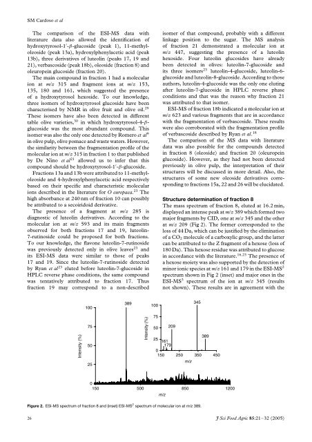
The mass spectrum <strong>of</strong> fraction 8, eluted at 16.2 min,<br />
displayed an intense peak at m/z 389 which formed two<br />
major fragments by CID, one at m/z 345 <strong>and</strong> the other<br />
at m/z 209 (Fig 2). The former corresponded to the<br />
loss <strong>of</strong> 44 Da, which can be justified by the elimination<br />
<strong>of</strong> a CO 2 molecule <strong>of</strong> a carboxylic group, <strong>and</strong> the latter<br />
can be attributed to the Z fragment <strong>of</strong> a hexose (loss <strong>of</strong><br />
180 Da). This hexose residue was attributed to glucose<br />
in accordance with the literature. 16,23 The presence <strong>of</strong><br />
a hexose moiety was also supported by the detection <strong>of</strong><br />
minor ionic species at m/z 161 <strong>and</strong> 179 in the ESI-MS 2<br />
spectrum shown in Fig 2 (inset) <strong>and</strong> major ones in the<br />
ESI-MS 3 spectrum <strong>of</strong> the ion at m/z 345 (results<br />
not shown). These results are in agreement with the<br />
100<br />
389<br />
100<br />
345<br />
Intensity (%)<br />
75<br />
50<br />
25<br />
Intensity (%)<br />
75<br />
50<br />
209<br />
25<br />
389<br />
161<br />
179<br />
0<br />
150 250 350 450<br />
m/z<br />
0<br />
150 500 850 1200<br />
m/z<br />
Figure 2. ESI-MS spectrum <strong>of</strong> fraction 8 <strong>and</strong> (inset) ESI-MS 2 spectrum <strong>of</strong> molecular ion at m/z 389.<br />
26 J Sci Food Agric 85:21–32 (2005)