Characterisation of phenolic extracts from olive pulp and ... - ESAC
Characterisation of phenolic extracts from olive pulp and ... - ESAC
Characterisation of phenolic extracts from olive pulp and ... - ESAC

You also want an ePaper? Increase the reach of your titles
YUMPU automatically turns print PDFs into web optimized ePapers that Google loves.
Phenolic compounds <strong>of</strong> <strong>olive</strong> pomace<br />
a<br />
100<br />
539<br />
75<br />
Intensity (%)<br />
50<br />
377<br />
701<br />
25<br />
307 162 Da 162 Da<br />
0<br />
300 400 500 600 700 800<br />
m/z<br />
b<br />
HO<br />
HO<br />
OH<br />
O<br />
OH<br />
Y<br />
O<br />
O<br />
O<br />
O<br />
O<br />
O<br />
Isomer I<br />
Y<br />
HO<br />
O<br />
OH<br />
O<br />
OH<br />
OH<br />
OH<br />
HO<br />
HO<br />
OH<br />
O<br />
OH<br />
Y 1<br />
O<br />
HO<br />
OH Y 2<br />
O<br />
O<br />
OH<br />
O<br />
Isomer II<br />
O<br />
O<br />
O<br />
O<br />
OH<br />
OH<br />
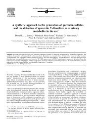
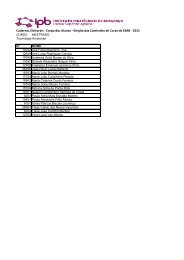
Figure 6. (a) ESI-MS 2 spectrum <strong>of</strong> molecular ion at m/z 701. (b) Structure <strong>of</strong> two diglucoside homologues <strong>of</strong> oleuropein.<br />
fragmentation pr<strong>of</strong>ile showed a main ionic species at<br />
m/z 539 that was formed by the loss <strong>of</strong> 162 Da, <strong>and</strong><br />
another intense peak at m/z 377 indicative <strong>of</strong> the<br />
elimination <strong>of</strong> another hexose unit (Fig 6a). These<br />
two main fragments correspond to oleuropein <strong>and</strong> its<br />
aglycone respectively <strong>and</strong> together they support the<br />
hypothesis <strong>of</strong> a hexose derivative <strong>of</strong> the oleuropein<br />
structure. To our knowledge, hexose derivatives <strong>of</strong><br />
oleuropein have never been described in <strong>olive</strong> fruit.<br />
However, angustifolioside A (isomer I represented in<br />
Fig 6b) was already described to occur in the family<br />
Oleaceae. 35 Also, De Nino et al 16 have proposed<br />
the presence <strong>of</strong> the same isomer in <strong>olive</strong> leaves,<br />
although their results did not allow the exact structural<br />
determination, <strong>and</strong> the exclusion <strong>of</strong> isomer II. In the<br />
present study the loss <strong>of</strong> 162 Da <strong>of</strong> the molecular<br />
ion at m/z 701 can fit for both isomer structures:<br />
the consecutive or simultaneous elimination <strong>of</strong> a Y-<br />
type hexose fragment would be possible for the two<br />
compounds, explaining the fragments at m/z 539 <strong>and</strong><br />
377. However, <strong>from</strong> the oleoside derivatives discussed<br />
in Figs 3 <strong>and</strong> 5, it can be expected that, if isomer II was<br />
present, the O-dihexosyl ion (at m/z 341) together with<br />
its fragments would have appeared in the ESI-MS 2<br />
spectrum <strong>of</strong> the molecular ion. Yet, the total absence<br />
<strong>of</strong> those species was confirmed, indicating that the<br />
isomer present in fraction 20 is angustifolioside A<br />
(isomer I in Fig 6b). These results together with those<br />
<strong>of</strong> De Nino et al 16 suggest that Oeuropaeahas the<br />
same glucoside derivative <strong>of</strong> oleuropein that is present<br />
in Fraxinus angustifolia. 35<br />
Structure determination <strong>of</strong> fraction 15a<br />
The MS analysis <strong>of</strong> fraction 15a showed a predominant<br />
[M − H] − signal at m/z 555. As for the oleoside<br />
derivatives, it was not possible to find any MS data in<br />
the literature about this compound. Alternatively, its<br />
structure elucidation was only based on its ESI-MS n<br />
analysis. Fig 7 shows the ESI-MS 2 spectrum <strong>of</strong> the<br />
ion at m/z 555. The main fragment ion represented in<br />
the spectrum was obtained by the loss <strong>of</strong> 18 Da (ion<br />
at m/z 537), suggesting that the compound has an OH<br />
group that is easily removed. Also, another two intense<br />
peaks at m/z 393 <strong>and</strong> 403 could be observed. The first<br />
corresponded to the aglycone (loss <strong>of</strong> 162 Da) <strong>and</strong><br />
the latter was equivalent to the mass <strong>of</strong> an 11-methyloleoside<br />
moiety. The aglycone at m/z 393 was already<br />
detected as 10-hydroxy-oleuropein aglycone in <strong>olive</strong><br />
oil using mass spectrometry, 36 which indicates that<br />
this compound should be 10-hydroxy-oleuropein. In<br />
this manner the fragments at m/z 537 <strong>and</strong> 393 arose<br />
<strong>from</strong> the loss <strong>of</strong> water <strong>from</strong> the 10-OH group <strong>and</strong><br />
<strong>from</strong> the Y-type cleavage <strong>of</strong> the molecule respectively.<br />
The fragment ion at m/z 403 could be originated by<br />
a cleavage X together with the loss <strong>of</strong> water. To our<br />
knowledge, 10-hydroxy-oleuropein has not previously<br />
been detected in any tissue <strong>of</strong> Oeuropaea.<br />
J Sci Food Agric 85:21–32 (2005) 29