Changes in zooxanthellae density, morphology, and mitotic index in ...
Changes in zooxanthellae density, morphology, and mitotic index in ...
Changes in zooxanthellae density, morphology, and mitotic index in ...

You also want an ePaper? Increase the reach of your titles
YUMPU automatically turns print PDFs into web optimized ePapers that Google loves.
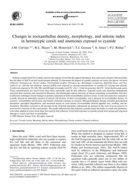
Mar<strong>in</strong>e Pollution Bullet<strong>in</strong> 46 (2003) 573–586<br />
www.elsevier.com/locate/marpolbul<br />
<strong>Changes</strong> <strong>in</strong> <strong>zooxanthellae</strong> <strong>density</strong>, <strong>morphology</strong>, <strong>and</strong> <strong>mitotic</strong> <strong>in</strong>dex<br />
<strong>in</strong> hermatypic corals <strong>and</strong> anemones exposed to cyanide<br />
J.M. Cerv<strong>in</strong>o a, *<br />
, R.L. Hayes b , M. Honovich c , T.J. Goreau d , S. Jones e , P.J. Rubec f<br />
a University of South Carol<strong>in</strong>a, Columbia, SC 29801, USA<br />
b Howard University, Wash<strong>in</strong>gton, DC, USA<br />
c B<strong>in</strong>ghamton University, B<strong>in</strong>ghamton, NY, USA<br />
d Global Coral Reef Alliance, New York, NY, USA<br />
e NY Aquarium for Wildlife Conservation, New York, NY, USA<br />
f International Mar<strong>in</strong>elife Alliance-USA, St Petersburg, FL, USA<br />
Abstract<br />
Sodium cyanide (NaCN) is widely used for the capture of reef fish throughout Southeast Asia <strong>and</strong> causes extensive fish mortality,<br />
but the effect of NaCN on reef corals rema<strong>in</strong>s debated. To document the impact of cyanide exposure on corals, the species Acropora<br />
millepora, Goniopora sp., Favites abdita, Trachyphyllia geoffrio, Plerogyra sp., Heliofungia act<strong>in</strong>formis, Euphyllia divisa, <strong>and</strong> Scarophyton<br />
sp., <strong>and</strong> the sea anemone Aiptasia pallida were exposed to vary<strong>in</strong>g concentrations of cyanide for vary<strong>in</strong>g time periods.<br />
Corals were exposed to 50, 100, 300, <strong>and</strong> 600 mg/l of cyanide ion (CN ) for 1–2 m<strong>in</strong> (<strong>in</strong> seawater, the CN forms hydrocyanic acid).<br />
These concentrations are much lower than those reportedly used by fish collectors. Exposed corals <strong>and</strong> anemones immediately<br />
retracted their tentacles <strong>and</strong> mesenterial filaments, <strong>and</strong> discharged copious amounts of mucus conta<strong>in</strong><strong>in</strong>g <strong>zooxanthellae</strong>. Gel electrophoreses<br />
techniques found changes <strong>in</strong> prote<strong>in</strong> expression <strong>in</strong> both <strong>zooxanthellae</strong> <strong>and</strong> host tissue. Corals <strong>and</strong> anemones exposed to<br />
cyanide showed an immediate <strong>in</strong>crease <strong>in</strong> <strong>mitotic</strong> cell division of their zooxenthellae, <strong>and</strong> a decrease <strong>in</strong> <strong>zooxanthellae</strong> <strong>density</strong>. In<br />
contrast, <strong>zooxanthellae</strong> cell division <strong>and</strong> <strong>density</strong> rema<strong>in</strong>ed constant <strong>in</strong> controls. Histopathological changes <strong>in</strong>cluded gastrodermal<br />
disruption, mesogleal degradation, <strong>and</strong> <strong>in</strong>creased mucus <strong>in</strong> coral tissues. Zooxanthellae showed pigment loss, swell<strong>in</strong>g, <strong>and</strong> deformation.<br />
Mortality occurred at all exposure levels. Exposed specimens experienced an <strong>in</strong>crease <strong>in</strong> the ratio of gram-negative to<br />
gram-positive bacteria on the coral surface. The results demonstrate that exposure cyanide causes mortality to corals <strong>and</strong> anemones,<br />
even when applied at lower levels than that used by fish collectors. Even brief exposure to cyanide caused slow-act<strong>in</strong>g <strong>and</strong> long-term<br />
damage to corals <strong>and</strong> their <strong>zooxanthellae</strong>.<br />
Ó 2003 Elsevier Science Ltd. All rights reserved.<br />
Keywords: Cyanide toxicity; Bleach<strong>in</strong>g; Mitotic <strong>in</strong>dices; Histopathology; Prote<strong>in</strong> synthesis; Chemical pollution<br />
1. Introduction<br />
S<strong>in</strong>ce the early 1960s, coral reefs have been <strong>in</strong>creas<strong>in</strong>gly<br />
exploited by fishermen who use cyanide to capture<br />
live reef fish <strong>and</strong> sell them to the restaurant <strong>and</strong><br />
aquarium trades (Rubec, 1986, 1997). Fishermen stun<br />
the live reef fish by squirt<strong>in</strong>g sodium cyanide (NaCN)<br />
* Correspond<strong>in</strong>g author. Present Address: USE: 117-20 5th Ave.,<br />
College Po<strong>in</strong>t, New York, NY 11356, USA. Tel.: +1-718-358-4946.<br />
E-mail addresses: cnidaria@earthl<strong>in</strong>k.net (J.M. Cerv<strong>in</strong>o), rhayes@fac.howard.edu(R.L.<br />
Hayes), bh11421@b<strong>in</strong>ghamton.edu(M.<br />
Honovich), goreau@bestweb.net (T.J. Goreau), samjones@idt.net (S.<br />
Jones), prubec@compuserve.com (P.J. Rubec).<br />
onto coral heads <strong>and</strong> <strong>in</strong>to crevices <strong>in</strong> coral reefs (Fig. 1).<br />
While this method is designed to capture liv<strong>in</strong>g fish, it<br />
results <strong>in</strong> high mortality rates. Approximately 50% of<br />
the NaCN-exposed aquarium fish die while still on the<br />
reef, <strong>and</strong> >80% of the rema<strong>in</strong>der die before they are sold<br />
by retailers <strong>in</strong> North America <strong>and</strong> Europe (Rubec, 1986;<br />
Rubec <strong>and</strong> Soundararajan, 1991). The NaCN tablets<br />
used by collectors <strong>in</strong> the Philipp<strong>in</strong>es <strong>and</strong> Indonesia<br />
weigh about 20 g each (Johannes <strong>and</strong> Riepen, 1995).<br />
Fishermen who are collect<strong>in</strong>g ornamental fish for<br />
aquarium tanks generally place one or two tablets of<br />
cyanide <strong>in</strong>to a 1-l plastic squirt bottle filled with seawater,<br />
while food-fish collectors use three to five tablets<br />
(Rubec et al., 2001). The tablets sequentially dissolve <strong>in</strong><br />
the squirt bottle as collectors proceed to spray the reefs,<br />
0025-326X/03/$ - see front matter Ó 2003 Elsevier Science Ltd. All rights reserved.<br />
doi:10.1016/S0025-326X(03)00071-7
574 J.M. Cerv<strong>in</strong>o et al. / Mar<strong>in</strong>e Pollution Bullet<strong>in</strong> 46 (2003) 573–586<br />
Fig. 1. Photo of a cyanide fisherman squirt<strong>in</strong>g cyanide from a squirt<br />
bottle to capture fish.<br />
mak<strong>in</strong>g it difficult to determ<strong>in</strong>e the cyanide ion (CN )<br />
concentrations be<strong>in</strong>g applied. It has been suggested that<br />
fish collectors use concentrations rang<strong>in</strong>g from 1500 to<br />
120,000 mg/l (Johannes <strong>and</strong> Riepen, 1995; Barber <strong>and</strong><br />
Pratt, 1998; Pet <strong>and</strong> Djohani, 1998; Jones et al., 1998).<br />
S<strong>in</strong>ce not all of the cyanide applied from squirt bottles<br />
dissolves, it is sometimes visible underwater as a whitish<br />
plume (Rubec et al., 2001). Not all fishermen limit<br />
themselves to squirt bottles. Reports from the Philipp<strong>in</strong>es<br />
assert that 55-gallon drums of cyanide have been<br />
dumped onto reefs to kill <strong>and</strong> capture food fish (del<br />
Norte et al., 1989; Johannes <strong>and</strong> Riepen, 1995).<br />
The various forms of cyanide fish<strong>in</strong>g contribute to<br />
degraded, lifeless coral reefs (Rubec, 1986; Johannes <strong>and</strong><br />
Riepen, 1995; Barber <strong>and</strong> Pratt, 1998) <strong>and</strong> force fishermen<br />
to relocate to more prist<strong>in</strong>e locations. Unfortunately,<br />
fishermen usually br<strong>in</strong>g cyanide techniques with<br />
them, thus repeat<strong>in</strong>g the cycle of destruction of the coral<br />
reefs <strong>and</strong> associated fish communities.<br />
Although illegal <strong>in</strong> most Southeast Asian countries,<br />
cyanide fish<strong>in</strong>g is widespread <strong>and</strong> is be<strong>in</strong>g <strong>in</strong>troduced to<br />
other regions (Johannes <strong>and</strong> Riepen, 1995; Barber <strong>and</strong><br />
Pratt, 1997).<br />
Fishermen <strong>in</strong> the Philipp<strong>in</strong>es, Indonesia, Malaysia,<br />
Vietnam, <strong>and</strong> Papua New Gu<strong>in</strong>ea have reported cyanide<br />
use (Rubec, 1986; Johannes <strong>and</strong> Riepen, 1995; Barber<br />
<strong>and</strong> Pratt, 1998). Squirt<strong>in</strong>g cyanide onto a reef is an easy<br />
way to force fish out of coral crevices. Stressed <strong>and</strong><br />
poisioned, they swim to open-water areas, where they<br />
are then captured. Although some fishermen acknowledge<br />
that cyanide fish<strong>in</strong>g kills corals, others deny that<br />
such damage occurs (Galvez et al., 1989; H<strong>in</strong>gco <strong>and</strong><br />
Rivera, 1991).<br />
In a field visit to devastated reefs <strong>in</strong> the Philipp<strong>in</strong>es,<br />
former cyanide fishermen showed the author <strong>in</strong>dividual<br />
coral heads that had died follow<strong>in</strong>g one application of<br />
cyanide (Cerv<strong>in</strong>o <strong>and</strong> Goreau, personal observation).<br />
Furthermore, an unpublished study by the Philipp<strong>in</strong>e<br />
Bureau of Fisheries <strong>and</strong> Aquatic Resources found that<br />
corals situated on test quadrants off Mactan Isl<strong>and</strong> near<br />
Cebudied after two applications of CN spaced 4<br />
months apart (Rubec, 1986, 1997).<br />
There have been several studies regard<strong>in</strong>g the deleterious<br />
effect of cyanide exposure to corals. Dr. Robert<br />
Richmond of the University of Guam found that Pocillopora<br />
damicornis colonies exposed to 4000 mg/l CN<br />
for 10 m<strong>in</strong> began to bleach with<strong>in</strong> 4 days (Johannes <strong>and</strong><br />
Riepen, 1995). N<strong>in</strong>e out of 10 Pocillopora specimens<br />
died with<strong>in</strong> 4 days. When exposed to CN concentrations<br />
of 100 mg/l for 30 m<strong>in</strong>, Pocillopora specimens<br />
bleached with<strong>in</strong> 3–4 days <strong>and</strong> tissue loss began after 9<br />
days. Likewise, Pocillopora damicornis <strong>and</strong> Porites lichen<br />
were exposed to a 5200 mg/l CN concentration caused<br />
100% mortality (Jones, 1997; Jones <strong>and</strong> Steven, 1997)<br />
with exposure times of 10, 20, or 30 m<strong>in</strong>. Tissues coat<strong>in</strong>g<br />
the skeletons of Pocillopora damicornis sloughed off<br />
with<strong>in</strong> 24 h. Porites lichen colonies became covered with<br />
a thick mucus-like tunic. After 7 days, the tunics of all<br />
the Porites colonies lifted off, leav<strong>in</strong>g bare skeletons.<br />
Shorter exposure times (1 or 5 m<strong>in</strong>) <strong>and</strong> lower CN<br />
concentrations (52 or 520 mg/l) <strong>in</strong>duced bleach<strong>in</strong>g (loss<br />
of pigment) <strong>in</strong> both coral species with<strong>in</strong> 7 days (Jones<br />
<strong>and</strong> Steven, 1997).<br />
Cyanide was also shown to <strong>in</strong>hibit photosynthesis<br />
<strong>and</strong> calcification rates of Acropora formosa <strong>and</strong> Acropora<br />
cervicornis <strong>in</strong> corals exposed to concentrations of 5<br />
mg/l (Chalker <strong>and</strong> Taylor, 1975). Photosynthesis <strong>and</strong><br />
calcification were not <strong>in</strong>hibited at the highest concentration<br />
tested (5 mg/l), suggest<strong>in</strong>g the existence of cyanide-resistant<br />
respiration <strong>in</strong> either the <strong>zooxanthellae</strong> or<br />
the host (Barnes, 1985). Research by Jones et al. (1998)<br />
showed that photo-<strong>in</strong>hibition from exposure to cyanide<br />
resulted <strong>in</strong> an almost complete cessation of photosynthetic<br />
electron transport from algae <strong>in</strong> tissues of Stylophora<br />
pistillata <strong>and</strong> Acropora aspera. Cyanide was<br />
applied to small branch tips of these species at concentrations<br />
estimated to occur dur<strong>in</strong>g cyanide fish<strong>in</strong>g.<br />
Pulse-amplitude-modulation (PAM) chlorophyll fluorescence<br />
was used to exam<strong>in</strong>e photo-<strong>in</strong>hibition <strong>and</strong><br />
photosynthetic electron transport <strong>in</strong> the symbiotic algae<br />
(<strong>zooxanthellae</strong> <strong>in</strong> the tissues of the corals). Jones <strong>and</strong><br />
Hoegh-Guldberg (1999) have suggested that cyanide<br />
acts as an <strong>in</strong>hibitor of the dark reactions of the Calv<strong>in</strong><br />
cycle; specifically, as an <strong>in</strong>hibitor of ribulose-1,5-biphosphate<br />
carboxylase/oxygenase.<br />
No known studies have been published <strong>in</strong>vestigat<strong>in</strong>g<br />
the direct effects on cyanide on symbiotic algae, the<br />
focus of this paper. Related work by Dr. R. Richmond<br />
<strong>in</strong>dicated that m<strong>in</strong>or concentrations of cyanide can have<br />
a deleterious effect on the relationship between symbiotic<br />
algae <strong>and</strong> host tissue <strong>in</strong> cnidarians, result<strong>in</strong>g <strong>in</strong><br />
death. But RichmondÕs <strong>in</strong> vitro experiment was limited<br />
to film<strong>in</strong>g the expulsion <strong>and</strong> behavior of <strong>zooxanthellae</strong><br />
from corals follow<strong>in</strong>g cyanide exposure (personal communication,<br />
1999).
J.M. Cerv<strong>in</strong>o et al. / Mar<strong>in</strong>e Pollution Bullet<strong>in</strong> 46 (2003) 573–586 575<br />
2. Materials <strong>and</strong> methods<br />
Corals were imported to the United States from the<br />
Indo-Pacific region packed <strong>in</strong> fresh seawater <strong>and</strong> kept at<br />
a hold<strong>in</strong>g facility. The samples were held <strong>in</strong> two 750-l<br />
tanks for 4 weeks prior to each of the three experiments.<br />
Dur<strong>in</strong>g the first segment of this experiment, 150 fragments<br />
(5 3 cm) of Acropora millipora were removed<br />
from larger parent colonies. The coral heads were<br />
fragmented 1 week prior to the experiment to allow the<br />
corals to recover from any damage/stress caused by the<br />
fragmentation process. Fragments that bleached <strong>and</strong>/or<br />
died from post-fragmentation damage/stress were not<br />
<strong>in</strong>cluded <strong>in</strong> the experiment.<br />
2.1. Water conditions for observation <strong>and</strong> exposure tanks<br />
Water quality was monitored <strong>and</strong> kept with<strong>in</strong> the<br />
follow<strong>in</strong>g ranges: temperature (28 2 °C, ammonia ¼<br />
0.1 mg/l, nitrite ¼ 0.2 mg/l, nitrate ¼ 3.5–5.0 mg/l, pH<br />
8.1–8.3, sal<strong>in</strong>ity 35 2 g/l, dissolved oxygen 8.0 0.1<br />
mg/l, <strong>and</strong> calcium 450 50 mg/l. Light<strong>in</strong>g was provided<br />
by metal halide lamps (175 W, 6500 K) emitt<strong>in</strong>g approximately<br />
17,000 lux. Filtration was conducted us<strong>in</strong>g<br />
biological/mechanical ‘‘box’’ type filters <strong>in</strong>oculated with<br />
nitrify<strong>in</strong>g bacteria from established aquaria.<br />
2.2. Dos<strong>in</strong>g concentration <strong>and</strong> rate<br />
This <strong>in</strong> vitro lab analysis was to determ<strong>in</strong>e if CN<br />
concentrations of 50, 100, 300, <strong>and</strong> 600 mg/l impaired<br />
normal cell physiology, lead<strong>in</strong>g to the impairment of<br />
coral symbiosis result<strong>in</strong>g <strong>in</strong> death. To prepare cyanide<br />
solutions, NaCN was dissolved <strong>in</strong> seawater. In seawater,<br />
the CN complexes form hydrocyanic acid (HCN) at<br />
pH values less than 8.5 (Leduc, 1984). The HCN molecule<br />
is very toxic to fish because it is rapidly absorbed<br />
across cell membranes (Duodoroff, 1980). Presumably,<br />
HCN is the form of cyanide toxic to corals <strong>in</strong> seawater.<br />
(To simplify, we refer to CN concentrations rather<br />
than HCN concentrations.)<br />
Exposures of the coral fragments to cyanide for 60 or<br />
120 s were conducted <strong>in</strong> 4 l aquaria situated <strong>in</strong>side a<br />
chemical fume hood. A stock solution of NaCN (94.2%<br />
pure) was prepared prior to exposure. Control tanks (4<br />
l) were set up with seawater accord<strong>in</strong>gly. The exposure<br />
tanks were set up by add<strong>in</strong>g 4 l of seawater conta<strong>in</strong><strong>in</strong>g<br />
various cyanide concentrations. Solutions of 600, 300,<br />
100, <strong>and</strong> 50 mg/l CN were created by respectively<br />
add<strong>in</strong>g 4.8, 2.4, 0.8, <strong>and</strong> 0.4 g of NaCN to 4 l of seawater.<br />
The corals were exposed to cyanide for 60 or 120<br />
s by directly dipp<strong>in</strong>g them <strong>in</strong>to cyanide solution, wear<strong>in</strong>g<br />
surgical gloves. Follow<strong>in</strong>g the cyanide dips, all specimens<br />
were lifted out of the exposure tank, r<strong>in</strong>sed <strong>in</strong><br />
seawater for 30 s, <strong>and</strong> immediately placed back <strong>in</strong>to the<br />
cyanide-free hold<strong>in</strong>g tank. Controls <strong>and</strong> cyanideexposed<br />
colonies were used (1 cm length tips) to extract<br />
tissue conta<strong>in</strong><strong>in</strong>g symbiotic algae. Fragments of<br />
Acropora millepora were exposed to 50 mg/l CN for<br />
60 or 120 s <strong>and</strong> observed for changes <strong>in</strong> <strong>zooxanthellae</strong><br />
densities <strong>and</strong> <strong>mitotic</strong> <strong>in</strong>dices <strong>in</strong> host tissue after<br />
24 h. The rema<strong>in</strong><strong>in</strong>g fragments were exam<strong>in</strong>ed after<br />
1 month.<br />
Dur<strong>in</strong>g the f<strong>in</strong>al stages of this experiment, exposed<br />
hard <strong>and</strong> soft whole-coral colonies of Acropora millepora,<br />
Goniopora sp., Favites abdita, Heliofungia act<strong>in</strong>formis,<br />
Euphyllia divisa, Trachyphyllia geoffrio, Plerogyra<br />
sp., Scarophyton sp., <strong>and</strong> the Caribbean sea anemone<br />
Aiptasia pallida. These species were exposed us<strong>in</strong>g the<br />
same methods (although at different doses) used for the<br />
Acropora fragments studied dur<strong>in</strong>g the first round of<br />
experiments. For accuracy, this procedure was conducted<br />
three times per species. The tables represent the<br />
numbers of corals used for each trial. Controls <strong>and</strong> cyanide-exposed<br />
animals were dipped for 120 s <strong>in</strong> solutions<br />
of 100, 300, or 600 mg/l CN (Tables 1 <strong>and</strong> 2).<br />
Table 1<br />
Responses of corals exposed to 50,100 & 300 ppm of NaCN for 1 to 2 m<strong>in</strong><br />
Species Dose (mg/l) Time after dose (weeks) Survival <strong>and</strong> morphological description<br />
Acropora millepora 9 colonies 50 4 4 died 4 lived after 4 weeks; high MI, lower alga <strong>density</strong>, mild<br />
bleach<strong>in</strong>g, tissue detachment, <strong>and</strong> swollen tissue<br />
Aiptasia pallida 10 animals 50 12 3 died, 7 rema<strong>in</strong>ed alive; all had high MI, lower alga <strong>density</strong>, (with<br />
abnormalities), mild bleach<strong>in</strong>g, <strong>and</strong> swollen tentacles<br />
Acropora millepora 6 colonies 100 3 4 died, 2 lived after 3 weeks; high MI, lower alga <strong>density</strong>, all exhibited<br />
mild bleach<strong>in</strong>g, slight tissue detachment, <strong>and</strong> swollen tissue<br />
Aiptasia pallida 10 animals 100 12 5 died, 5 survived; high MI, lower alga <strong>density</strong>, mild bleach<strong>in</strong>g, <strong>and</strong><br />
swollen tentacles<br />
Acropora millepora 9 colonies 300 3 7 died, 2 survived; high MI, lower alga <strong>density</strong>, severe bleach<strong>in</strong>g, <strong>and</strong><br />
detachment<br />
Aiptasia pallida 10 animals 300 6 10 died after 6 weeks; high MI, lower alga <strong>density</strong>; all died tissue<br />
exploded all tentacles retracted, <strong>and</strong> tissue necrosis<br />
Goniopora sp. 9 colonies 300 8 2 died, 2 are alive with polyps retracted, <strong>and</strong> 5 rema<strong>in</strong>ed alive; high MI,<br />
lower alga <strong>density</strong>, bleached slowly, tentacles retracted <strong>and</strong> exp<strong>and</strong>ed<br />
dur<strong>in</strong>g different time of the day, 1 survived; partial detachment was<br />
evident before death
576 J.M. Cerv<strong>in</strong>o et al. / Mar<strong>in</strong>e Pollution Bullet<strong>in</strong> 46 (2003) 573–586<br />
Table 2<br />
Responses to corals exposed to 600 ppm of NaCN for 1 to 2 m<strong>in</strong><br />
Species Dose (mg/l) Time after dose (weeks) Survival <strong>and</strong> morphological description<br />
Plerogyra sp. 4 animals 600 9 2 died, (with abnormalities), extreme swollen tissue, mild bleach<strong>in</strong>g<br />
<strong>and</strong> detachment <strong>in</strong> two specimens exposed; 2 survived <strong>and</strong> appear<br />
healthy at 9 weeks<br />
Heliofungia 9 animals 600 2 9 died, no bleach<strong>in</strong>g, tissue detachment <strong>and</strong> retracted tentacles,<br />
mucuc production <strong>and</strong> swollen tissues; all were <strong>in</strong>fected <strong>and</strong><br />
showed signs of necrotic lesions<br />
Trachyphyllia geoffrio 9 animals 600 5 9 died tissue detachment, swollen tissue, constant expansion <strong>and</strong><br />
contraction, swollen tissue; 2 survived for 4 weeks, followed by<br />
mild bleach<strong>in</strong>g <strong>and</strong> detachment<br />
Euphyllia divisa 9 animals 600 9 5 died, with tissue detachment, all exhibit swollen tissue <strong>and</strong> excess<br />
mucus production; 4 rema<strong>in</strong>ed swollen until the 9th week, death<br />
was due to <strong>in</strong>fection of an overly<strong>in</strong>g microbial mass<br />
Goniopora sp. 9 animals 600 9 5 died after 9 weeks; 3 died of tissue detachment, all exhibit<br />
swollen tissue, mild bleach<strong>in</strong>g took place followed by death; 1<br />
appeared to rema<strong>in</strong> healthy after the 9th week<br />
Scarophyton 4 animals 600 9 3 died, tissue pigment darkened, 50% tentacle retracted tentacles,<br />
swollen tissue; 1 appears to rema<strong>in</strong>s healthy as of the 9th week<br />
Favites abdita 4 animals 600 9 2 died 2 rema<strong>in</strong> alive, swollen tissue, mild bleach<strong>in</strong>g, <strong>and</strong> pigment<br />
change<br />
Acropora millepora 9 animals 600 1 4 died with<strong>in</strong> the first 48 h, 5 died with<strong>in</strong> the first week; tissue<br />
detachment, bleach<strong>in</strong>g, <strong>and</strong> slight microbial <strong>in</strong>fection near lesion<br />
Aiptasia pallida 20 animals 600 6 4 died after 1 week, 16 died after 6 weeks, mild bleach<strong>in</strong>g, extreme<br />
swollen tissue, constant expansion <strong>and</strong> contraction, excessive<br />
mucus production<br />
Methods to determ<strong>in</strong>e cellular impairment <strong>and</strong> necrosis<br />
<strong>in</strong>cluded: (a) gel electrophoresis to determ<strong>in</strong>e<br />
changes <strong>in</strong> prote<strong>in</strong> synthesis; (b) histology to observe<br />
visual tissue damage to the cellular structure <strong>and</strong> <strong>in</strong>tegrity<br />
of host <strong>and</strong> symbiotic zoozanthellae; <strong>and</strong> (c)<br />
enumeration of densities of <strong>zooxanthellae</strong> <strong>and</strong> <strong>mitotic</strong><br />
<strong>in</strong>dices (cell division of the <strong>zooxanthellae</strong>). The <strong>mitotic</strong><br />
<strong>in</strong>dex has been measured previously with corals subjected<br />
to elevated or reduced temperatures (Suharsono<br />
<strong>and</strong> Brown, 1992; Wilkerson et al., 1983), <strong>and</strong> <strong>in</strong> toxicity<br />
tests us<strong>in</strong>g elevated dosages of copper on Acropora<br />
formosa (Jones, 1997).<br />
2.3. Preparation of corals to determ<strong>in</strong>e <strong>mitotic</strong> <strong>in</strong>dices <strong>and</strong><br />
prote<strong>in</strong> analysis<br />
To estimate the rate of cell division of <strong>zooxanthellae</strong><br />
for the coral <strong>and</strong> anemone species <strong>in</strong> the present study,<br />
we used a Pasteur pipette <strong>and</strong> light suction to capture<br />
<strong>zooxanthellae</strong> expelled from the gastrovascular cavity.<br />
Mitotic <strong>in</strong>dices <strong>and</strong> <strong>zooxanthellae</strong> densities were analyzed<br />
for the corals Acropora millepora, Goniopora sp.,<br />
Favites abdita, Heliofungia act<strong>in</strong>formis, Trachyphyllia<br />
geoffrio, Euphyllia divisa <strong>and</strong> the Caribbean sea anemone<br />
Aiptasia pallida at 24 h, 30 <strong>and</strong> 60 days follow<strong>in</strong>g<br />
cyanide exposure. Trachyphyllia geoffrio, Euphyllia sp.,<br />
Acropora sp., Aiptasia sp. <strong>and</strong> Heliofungia sp. were also<br />
exam<strong>in</strong>ed 1 <strong>and</strong> 2 months after exposure to CN .<br />
Corals for histology <strong>and</strong> <strong>mitotic</strong> <strong>in</strong>dices were preserved<br />
<strong>in</strong> a 10% gluteraldehyde filtered seawater (FSW)<br />
solution. The corals were placed <strong>in</strong> sterile 100 ml polyethylene<br />
bottles. The samples were kept <strong>in</strong> ice coolers<br />
until the beg<strong>in</strong>n<strong>in</strong>g of the experiment. Specimens of<br />
Acropora millepora were used for prote<strong>in</strong> analysis 24 h<br />
after exposure. Samples placed <strong>in</strong> sterile 100 ml polyethylene<br />
bottles conta<strong>in</strong><strong>in</strong>g FSW were stored <strong>in</strong> a freezer<br />
()4 °C) until tissue process<strong>in</strong>g was conducted. The coral<br />
tissue was removed from all specimens us<strong>in</strong>g a Water<br />
Pik (Johannes <strong>and</strong> Wiebe (1970). The liquid portion<br />
conta<strong>in</strong><strong>in</strong>g tissue <strong>and</strong> <strong>zooxanthellae</strong> was collected <strong>and</strong><br />
<strong>in</strong>serted <strong>in</strong>to 2 ml Eppendorf tubes <strong>and</strong> centrifuged<br />
(5000 rpm Vari Hi-Speed Centricone; Precision Scientific)<br />
to separate host tissue from <strong>zooxanthellae</strong>. After<br />
centrifug<strong>in</strong>g, the supernatant was discarded <strong>and</strong> a 20–30<br />
lg pellet was extracted <strong>and</strong> homogenized on ice. A 500<br />
ll dilution of FSW was added to the rema<strong>in</strong><strong>in</strong>g pellet for<br />
<strong>mitotic</strong> <strong>in</strong>dex <strong>and</strong> prote<strong>in</strong> analysis. Mitotic <strong>in</strong>dices were<br />
observed under a phase contrast microscope 40 <strong>and</strong><br />
100 (Meiji Scientific, Japan), <strong>and</strong> counted us<strong>in</strong>g a<br />
Neubauer rul<strong>in</strong>g hemocytometer (Levy Scientific).<br />
For prote<strong>in</strong> analysis of corals exposed to 50 <strong>and</strong> 100<br />
mg/l CN , observations were made 24 h post-exposure.<br />
The corals were collected <strong>and</strong> immediately prepared for<br />
prote<strong>in</strong> analysis. A 5 ll of beta mercaptoethanol <strong>and</strong> a<br />
14 ll of load<strong>in</strong>g buffer was added to the sample conta<strong>in</strong><strong>in</strong>g<br />
the 20–30 lg pellet. St<strong>and</strong>ard low molecular<br />
weight markers (Sigma) were used to compare molecular<br />
weights of prote<strong>in</strong>s <strong>in</strong> controls <strong>and</strong> cyanide-exposed<br />
corals. A 2-sample treatment buffer (0.5 Tris–HCl, pH<br />
6.8, 10% w/v SDS, glycerol, 5 ll beta mercaptoethanol,<br />
0.1% bromophenol blue, pH 6.8) was added to the 20–30<br />
lg sample. Precast 4–20% tris–glyc<strong>in</strong>e gels (Novex) were
J.M. Cerv<strong>in</strong>o et al. / Mar<strong>in</strong>e Pollution Bullet<strong>in</strong> 46 (2003) 573–586 577<br />
used for gel electrophoresis to recognize the relative<br />
abundances of expressed prote<strong>in</strong>s <strong>in</strong> corals before <strong>and</strong><br />
after exposure to cyanide. Homogenized pellet samples<br />
were placed <strong>in</strong> boil<strong>in</strong>g water for 5 m<strong>in</strong> <strong>and</strong> cooled before<br />
load<strong>in</strong>g. Each of the 10 gel lanes was loaded with 14 ll<br />
of sample (control or CN), <strong>and</strong> the rema<strong>in</strong><strong>in</strong>g sample<br />
was reused. The follow<strong>in</strong>g samples were loaded: (CN)<br />
cyanide-dosed (100 mg/l CN ); (C) control (0 mg/l<br />
CN ); <strong>and</strong> (M) Marker (known low-molecular weights).<br />
Upon completion, a runn<strong>in</strong>g buffer (0.025 M Tris, 0.192<br />
M glyc<strong>in</strong>e, 0.1% SDS, pH 8.3) was poured over the gels.<br />
The gels were run on a Hoffer Mighty Small II, SE 250/<br />
260 electrophoresis apparatus at 155 V for 1 h. The gel<br />
was removed <strong>and</strong> placed overnight <strong>in</strong> Coomassie Brilliant<br />
Blue R (Sigma) sta<strong>in</strong> solution. The <strong>in</strong>itial desta<strong>in</strong><strong>in</strong>g<br />
process took place 12 h later us<strong>in</strong>g 40% methanol<br />
<strong>and</strong> 7% acetic acid. Desta<strong>in</strong> II conta<strong>in</strong><strong>in</strong>g 7% acetic acid<br />
<strong>and</strong> 5% methanol was then poured onto the gels. The<br />
same procedure was conducted with host tissue samples<br />
lack<strong>in</strong>g symbiotic <strong>zooxanthellae</strong>, prepared by centrifugation,<br />
<strong>and</strong> then homogenized.<br />
2.4. Preparation of corals for light microscopic histology<br />
All tissues exam<strong>in</strong>ed us<strong>in</strong>g light microscopic histology<br />
were fixed <strong>in</strong> a 3% solution of glutaraldehyde <strong>in</strong> FSW.<br />
Fixation was extended for several days, <strong>and</strong> the tissues<br />
were then transferred to 70% ethanol. The tissues were<br />
left <strong>in</strong> alcohol for several more days <strong>and</strong> then decalcified<br />
<strong>in</strong> 70% alcohol mixed with a solution conta<strong>in</strong><strong>in</strong>g ethylene-diam<strong>in</strong>e<br />
tetra-acetic acid (EDTA), tannic acid <strong>and</strong><br />
hydrochloric acid. Decalcification was cont<strong>in</strong>ued until<br />
release of gaseous carbon dioxide from the tissues had<br />
term<strong>in</strong>ated. The tissues were then dehydrated through a<br />
graded series of alcohols to absolute ethanol. After<br />
gradual transfer to propylene oxide, tissues were embedded<br />
<strong>in</strong> low viscosity Spurr plastic res<strong>in</strong>. The plastic<br />
blocks were cured at 60 °C for 48 h before the specimens<br />
were trimmed <strong>and</strong> prepared for section<strong>in</strong>g. One-to-two<br />
micron thick sections were cut on an LKB microtome<br />
us<strong>in</strong>g a diamond knife. The sections were adhered to<br />
clean glass microscope slides, sta<strong>in</strong>ed with aqueous<br />
Toluid<strong>in</strong>e blue. Then, a cover slip was mounted over the<br />
section before photomicroscopy. Pictures were taken on<br />
a B&L Balplan microscope with 35 mm photographic<br />
attachment. Fuji pr<strong>in</strong>t film (ASA 100) was used to<br />
generate the images shown.<br />
3. Results<br />
3.1. Visual observations<br />
We found cyanide to be lethal to hermatypic corals,<br />
both <strong>in</strong> situ <strong>and</strong> <strong>in</strong> vitro. Immediately upon exposure to<br />
50, 100, 300, or 600 mg/l CN solutions, all corals secreted<br />
a substantial amount of mucus while retract<strong>in</strong>g<br />
their tissue <strong>and</strong> tentacles. Mucous production at much<br />
lower levels was also evident from cyanide-free (controls)<br />
corals, <strong>and</strong> ended 6 h after exposure to FSW. This<br />
mucus was a stress response to the h<strong>and</strong>l<strong>in</strong>g of corals.<br />
All of the cyanide-exposed corals, with the exception<br />
of Favites abdita <strong>and</strong> Plerogyra sp., released mucus on a<br />
daily basis throughout the experiment. Mucus production<br />
<strong>in</strong> Plerogyra sp. <strong>and</strong> Favites abdita ceased after 1<br />
month, <strong>and</strong> some colonies survived 7 months after the<br />
cyanide exposure (Table 2). Although most Plerogyra<br />
sp. <strong>and</strong> Favites abdita specimens survived exposure,<br />
three of the 12 colonies tested revealed necrotic tissue<br />
that sloughed off small portions of the coral head. Acropora<br />
millepora <strong>and</strong> Scarophyton sp. appeared lighter <strong>in</strong><br />
color after 24 h, cont<strong>in</strong>ued produc<strong>in</strong>g mucus, <strong>and</strong> never<br />
fully extended their polyps. Goniopora sp., Favites abdita,<br />
Trachyphyllia geoffrio, Plerogyra sp., Heliofungia<br />
act<strong>in</strong>formis, Euphyllia divisa, <strong>and</strong> the Caribbean sea<br />
anemone Aiptasia pallida all appeared darker <strong>in</strong> color<br />
after exposure <strong>and</strong> were engulfed with mucus. Their<br />
tentacles were fully extended for the majority of the day<br />
<strong>and</strong> even<strong>in</strong>g hours. In some cases, mucus with <strong>zooxanthellae</strong><br />
dislodged from the oral cavity <strong>and</strong> hung on the<br />
tips of the tentacles. Over time, the tissues of Aiptasia<br />
pallida, Trachyphyllia geoffrio <strong>and</strong> Helifungia sp. became<br />
somewhat lighter <strong>in</strong> color, but appeared to have recovered<br />
after 30 days, except for a slight residual pal<strong>in</strong>g.<br />
Some corals exposed to CN concentrations rang<strong>in</strong>g<br />
from 50 to 600 mg/l died <strong>and</strong> exhibited tissue detachment<br />
from the skeleton (Tables 1 <strong>and</strong> 2). Gram sta<strong>in</strong><strong>in</strong>g<br />
revealed a higher overall percentage of gram-negative<br />
bacteria with<strong>in</strong> cyanide-exposed coral samples compared<br />
to unexposed samples. Euphyllia divisa, Heliofungia<br />
act<strong>in</strong>formis <strong>and</strong> Goniopora sp. showed signs of<br />
<strong>in</strong>fection, <strong>in</strong> some cases exhibit<strong>in</strong>g a brown microbial<br />
mat before death occurred. Comparison of controls <strong>and</strong><br />
cyanide-treated corals <strong>in</strong>dicated severe morphological<br />
changes (Fig. 2a–f).<br />
3.2. Zooxanthellae abundance <strong>and</strong> <strong>mitotic</strong> <strong>in</strong>dex<br />
Our results reveal higher than normal <strong>mitotic</strong> <strong>in</strong>dices<br />
(MI) <strong>and</strong> decreased <strong>zooxanthellae</strong> densities upon exposure<br />
to CN concentrations of 50, 100, 300, or 600 mg/l.<br />
Acropora sp. Control samples had higher counts of<br />
symbiotic algae cells (n ¼ 4972; P < 0:05) with<strong>in</strong> host<br />
tissue compared to test samples. In samples exposed to<br />
50 mg/l CN , the <strong>density</strong> was (n ¼ 4424) per 2.5 sq cm.<br />
This represents an 11% decrease compared to control<br />
after 24 h (Figs. 3a,b <strong>and</strong> 4). The MI <strong>in</strong>creased from<br />
2.3% <strong>in</strong> controls to 3.8% <strong>in</strong> exposed samples (P < 0:05).<br />
Slight bleach<strong>in</strong>g was evident. Acropora tips exposed to<br />
CN for 1–2 m<strong>in</strong> <strong>and</strong> analyzed after 30 days, showed a<br />
<strong>zooxanthellae</strong> <strong>density</strong> of (n ¼ 977) compared to<br />
(n ¼ 1395) <strong>in</strong> controls, a 30% decrease. The MI was
578 J.M. Cerv<strong>in</strong>o et al. / Mar<strong>in</strong>e Pollution Bullet<strong>in</strong> 46 (2003) 573–586<br />
Fig. 2. All are exposed to 600 mg/l of CN: (a) Acropora millepora control; (b) Acropora millepora 24 h after exposure; (c) Heliofungia control; (d)<br />
Heliofungi 72 h after exposure; (e) Trachyphyllia geoffrio control; (f) Trachyphyllia geoffrio fully bleached 3 weeks after exposure.<br />
2.7% <strong>in</strong> cyanide-exposed samples, compared to 2.0% <strong>in</strong><br />
controls.<br />
Favites abdita. Control samples had (n ¼ 864;<br />
P < 0:05) symbiotic algae with<strong>in</strong> host tissue. The <strong>density</strong><br />
of <strong>zooxanthellae</strong> <strong>in</strong> samples exposed to 600 mg/l CN<br />
was (n ¼ 1713) after 24 h, a 50% <strong>in</strong>crease. The MI <strong>in</strong>creased<br />
from 1.16% <strong>in</strong> controls to 1.44% (P < 0:05).<br />
Mild bleach<strong>in</strong>g was evident (Fig. 5a <strong>and</strong> b). The animals<br />
that survived rema<strong>in</strong>ed healthy 7 months post-exposure.<br />
Heliofungia act<strong>in</strong>formis. Controls had (n ¼ 4532;<br />
P < 0:05) symbiotic algae with<strong>in</strong> host tissue. The <strong>density</strong><br />
of <strong>zooxanthellae</strong> <strong>in</strong> samples exposed to 600 mg/l CN<br />
was (n ¼ 2590) after 24 h, a 43% decrease. The MI <strong>in</strong>creased<br />
from 0.7% <strong>in</strong> control samples to 1.00% (P <<br />
0:05) with cyanide-exposed samples (Fig. 6a <strong>and</strong> b). The<br />
same sample, 30 days post-exposure, showed the <strong>zooxanthellae</strong><br />
<strong>density</strong> was (n ¼ 818; P < 0:05), an 82%<br />
decrease. The MI for the 30-day-exposure sample <strong>in</strong>creased<br />
from 0.7% <strong>in</strong> controls to 3.67% <strong>in</strong> CN exposed<br />
specimens.<br />
Euphyllia divisa. Control samples had counts of<br />
(n ¼ 2934) symbiotic algae with<strong>in</strong> host tissue. Zooxanthellae<br />
<strong>density</strong> <strong>in</strong> test samples exposed to 600 mg/l CN<br />
was (n ¼ 1314) after 24 h (55% decrease). The MI<br />
<strong>in</strong>creased from 1.53% <strong>in</strong> controls to 5.03% <strong>in</strong> test corals.<br />
The same sample, 8 weeks post-exposure, showed the<br />
<strong>zooxanthellae</strong> <strong>density</strong> had decreased to (n ¼ 797:6), a<br />
73% decrease. The MI <strong>in</strong>creased from 1.53% <strong>in</strong> controls<br />
to 8.06% <strong>in</strong> test corals (Fig. 7a <strong>and</strong> b).<br />
Trachyphyllia geoffrio. Control samples had (n ¼<br />
2821) symbiotic algae with<strong>in</strong> host tissue. The <strong>zooxanthellae</strong><br />
<strong>density</strong> <strong>in</strong> test samples exposed to 600 mg/l CN<br />
was (n ¼ 1460), a 48% decrease. The MI <strong>in</strong>creased from<br />
2.0% <strong>in</strong> control samples to 2.3% <strong>in</strong> test corals. Eight
J.M. Cerv<strong>in</strong>o et al. / Mar<strong>in</strong>e Pollution Bullet<strong>in</strong> 46 (2003) 573–586 579<br />
Fig. 3. (a) Zooxanthellae densities <strong>in</strong> Acropora millepora exposed to 50<br />
mg/l of CN (P < 0:05). (b) Mitotic <strong>in</strong>dex (MI) of Acropora millepora<br />
exposed to 50 mg/l of CN. Twenty four hours after exposure at 120 s<br />
(P < 0:05).<br />
weeks post-exposure, the <strong>zooxanthellae</strong> <strong>density</strong> with the<br />
same test sample was (n ¼ 884), a 69% decrease. The MI<br />
<strong>in</strong>creased to 6.7% compared to controls. (Fig. 8a <strong>and</strong> b).<br />
Aiptasia pallida. Twenty four hours control samples<br />
had (n ¼ 42,740; P < 0:05) symbiotic algae with<strong>in</strong> host<br />
tissue. The <strong>zooxanthellae</strong> <strong>density</strong> <strong>in</strong> samples exposed to<br />
600 mg/l CN was (n ¼ 2267), a 47% decrease. The MI<br />
<strong>in</strong>creased to 2.7% compared to 1.2% <strong>in</strong> control samples.<br />
The same test sample, 8 weeks post-exposure, showed<br />
<strong>zooxanthellae</strong> <strong>density</strong> was (n ¼ 1397; P < 0:05), a 67%<br />
decrease. The MI <strong>in</strong>creased to 4.4% compared to controls<br />
(Fig. 9a <strong>and</strong> b).<br />
Fig. 4. (a) Zooxanthellae densities <strong>in</strong> Acropora millepora exposed to 50<br />
mg/l of CN after 30 days. (b) MI of Acropora millepora exposed to 50<br />
mg/l of CN after 30 days.<br />
3.3. Prote<strong>in</strong> electrophoresis<br />
Gel electrophoresis was used to exam<strong>in</strong>e whether<br />
Acropora millepora exposed to 52 mg/l CN underwent<br />
changes <strong>in</strong> prote<strong>in</strong> expression compared to control<br />
samples. Results show that expression of low molecular<br />
weight prote<strong>in</strong>s is <strong>in</strong>hibited <strong>in</strong> cyanide-exposed samples,<br />
<strong>and</strong> expression of other prote<strong>in</strong>s is <strong>in</strong>creased (Fig. 10).<br />
3.4. Histology<br />
Histological exam<strong>in</strong>ation <strong>in</strong>dicated that dur<strong>in</strong>g exposure<br />
to 50 <strong>and</strong> 100 mg/l CN , cellular damage occured<br />
<strong>in</strong> Acropora millepora (Fig. 11a–c). The control<br />
tissue revealed darkly pigmented <strong>zooxanthellae</strong> with<br />
dist<strong>in</strong>ct organelles. Light microscopy revealed retraction<br />
Fig. 5. (a) Zooxanthellae densities <strong>in</strong> Favites abdita exposed to 600 mg/l<br />
of CN (P < 0:05). (b) MI of Favites abdita exposed to 600 mg/l CN<br />
(P < 0:05).
580 J.M. Cerv<strong>in</strong>o et al. / Mar<strong>in</strong>e Pollution Bullet<strong>in</strong> 46 (2003) 573–586<br />
Fig. 6. (a) Zooxanthellae densities <strong>in</strong> Heliofungia act<strong>in</strong>iformis exposed<br />
to 600 mg/l CN (P < 0:05). (b) MI of Heliofungi act<strong>in</strong>iformis after<br />
exposure to 600 mg/l CN (P < 0:05).<br />
% Mitotic Index gm/0.1mL N=Zooxanthellae gm/0.1mL<br />
(a)<br />
(b)<br />
3500<br />
3000<br />
2500<br />
2000<br />
1500<br />
1000<br />
500<br />
0<br />
12.0%<br />
10.0%<br />
8.0%<br />
6.0%<br />
4.0%<br />
2.0%<br />
0.0%<br />
2821 2460<br />
884<br />
Control NaCN 24 Hrs. NaCN 8 Weeks<br />
Time<br />
6.7%<br />
2.3%<br />
2.0%<br />
Control NaCN 24 Hrs. NaCN 8 Weeks<br />
Time<br />
% Mitotic Index gm/0.1mL N=Zooxanthellae gm/0.1mL<br />
(a)<br />
(b)<br />
3500<br />
3000<br />
2500<br />
2000<br />
1500<br />
1000<br />
500<br />
0<br />
9.0%<br />
8.0%<br />
7.0%<br />
6.0%<br />
5.0%<br />
4.0%<br />
3.0%<br />
2.0%<br />
1.0%<br />
0.0%<br />
2934<br />
1.53%<br />
1314<br />
5.03%<br />
797<br />
Control 24 Hr. NaCN 8 Weeks NaCN<br />
Time<br />
8.06 %<br />
Control 24 Hr. NaCN 8 Weeks NaCN<br />
Time<br />
Fig. 7. (a) Zooxanthellae densities <strong>in</strong> Euphyllia divisa exposed to 600<br />
mg/l CN. (b) MI of Euphyllia divisa exposed to 600 mg/l CN.<br />
Fig. 8. (a) Zooxanthellae densities <strong>in</strong> Trachyphyllia geoffrio exposed to<br />
600 mg/l of CN. (b) MI of Trachyphyllia geoffrio exposed to 600 mg/l<br />
CN.<br />
of mesenterial filaments, gastrodermal disruption, excess<br />
mucus production, <strong>and</strong> loss of pigment as well as<br />
swell<strong>in</strong>g <strong>and</strong> disfigurement of the <strong>zooxanthellae</strong> cells.<br />
M<strong>in</strong>imal doses of CN caused an immediate stress response<br />
by Acropora millepora. The slight loss of pigment<br />
<strong>and</strong> excess mucus release are the first signs of a stressresponse<br />
(Hayes <strong>and</strong> Goreau, 1998). Swell<strong>in</strong>g of the<br />
<strong>zooxanthellae</strong> cells was visible macroscopically. Cyanide-exposed<br />
corals <strong>and</strong> anemones showed changes <strong>in</strong><br />
cellular <strong>morphology</strong> compared to control samples. The<br />
average diameter of cells <strong>in</strong> control tissues was approximately<br />
1.14 cm at a pr<strong>in</strong>t magnification of 1250.<br />
This means that the unit diameter of the <strong>zooxanthellae</strong><br />
was about 0.01 mm. At 48 h after cyanide dos<strong>in</strong>g for 60<br />
s, the <strong>zooxanthellae</strong> had lost some of their sta<strong>in</strong><strong>in</strong>g <strong>in</strong>tensity<br />
<strong>and</strong> the cytoplasm appeared disorganized. At 48<br />
h after a 120-s exposure, the same trend was apparent.<br />
The cytoplasm was poorly sta<strong>in</strong>ed <strong>and</strong> the chloroplasts<br />
appeared less dense. At 48 h after the 120-s exposure, the<br />
cytoplasm appeared vacuolated <strong>and</strong> the chloroplasts<br />
appeared less dense. No changes <strong>in</strong> the <strong>morphology</strong> of<br />
the pyrenoid body or the nucleus were apparent. The<br />
average diameter of the <strong>zooxanthellae</strong> cells seemed to be<br />
reduced by 5–9% <strong>in</strong> cross-sectional area. This result may
J.M. Cerv<strong>in</strong>o et al. / Mar<strong>in</strong>e Pollution Bullet<strong>in</strong> 46 (2003) 573–586 581<br />
N=Zooxanthellae gm/0.1mL<br />
(a)<br />
% Mitotic Index gm/0.1mL<br />
(b)<br />
6000<br />
5000<br />
4000<br />
3000<br />
2000<br />
1000<br />
0<br />
6.0%<br />
5.0%<br />
4.0%<br />
3.0%<br />
2.0%<br />
1.0%<br />
0.0%<br />
4274<br />
be attributed to reduced cell volume or to changes <strong>in</strong><br />
roundness of the cells dur<strong>in</strong>g cyanide exposure.<br />
Corals exposed to CN for either 60 or 120 s were<br />
exam<strong>in</strong>ed 15 days post-exposure. The <strong>zooxanthellae</strong><br />
226<br />
139<br />
Control 24 Hr NaCN 8 Weeks NaCN<br />
Time<br />
1.2%<br />
2.7%<br />
4.4%<br />
Control 24 Hr NaCN 8 Weeks NaCN<br />
Time<br />
Fig. 9. (a) Zooxanthellae densities <strong>in</strong> Aiptasia pallida exposed to 600<br />
mg/l of CN (P > 0:46). (b) MI of Acropora pallida exposed to 600 mg/l<br />
CN (P < 0:05).<br />
Fig. 10. Results of gel electrophoresis on the symbiotic algae <strong>in</strong> Acropora<br />
millepora exposed to 100 ppm of CN. Lanes are labeled accord<strong>in</strong>gly;<br />
control for non-exposed <strong>and</strong> CN for cyanide-exposed<br />
samples. M is for the low molecular weight marker. Expressed arrows<br />
<strong>in</strong>dicate a production of prote<strong>in</strong>s that are not evident <strong>in</strong> the control<br />
lanes. Inhibited arrows <strong>in</strong>dicate a loss of prote<strong>in</strong> content or a failure of<br />
enzyme activity that is evident <strong>in</strong> the control samples.<br />
from the corals exposed for 60 s had cross-sectional<br />
areas close to control values, but those exposed for 120 s<br />
were about 7% reduced <strong>in</strong> size <strong>in</strong> comparison to controls.<br />
After 7 days, the <strong>in</strong>tegrity of the gastrodermis<br />
began to disappear <strong>in</strong> the 60- <strong>and</strong> 120-s exposures (Fig.<br />
11b <strong>and</strong> c). The gastrodermal tissue exposed for 60 s<br />
showed irregularity of the lum<strong>in</strong>al border as well as<br />
significant release of mucus, leav<strong>in</strong>g a vacuolated cytoplasm.<br />
Blebb<strong>in</strong>g of the lum<strong>in</strong>al membrane of the gastrodermal<br />
cells may account for the release of<br />
<strong>zooxanthellae</strong> <strong>in</strong>to the gastrocoele. Disruption was more<br />
obvious <strong>in</strong> the 120-s exposure. Zooxanthellae were liberated<br />
from the gastrodermis <strong>and</strong> the <strong>in</strong>tegrity of the<br />
epithelial sheet was further advanced (Fig. 11c). The<br />
same structural change was apparent <strong>in</strong> the mesenterial<br />
filament, which discharged their <strong>zooxanthellae</strong> <strong>and</strong> evidenced<br />
filament disruption. After 15 days, the 60-sexposed<br />
tissues showed further dis<strong>in</strong>tegration of the<br />
gastroderm <strong>and</strong> persistent loss of mucus. The 120-sexposed<br />
tissues showed disruption of the gastrodermis,<br />
both on the cellular <strong>and</strong> tissue levels. A 300 magnification<br />
image of the cyanide-exposed Heliofungia act<strong>in</strong>formis<br />
showed three cross-sections of the epidermis,<br />
mesogleoa <strong>and</strong> gastrodermis (Fig. 12a). Unusual configurations<br />
of <strong>zooxanthellae</strong> with<strong>in</strong> gastrodermal tissue<br />
were evident. Images taken after two days showed<br />
<strong>zooxanthellae</strong> be<strong>in</strong>g released from the gastrodermis<br />
(Fig. 12b). Heliofungia act<strong>in</strong>formis showed ghosts of<br />
<strong>zooxanthellae</strong>, <strong>and</strong> the gastrodermis was observed to<br />
release mucus (Fig. 12b). The effect <strong>in</strong>creased with duration<br />
of exposure to cyanide, as well as dosage level.<br />
We observed a gradual loss of gastrodermal <strong>in</strong>tegrity<br />
over time.<br />
Apical blebb<strong>in</strong>g or vesiculation of the epidermis was<br />
also evident. In comparison to control tissues, the 72-h<br />
exposure to CN appeared to result <strong>in</strong> disruption of the<br />
normal <strong>in</strong>tegrity of the apical epidermal cells. Not only<br />
were mucous contents released from these cells, but the<br />
epidermal border <strong>and</strong> the cell surfaces were irregular<br />
<strong>and</strong> lack<strong>in</strong>g <strong>in</strong> normal membrane projections (Fig. 13).<br />
Follow<strong>in</strong>g cyanide exposure, the <strong>zooxanthellae</strong> <strong>in</strong> the<br />
tissue are pale. Some appear homogeneous <strong>and</strong> empty of<br />
organelles, while other <strong>zooxanthellae</strong> are distorted or<br />
irregular <strong>in</strong> shape (configuration). Based upon the histopathology<br />
of Heliofungia act<strong>in</strong>formis, CN exposure<br />
has severely damaged tissue <strong>morphology</strong> <strong>and</strong> function.<br />
Also, the <strong>zooxanthellae</strong> with<strong>in</strong> the gastrodermis appear<br />
abnormal. The other coral genera appear to be similarly<br />
degenerated follow<strong>in</strong>g cyanide exposure, with the worst<br />
effects <strong>in</strong> corals exposed for the longest duration. Even<br />
brief exposure to low cyanide levels resulted <strong>in</strong> longterm<br />
<strong>and</strong> irreversible damage to corals. In Plerogyra sp.<br />
<strong>and</strong> Goniopora sp., slight bleach<strong>in</strong>g was evident follow<strong>in</strong>g<br />
exposure, <strong>and</strong> polyps rema<strong>in</strong>ed retracted on certa<strong>in</strong><br />
areas on the colony while fully extended <strong>in</strong> other areas.<br />
Plerogyra sp. <strong>and</strong> Favites abdita survived the longest
582 J.M. Cerv<strong>in</strong>o et al. / Mar<strong>in</strong>e Pollution Bullet<strong>in</strong> 46 (2003) 573–586<br />
Fig. 11. Acropora millepora exposed to 50 <strong>and</strong> 100 mg/l. Photo on the upper left is the control, middle is 50 mg/l, <strong>and</strong> far right is 100 mg/l exposures.<br />
50 <strong>and</strong> 100 mg/l exposures images reveal a complete loss of <strong>in</strong>tegrity with<strong>in</strong> the epidermis, <strong>in</strong>crease production of mucocytes, swell<strong>in</strong>g of the mesoglea,<br />
<strong>and</strong> abnormal <strong>zooxanthellae</strong> released <strong>in</strong>to the gastroderm. The average diameter of a cells <strong>in</strong> control tissues is approximately 1.14 cm at a pr<strong>in</strong>t<br />
magnification of 1250. This means that the unit diameter of the <strong>zooxanthellae</strong> is about 0.01 mm. The average diameter of the cells, however, seems<br />
to be reduced by 5–9% <strong>in</strong> cross-sectional area.<br />
after exposure, whereas Acropora millepora bleached<br />
with<strong>in</strong> the first week. Heliofungia act<strong>in</strong>formis detached<br />
with<strong>in</strong> 1 week after exposure, <strong>and</strong> Euphyllia divisa detached<br />
between 2 <strong>and</strong> 4 weeks post-exposure. Scarophyton<br />
sp. lost the ability to extend its tentacles fully<br />
compared to controls. The polyps rema<strong>in</strong>ed fully retracted<br />
or partially extended until necrosis. The edges of<br />
Scarophyton after exposure were severely necrotic, develop<strong>in</strong>g<br />
a granular appearance as well as a loss of polyp<br />
extensions. Some species with thicker tissues such as<br />
Goniopora sp., Plerogyra sp. <strong>and</strong> Euphyllia divisa seemed<br />
hardier <strong>and</strong> better able to survive.<br />
4. Discussion<br />
This is the first medium-term study to monitor sublethal<br />
affects on corals from cyanide exposure. Corals<br />
<strong>and</strong> anemones exposed with 50–600 mg/l CN showed<br />
cellular impairment that dissociated the symbiotic relationship<br />
between host coral <strong>and</strong> resident alga (<strong>zooxanthellae</strong>).<br />
The CN dosage used by fishermen can be far<br />
greater, <strong>and</strong> therefore far more devastat<strong>in</strong>g to corals<br />
reefs. Estimates of the CN concentration used by<br />
fishermen range from 1500 to 120,000 mg/l (Johannes<br />
<strong>and</strong> Riepen, 1995; Pet <strong>and</strong> Djohani, 1998; Jones <strong>and</strong><br />
Hoegh-Guldberg, 1999). This <strong>in</strong> vitro experiment <strong>in</strong>dicates<br />
that m<strong>in</strong>imal concentrations of cyanide on corals<br />
<strong>in</strong>duce cellular damage <strong>and</strong> death.<br />
Species of Acropora appear to be particularly targeted<br />
by cyanide fishermen because fish hide <strong>in</strong> Acropora<br />
branches. Our experiments <strong>in</strong>dicated that Acropora was<br />
the genus most susceptible to cyanide, show<strong>in</strong>g more<br />
rapid signs of stress <strong>and</strong> loss of <strong>zooxanthellae</strong> (Acropora<br />
sp. died <strong>in</strong> 24 h of exposre) compared to the other coral<br />
genera tested. The results showed that coral species with<br />
thicker tissue, such as Plerogyra sp., Goniopora sp., <strong>and</strong><br />
Favites abdita, respond more slowly to cyanide exposure.<br />
Favites <strong>and</strong> Plerogyra sp. survived the longest (up<br />
to 7 months) after CN exposure. Swollen tissue <strong>in</strong> the<br />
subject corals was evident throughout the experiment<br />
<strong>and</strong> bleach<strong>in</strong>g rema<strong>in</strong>ed evident after 4 months <strong>in</strong> two of
J.M. Cerv<strong>in</strong>o et al. / Mar<strong>in</strong>e Pollution Bullet<strong>in</strong> 46 (2003) 573–586 583<br />
Fig. 13. In comparison to control tissues, exposure to CN appears to<br />
result <strong>in</strong> disruption of the normal <strong>in</strong>tegrity <strong>in</strong>dicates apical blebb<strong>in</strong>g of<br />
epidermal cells after 48 h. Not only is there release of mucous contents<br />
from these cells, but the microvillous border <strong>and</strong> the cell surfaces are<br />
irregular <strong>and</strong> lack<strong>in</strong>g <strong>in</strong>normal membrane projections. 48 h after CN<br />
exposure shows abnormal <strong>zooxanthellae</strong> with<strong>in</strong> the gastrodermis (notice<br />
the fragmented degenerative alga). At 8 weeks, the epidermis is<br />
abnormal, however the gastrodermis is still affected.<br />
Fig. 12. (a) Histology of Heliofungia shows three profiles hours after<br />
exposure to 600 mg/l CN: the histological section Heliofungia act<strong>in</strong>formis<br />
(300 magnification) depicts <strong>zooxanthellae</strong> <strong>in</strong> the basal gastrodermis<br />
show<strong>in</strong>g signs of abnormal configuration. The mesoglea is<br />
swollen <strong>and</strong> irregular at 2 days after 120 s exposures. The changes are<br />
most evident <strong>in</strong> the coral gastrodermis <strong>and</strong> <strong>in</strong> the <strong>zooxanthellae</strong>.<br />
However, changes are also evident <strong>in</strong> the epidermis <strong>and</strong> <strong>in</strong> the mesoglea<br />
as well. (b) Image <strong>in</strong>dicates swell<strong>in</strong>g of the gastrodermal cells,<br />
lead<strong>in</strong>g to their progressive ruptur<strong>in</strong>g <strong>and</strong> the ultimate releas<strong>in</strong>g of<br />
<strong>zooxanthellae</strong>. The swell<strong>in</strong>g of the vacuoles encompass<strong>in</strong>g the algal cell<br />
results <strong>in</strong> the loss of membrane <strong>in</strong>tegrity. There also appears to be the<br />
vacuolization of the algae themselves, as is seen after the 100 mg exposure.<br />
the specimens tested. Our results <strong>in</strong>dicate that changes <strong>in</strong><br />
prote<strong>in</strong> expression occur follow<strong>in</strong>g cyanide stress. Some<br />
prote<strong>in</strong>s showed <strong>in</strong>creased <strong>density</strong>, while others decreased.<br />
Further, results show that prolonged exposure<br />
to low levels of cyanide may dissociate or alter the<br />
conformation of <strong>in</strong>tegral membrane prote<strong>in</strong>s.<br />
Cyanide is one of the most rapidly act<strong>in</strong>g, toxic, <strong>and</strong><br />
devastat<strong>in</strong>g poisons to biological systems. It acts by<br />
abruptly term<strong>in</strong>at<strong>in</strong>g cellular respiration through <strong>in</strong>hibition<br />
of key enzymes (Buchel <strong>and</strong> Garab, 1995). Electron<br />
transport <strong>in</strong> mitochondria is <strong>in</strong>hibited by cyanide<br />
due to disruption of the cytochrome oxidase enzyme<br />
system. Cyanide b<strong>in</strong>ds to the trivalent iron atom <strong>in</strong> the<br />
heme molecule of the cytochrome a, a3 component<br />
dur<strong>in</strong>g electron transport, prevent<strong>in</strong>g oxygen from react<strong>in</strong>g<br />
with this term<strong>in</strong>al receptor (Buchel <strong>and</strong> Garab,<br />
1995; Devl<strong>in</strong>, 1997). The result<strong>in</strong>g loss of mitochondrial<br />
respiration <strong>and</strong> energy production are responsible for<br />
cell death follow<strong>in</strong>g cyanide exposure (Devl<strong>in</strong>, 1997). It<br />
is <strong>in</strong>terest<strong>in</strong>g to note that the <strong>zooxanthellae</strong>, which are<br />
symbiotic <strong>in</strong> corals, are so susceptible to cyanide.<br />
Cyanide is also known to <strong>in</strong>hibit the normal function<br />
of carbonic anhydrase, the enzyme responsible for generat<strong>in</strong>g<br />
the carbonate ions required for the production<br />
of coral skeletal aragonite (Hayes <strong>and</strong> Goreau, 1977).<br />
Both carbonic anhydrase <strong>and</strong> cytochrome oxidase are<br />
membrane-bound enzymes. Cytochrome oxidase is an<br />
<strong>in</strong>tegral component of the <strong>in</strong>ner mitochondrial membrane<br />
<strong>in</strong> animal cells <strong>and</strong> of the thylakoid membrane of<br />
the chloroplast <strong>in</strong> plant cells. Carbonic anhydrase <strong>in</strong> the<br />
coral epidermal cell associates with the plasma-lemmal<br />
membrane <strong>and</strong> associated cytoplasmic vesicles.<br />
Cyanide appears to be disrupt<strong>in</strong>g the specialized deposition<br />
of calcium salts <strong>in</strong> epidermal surfaces contact<strong>in</strong>g<br />
substrate skeleton. Anchor<strong>in</strong>g desmocytes are cells<br />
that attach the calicoblastic epithelium to the skeleton.<br />
This dynamic adhesion is jo<strong>in</strong>ed by fibers that pass<br />
through the plasma membrane attach<strong>in</strong>g to the skeleton<br />
(Muscat<strong>in</strong>e et al., 1997). Cyanide may cause corals to<br />
lose the capacity to attach to the calcareous skeleton,<br />
lead<strong>in</strong>g to detachment of the tegument over time. Our<br />
results found that cyanide-exposed Heliofungia act<strong>in</strong>formis,<br />
Euphyllia divisa, Goniopora sp., <strong>and</strong> Trachyphyllia<br />
geoffrio frequently exhibited detachment of tissue<br />
from skeleton (Fig. 14).<br />
Increases <strong>in</strong> cell division (reproduction) of the <strong>zooxanthellae</strong><br />
<strong>and</strong> lower <strong>zooxanthellae</strong> densities were evident<br />
even when the mildest dose of CN was used (50<br />
mg/l). Those responses were most severe <strong>in</strong> corals
584 J.M. Cerv<strong>in</strong>o et al. / Mar<strong>in</strong>e Pollution Bullet<strong>in</strong> 46 (2003) 573–586<br />
exposed to the highest dose (600 mg/l). Overall, <strong>zooxanthellae</strong><br />
<strong>in</strong> controls divided approximately two times less<br />
frequently than those <strong>in</strong> corals exposed to 50–600 mg/l<br />
CN . Rapid <strong>in</strong>creases <strong>in</strong> mitosis lead<strong>in</strong>g to expulsion of<br />
<strong>zooxanthellae</strong> were also evident dur<strong>in</strong>g our experiment.<br />
This can leave the coral with reduced photosynthetic<br />
capabilities. Cont<strong>in</strong>uous cell division of algal populations<br />
may result from lower densities of <strong>zooxanthellae</strong>, a<br />
condition that may <strong>in</strong>duce <strong>density</strong>-dependant division <strong>in</strong><br />
order to restore orig<strong>in</strong>al algal concentrations (Muscat<strong>in</strong>e<br />
<strong>and</strong> Neckelmann, 1981; Hoegh-Guldberg et al., 1987;<br />
Jones <strong>and</strong> Yellowlees, 1997; Falkowski et al., 1993;<br />
Cerv<strong>in</strong>oÕs Masters Thesis, 1995).<br />
Our results showed <strong>in</strong>creased division of <strong>zooxanthellae</strong><br />
despite decreased <strong>zooxanthellae</strong> densities. A<br />
similar outcome occurred <strong>in</strong> other experiments, when<br />
corals were exposed to various concentrations of copper<br />
(Jones <strong>and</strong> Yellowlees, 1997). In our experiment, only<br />
Favites abdita showed an <strong>in</strong>crease <strong>in</strong> algal <strong>density</strong> <strong>in</strong><br />
comb<strong>in</strong>ation with a higher rate of cell division upon<br />
exposure to CN . The failure of Favites to expel the<br />
symbiotic algae, <strong>and</strong> the presence of thicker tissue deep<br />
<strong>in</strong>to the skeletal matrix, may expla<strong>in</strong> this occurrence.<br />
Why this coral species behaves differently than the<br />
others is unknown <strong>and</strong> requires further <strong>in</strong>vestigation.<br />
However, it has been hypothesized that <strong>density</strong>-dependant<br />
release factors dur<strong>in</strong>g the recovery period follow<strong>in</strong>g<br />
bleach<strong>in</strong>g may account for the balance of host cells <strong>and</strong><br />
symbiotic alga (Jones <strong>and</strong> Yellowlees, 1997). Follow<strong>in</strong>g<br />
exposure to cyanide, Favites samples were observed to<br />
have symbiotic algae along the surface of the oral cavity<br />
packaged <strong>in</strong> mucus. The alga was be<strong>in</strong>g slowly released<br />
while rarely show<strong>in</strong>g signs of tissue-bleach<strong>in</strong>g. Postexposure,<br />
we noticed the slowly expelled alga <strong>in</strong> good<br />
condition. Jones <strong>and</strong> Yellowlees (1997) hypothesized<br />
that the presence of normal <strong>zooxanthellae</strong> <strong>in</strong> the coelenteron<br />
may act as a readily obta<strong>in</strong>able supply for algalfree<br />
host cells to replenish themselves <strong>in</strong> the actively<br />
grow<strong>in</strong>g areas. In the case of our experiment, dur<strong>in</strong>g<br />
cyanide exposure, Favites may have been accumulat<strong>in</strong>g<br />
symbionts. Dur<strong>in</strong>g cyanide exposure, algal division frequency<br />
was <strong>in</strong>versely correlated with algal <strong>density</strong>. Jones<br />
<strong>and</strong> Yellowlees (1997) reported a similar response<br />
dur<strong>in</strong>g the bleach<strong>in</strong>g recovery period. The highest algal<br />
division frequency was <strong>in</strong> the grow<strong>in</strong>g white tips of<br />
Acropora formosa colonies. In those regions, the algal<br />
densities were at their lowest. In the segments adjacent<br />
to the tips, the <strong>zooxanthellae</strong> <strong>density</strong> sequentially <strong>in</strong>creases<br />
<strong>and</strong> <strong>mitotic</strong> <strong>in</strong>dex decreases. Tissue samples<br />
conta<strong>in</strong><strong>in</strong>g symbiotic algae below the tips showed consistently<br />
higher MI <strong>and</strong> lower densities post-cyanide<br />
exposure. This may be due to an impairment of key<br />
enzymes that play a role <strong>in</strong> cell division. The control <strong>and</strong><br />
regulation of algal division frequency <strong>and</strong> <strong>density</strong> may<br />
be due to mitogenic factors (Muscat<strong>in</strong>e <strong>and</strong> Pool, 1979).<br />
The disruption of the gastrodermis may also account<br />
for expulsion of <strong>zooxanthellae</strong> from the gastrodermis,<br />
<strong>and</strong> thus for the bleach<strong>in</strong>g response of cyanide-treated<br />
tissues. Experiments conducted by Jones <strong>and</strong> Hoegh-<br />
Guldberg (1999) are consistent with our studies, which<br />
have shown that cyanide exposure will <strong>in</strong>duce bleach<strong>in</strong>g.<br />
This response is similar to the expulsion of <strong>zooxanthellae</strong><br />
result<strong>in</strong>g from temperature-related coral bleach<strong>in</strong>g<br />
events (Gates et al., 1992, 1995; Goreau<strong>and</strong> Hayes,<br />
1994).<br />
Gram-sta<strong>in</strong><strong>in</strong>g of corals <strong>and</strong> anemones subjected to<br />
CN had higher levels of gram negative bacteria on the<br />
coral surface microlayer (CSM) than controls. The CSM<br />
is heavily colonized by bacteria (DiSalvo, 1971; Ducklow<br />
<strong>and</strong> Mitchell, 1979a,b) <strong>and</strong> by other microorganisms<br />
(Lyons et al., 1998). The bacterial component on<br />
the CSM is important <strong>in</strong> nutrient recycl<strong>in</strong>g (Lyons et al.,<br />
1998), <strong>and</strong> responds to stresses applied to its coral host<br />
(Ducklow <strong>and</strong> Mitchell, 1979a,b).<br />
5. Conclusion<br />
Fig. 14. Trachyphyllia geoffrio tissue detachment from the skeleton 7<br />
days after exposure.<br />
Our results showed that a s<strong>in</strong>gle exposure to cyanide<br />
at a concentration lower than that used by cyanide<br />
fisherman (Rubec et al., 2001) kills or severely impairs<br />
corals. Mortality was seen <strong>in</strong> all species tested at all<br />
CN concentrations used. We have shown that zooxanthallae<br />
densities decrease upon exposure, lead<strong>in</strong>g to<br />
bleach<strong>in</strong>g or immediate death of the <strong>zooxanthellae</strong>, but<br />
that the rate of cell division <strong>in</strong>creases. <strong>Changes</strong> occur <strong>in</strong><br />
the structure of the <strong>zooxanthellae</strong>, coral host tissues,<br />
<strong>and</strong> <strong>in</strong> bacterial flora on the surfaces of the corals.<br />
Follow<strong>in</strong>g cyanide exposure <strong>in</strong> this study, the coral<br />
species exposed to CN exhibited various cellular re-
J.M. Cerv<strong>in</strong>o et al. / Mar<strong>in</strong>e Pollution Bullet<strong>in</strong> 46 (2003) 573–586 585<br />
sponses that <strong>in</strong>volved changes <strong>in</strong> prote<strong>in</strong> synthesis, a<br />
process dependant upon activation of transcription or<br />
repression of certa<strong>in</strong> genes (Hayes <strong>and</strong> K<strong>in</strong>g, 1995;<br />
Black et al., 1995). Further molecular tests are necessary<br />
to identify the specific prote<strong>in</strong>s be<strong>in</strong>g <strong>in</strong>hibited or<br />
expressed.<br />
This experiment was unique from past <strong>in</strong> vitro experiments<br />
regard<strong>in</strong>g CN exposure, due to the time<br />
frame of analysis <strong>and</strong> monitor<strong>in</strong>g follow<strong>in</strong>g exposure.<br />
Previous <strong>in</strong> vitro experiments were conducted for significantly<br />
shorter time periods than used <strong>in</strong> this study.<br />
We attempted to lengthen the experiment to monitor<br />
medium-term effects follow<strong>in</strong>g exposure (R. Jones, personal<br />
communication, 2000).<br />
Cyanide fishermen frequently revisit the same reefs.<br />
We conclude that even a s<strong>in</strong>gle m<strong>in</strong>or dose of cyanide<br />
kills corals or <strong>in</strong>duces long term cellular impairments <strong>in</strong><br />
surviv<strong>in</strong>g colonies. Corals are susceptible to microbial<br />
<strong>in</strong>fection (Tables 1 <strong>and</strong> 2) <strong>and</strong>unable to control algal<br />
population densities, thereby lead<strong>in</strong>g to bleach<strong>in</strong>g. We<br />
observed progressive tissue detachment with Goniopora<br />
sp., Trachyphyllia geoffrio <strong>and</strong> Euphyllia divisa until the<br />
tegument completely sloughed off from the skeleton,<br />
lead<strong>in</strong>g to death. Our results refute widespread claims by<br />
collectors of aquarium fish <strong>and</strong> live food fish that their<br />
cyanide use has little or no effect on corals. In fact, cyanide<br />
blocks respiratory electron transport, <strong>and</strong> impairs<br />
the dynamic between host corals <strong>and</strong> symbiotic <strong>zooxanthellae</strong>,<br />
as this study proves. Hence, cyanide fish<strong>in</strong>g<br />
is detrimental to coral reefs. There is an urgent need for<br />
governments throughout the world to develop strategies<br />
to stop destructive fish<strong>in</strong>g practices <strong>in</strong>clud<strong>in</strong>g the use of<br />
cyanide.<br />
Acknowledgements<br />
The research was partially funded through a grant<br />
from the International Mar<strong>in</strong>elife Alliance based <strong>in</strong><br />
Honolulu. We also want to thank Anthony Barricelli<br />
<strong>and</strong> Maryanne Spitzajaric of St Francis <strong>in</strong> NY for laboratory<br />
use <strong>and</strong> assistance dur<strong>in</strong>g this experiment. We<br />
also want to thank Ove Hoegh-Guldberg <strong>and</strong> Ross<br />
Jones of the University of Queensl<strong>and</strong>, Bob Trench, Bob<br />
Richmond <strong>and</strong> Len Muscat<strong>in</strong>e for scientific feedback<br />
dur<strong>in</strong>g the years of this experiment. We also would like<br />
to thank Kathryn W<strong>in</strong>iarski of the New York Academy<br />
of Medic<strong>in</strong>e for editorial comments on the manuscript<br />
<strong>and</strong> Fishy Bus<strong>in</strong>ess (SC USA) for provid<strong>in</strong>g some of the<br />
corals for this experiment.<br />
References<br />
Barber, C.V., Pratt, V.R., 1997. Sullied Seas: Strategies for Combat<strong>in</strong>g<br />
Cyanide Fish<strong>in</strong>g <strong>in</strong> Southeast Asia <strong>and</strong> Beyond. World Resources<br />
Institute <strong>and</strong> International Mar<strong>in</strong>elife Alliance, Wash<strong>in</strong>gton, DC,<br />
57 pp.<br />
Barber, C.V., Pratt, V.R., 1998. Poison <strong>and</strong> profits: cyanide fish<strong>in</strong>g <strong>in</strong><br />
the Indo-Pacific. Environment 40 (8), 4–9, <strong>and</strong> 28–34.<br />
Barnes, D.J., 1985. The effects of photosynthetic <strong>and</strong> respiratory<br />
<strong>in</strong>hibitors upon calcification <strong>in</strong> the staghorn coral Acropora<br />
formosa. Proceed<strong>in</strong>gs of the 5th International Coral Reef Congress<br />
6, 161–166.<br />
Black, N.A., Voellmy, R., Szmant, A.M., 1995. Heat shock prote<strong>in</strong><br />
<strong>in</strong>duction <strong>in</strong> Montastraea faveolata <strong>and</strong> Aiptasia pallida exposed<br />
to elevated temperatures. Biological Bullet<strong>in</strong> (Woods Hole) 188,<br />
234–240.<br />
Buchel, C., Garab, G., 1995. Evidence for the operation of a cyanidesensitive<br />
oxidase <strong>in</strong> chlororespiration <strong>in</strong> the thylakoids of the<br />
chlorophyll c conta<strong>in</strong><strong>in</strong>g alga pleurichloris meir<strong>in</strong>gensis (Xanthophyceae).<br />
Planta 197, 69–75.<br />
Cerv<strong>in</strong>o, 1995. Masters Thesis, Boston University Mar<strong>in</strong>e Program<br />
Woods Hole, MA.<br />
Chalker, B.E., Taylor, D.L., 1975. Light-enhanced calcification, <strong>and</strong><br />
the role of oxidative phosphorylation <strong>in</strong> calcification of the coral<br />
Acropora cervicornis. Proceed<strong>in</strong>gs of the Royal Society of London<br />
Series B 190, 323–331.<br />
del Norte, A.G.C., Nanola, C.L., McManus, J.W., Reyes, R.B.,<br />
Campos, W.L., Cabansag, J.B.P., 1989. Overfish<strong>in</strong>g on a Philipp<strong>in</strong>e<br />
coral reef: a glimpse <strong>in</strong>to the future. In: Magoon, O.T., Converse,<br />
H., M<strong>in</strong>er, D., Tob<strong>in</strong>, L.T., Clark, D. (Eds.), Coastal Zone Õ89. In:<br />
Proceed<strong>in</strong>gs of the Sixth Symposium on Coastal <strong>and</strong> Ocean<br />
Management, vol. 4, pp. 3089–3097.<br />
Devl<strong>in</strong>, T.M., 1997. Textbook of Biochemistry. John Wiley, New<br />
York, NY.<br />
DiSalvo, L.H., 1971. Regenerative functions <strong>and</strong> microbial ecology of<br />
coral reefs: labeled bacteria <strong>in</strong> a coral reef microcosm. Journal of<br />
Experimental Mar<strong>in</strong>e Biology <strong>and</strong> Ecology 7, 123–136.<br />
Ducklow, H.W., Mitchell, R., 1979a. Composition of mucus released<br />
by coral reef coelenterates. Limnology <strong>and</strong> Oceanography 24, 706–<br />
714.<br />
Ducklow, H.W., Mitchell, R., 1979b. Bacterial populations <strong>and</strong><br />
adaptations <strong>in</strong> mucus layers on liv<strong>in</strong>g corals. Limnology <strong>and</strong><br />
Oceanography 24, 715–725.<br />
Duodoroff, P., 1980. A Critical Review of Recent Literature on the<br />
Toxicity of Cyanides on Fish. American Petroleum Institute,<br />
Wash<strong>in</strong>gton, DC, 71 pp.<br />
Falkowski, P., Dub<strong>in</strong>ski, Z., Muscat<strong>in</strong>e, L., McCloskey, L.R., 1993.<br />
Population control <strong>in</strong> symbiotic corals. BioScience 43, 606–<br />
611.<br />
Galvez, R., Rivera, T.G., Bautista, C., Tungpalan, M.T., 1989.<br />
Sociocultural dynamics of blast fish<strong>in</strong>g <strong>and</strong> sodium cyanide<br />
fish<strong>in</strong>g <strong>in</strong> two fish<strong>in</strong>g villages <strong>in</strong> the L<strong>in</strong>gayen Gulf area. In:<br />
Silvestre, G., Miclat, E., Chua, T.-E. (Eds.), Towards Susta<strong>in</strong>able<br />
Development <strong>in</strong> the L<strong>in</strong>gayen Gulf Area. In: ICLARM Conference<br />
Proceed<strong>in</strong>gs, vol. 17. Philipp<strong>in</strong>e Council for Aquatic <strong>and</strong><br />
Mar<strong>in</strong>e Research <strong>and</strong> Development & International Center for<br />
Liv<strong>in</strong>g Aquatic Resources Management, Los Baos & Makati,<br />
Philipp<strong>in</strong>es, pp. 43–62.<br />
Gates, R.D., Baghdasarian, G., Muscat<strong>in</strong>e, L., 1992. Temperature<br />
stress causes host cell detachment <strong>in</strong> symbiotic cnidarians: implications<br />
for coral bleach<strong>in</strong>g. Bioliogical Bullet<strong>in</strong> (Woods Hole) 182,<br />
324–332.<br />
Gates, R.D., Hoegh-Guldberg, O., Mcfall-Ngai, M.J., Bil, K.Y.,<br />
Muscat<strong>in</strong>e, L., 1995. Free am<strong>in</strong>o acids exhibit anthozoans, A host<br />
factor activity: they <strong>in</strong>duce the release of photosynthate from<br />
symbiotic d<strong>in</strong>o flagellates <strong>in</strong> vitro. Proceed<strong>in</strong>gs National Academy<br />
of Science 92, 7430–7434.<br />
Goreau, T.J., Hayes, R.L., 1994. Coral bleach<strong>in</strong>g <strong>and</strong> ocean ‘‘hot<br />
spots’’. Ambio 23, 176–180.<br />
Hayes, R.L., Goreau, N.I., 1977. Cytodynamics of coral calcification.<br />
Proceed<strong>in</strong>gs 3rd International Coral Reef Symposium 2, 434–439.
586 J.M. Cerv<strong>in</strong>o et al. / Mar<strong>in</strong>e Pollution Bullet<strong>in</strong> 46 (2003) 573–586<br />
Hayes, R.L., K<strong>in</strong>g, C.M., 1995. Induction of 70-kD heat shock prote<strong>in</strong><br />
<strong>in</strong> scleract<strong>in</strong>ian corals by elevated temperature: significance for<br />
coral bleach<strong>in</strong>g. Molecular Mar<strong>in</strong>e Biology <strong>and</strong> Biotechnology 4,<br />
36–42.<br />
Hayes, R.L., Goreau, N.I., 1998. The significance of emerg<strong>in</strong>g diseases<br />
<strong>in</strong> the tropical coral reef ecosystem. Revista de Biologia Tropical 46<br />
(Suppl. 5), 173–185.<br />
H<strong>in</strong>gco, T.G., Rivera, R., 1991. Aquarium fish <strong>in</strong>dustry <strong>in</strong> the<br />
Philipp<strong>in</strong>es: towards development or destruction? In: Chou,<br />
L.M., Khoo, H.W., Lim, P.E., Paw, J.N., Silvestre, G.T., Valencia,<br />
M.J., White, A.J., Wong, P.K. (Eds.), Towards an Integrated<br />
Management of Tropical Coastal Resources. In: ICLARM Conference<br />
Proceed<strong>in</strong>gs, vol. 22. National University of S<strong>in</strong>gapore &<br />
International Center for Liv<strong>in</strong>g Aquatic Resources Management,<br />
S<strong>in</strong>gapore & Makati, Philipp<strong>in</strong>es, pp. 249–253.<br />
Hoegh-Guldberg, O., McCloskey, L.R., Muscat<strong>in</strong>e, L., 1987. Expulsion<br />
of <strong>zooxanthellae</strong> by symbiotic cnidarians from the Red Sea.<br />
Coral Reefs 5, 201–204.<br />
Johannes, R.E., Riepen, M., 1995. Environmental, Economic <strong>and</strong><br />
Social Implications of the Live Reef Fish Trade <strong>in</strong> Asia <strong>and</strong> the<br />
Western Pacific. South Pacific Commission, Forum Fisheries<br />
Agency, Report to the Nature Conservancy, 83 pp.<br />
Johannes, R.E., Wiebe, W.J., 1970. A method for determ<strong>in</strong>ation of<br />
coral tissue biomass <strong>and</strong> composition. Limnology <strong>and</strong> Oceanography<br />
21, 540–547.<br />
Jones, R.J., 1997. Zooxanthellae loss as a bioassay for assess<strong>in</strong>g stress<br />
<strong>in</strong> corals. Mar<strong>in</strong>e Ecology Progress Series 149, 163–171.<br />
Jones, R.J., Steven, A.L., 1997. Effects of cyanide on corals <strong>in</strong> relation<br />
to cyanide fish<strong>in</strong>g on reefs. Mar<strong>in</strong>e <strong>and</strong> Freshwater Research 48,<br />
517–522.<br />
Jones, R.J., Hoegh-Guldberg, O., 1999. Effects of cyanide on<br />
coral photosynthesis: implications for identify<strong>in</strong>g the cause of<br />
coral bleach<strong>in</strong>g <strong>and</strong> for assess<strong>in</strong>g the environmental effects<br />
of cyanide fish<strong>in</strong>g. Mar<strong>in</strong>e Ecology Progress Series 177, 83–<br />
91.<br />
Jones, R.J., Kildea, T., Hoegh-Guldberg, O., 1998. PAM chlorophyll<br />
flourometry: a new <strong>in</strong> situ technique for stress assessment<br />
<strong>in</strong> scleract<strong>in</strong>ian corals, used to exam<strong>in</strong>e the effects of cyanide<br />
from cyanide fish<strong>in</strong>g. Mar<strong>in</strong>e Pollution Bullet<strong>in</strong> 38, 864–<br />
874.<br />
Jones, R.J., Yellowlees, D., 1997. Regulation <strong>and</strong> control of <strong>in</strong>tracellular<br />
algae ( ¼ <strong>zooxanthellae</strong>) <strong>in</strong> hard corals. Phil. Trans. Royal<br />
Society London 352, 457–468.<br />
Leduc, G., 1984. Cyanides <strong>in</strong> water: toxicological significance. In:<br />
Weber, L.J. (Ed.), Aquatic Toxicology, vol. 2. Raven Press, New<br />
York, pp. 153–224.<br />
Lyons, M.M., Aas, P., Pakulski, J.D., Van Waasbergen, L., Miller,<br />
R.V., Mitchell, D.L., Jeffery, W.H., 1998. DNA damage <strong>in</strong>duced<br />
by ultraviolet radiation <strong>in</strong> coral-reef microbial communities.<br />
Mar<strong>in</strong>e Biology 130, 537–543.<br />
Muscat<strong>in</strong>e, L., Pool, R.R., 1979. Regulation of numbers of <strong>in</strong>tracellular<br />
algae. Proceed<strong>in</strong>gs of the Royal Society of London Series B<br />
204, 13–139.<br />
Muscat<strong>in</strong>e, L., Neckelmann, N., 1981. Regulation of numbers of algae<br />
<strong>in</strong> the Hydra-Chlorella symbiosis. Berichte der Deutschen Botanischen<br />
Gesellschaft 94, 571–582.<br />
Muscat<strong>in</strong>e, L., Tambutte, E., Allem<strong>and</strong>, D., 1997. Morphology of<br />
coral desmocytes, cells that anchor the calicoblastic epithelium to<br />
the skeleton. Coral Reefs 16, 205–213.<br />
Pet, J.S., Djohani, R.H., 1998. Combat<strong>in</strong>g destructive fish<strong>in</strong>g practices<br />
<strong>in</strong> Komodo National Park: ban the hookah compressor! South<br />
Pacific Commission. Live Reef Fish Information Bullet<strong>in</strong> 4 (April),<br />
17–28.<br />
Rubec, P.J., 1986. The effects of sodium cyanide on coral reefs <strong>and</strong><br />
mar<strong>in</strong>e fish <strong>in</strong> the Philipp<strong>in</strong>es. In: Maclean, J.L., Dizon, L.B.,<br />
Hosillos, L.V. (Eds.), The First Asian Fisheries Forum, vol. 1.<br />
Asian Fisheries Society, Manila, Philipp<strong>in</strong>es, pp. 297–302.<br />
Rubec, P.J., 1997. Testimony for US Subcommittee on Fisheries<br />
Conservation, Wildlife <strong>and</strong> Oceans Concern<strong>in</strong>g House Resolution<br />
87, Wash<strong>in</strong>gton, DC. Available from .<br />
Rubec, P.J., Cruz, F., Pratt, V., Oellers, R., McCullough, B., Lallo, F.,<br />
2001. Cyanide-free net-caught fish for the mar<strong>in</strong>e aquarium trade.<br />
Aquarium Sciences <strong>and</strong> Conservation 3, 37–51.<br />
Rubec, P.J., Soundararajan, R., 1991. Chronic toxic effects of cyanide<br />
on tropical mar<strong>in</strong>e fish. In: Chapman, P. et al. (Eds.), Proceed<strong>in</strong>gs<br />
of the Seventeenth Annual Toxicity Workshop, 5–7 November<br />
1990. In: Canadian Technical Report of Fisheries <strong>and</strong> Aquatic<br />
Sciences, vol. 1774. Vancouver, BC, pp. 243–251.<br />
Suharsono, Brown, B.E., 1992. Comparative measurements of <strong>mitotic</strong><br />
<strong>in</strong>dex <strong>in</strong> <strong>zooxanthellae</strong> from a symbiotic cnidarian subject to<br />
temperature <strong>in</strong>crease. Journal of Experimental Mar<strong>in</strong>e Biology <strong>and</strong><br />
Ecology 158, 179–188.<br />
Wilkerson, F.P., Muller-Parker, G., Muscat<strong>in</strong>e, L., 1983. Temporal<br />
patterns of cell division <strong>in</strong> natural populations of endosymbiotic<br />
algae. Limnology <strong>and</strong> Oceanography 28, 1009–1014.