Changes in zooxanthellae density, morphology, and mitotic index in ...
Changes in zooxanthellae density, morphology, and mitotic index in ...
Changes in zooxanthellae density, morphology, and mitotic index in ...

Create successful ePaper yourself
Turn your PDF publications into a flip-book with our unique Google optimized e-Paper software.
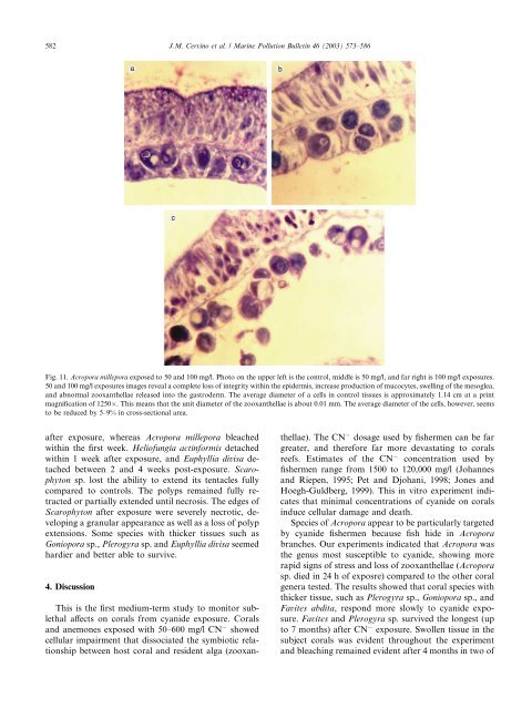
582 J.M. Cerv<strong>in</strong>o et al. / Mar<strong>in</strong>e Pollution Bullet<strong>in</strong> 46 (2003) 573–586<br />
Fig. 11. Acropora millepora exposed to 50 <strong>and</strong> 100 mg/l. Photo on the upper left is the control, middle is 50 mg/l, <strong>and</strong> far right is 100 mg/l exposures.<br />
50 <strong>and</strong> 100 mg/l exposures images reveal a complete loss of <strong>in</strong>tegrity with<strong>in</strong> the epidermis, <strong>in</strong>crease production of mucocytes, swell<strong>in</strong>g of the mesoglea,<br />
<strong>and</strong> abnormal <strong>zooxanthellae</strong> released <strong>in</strong>to the gastroderm. The average diameter of a cells <strong>in</strong> control tissues is approximately 1.14 cm at a pr<strong>in</strong>t<br />
magnification of 1250. This means that the unit diameter of the <strong>zooxanthellae</strong> is about 0.01 mm. The average diameter of the cells, however, seems<br />
to be reduced by 5–9% <strong>in</strong> cross-sectional area.<br />
after exposure, whereas Acropora millepora bleached<br />
with<strong>in</strong> the first week. Heliofungia act<strong>in</strong>formis detached<br />
with<strong>in</strong> 1 week after exposure, <strong>and</strong> Euphyllia divisa detached<br />
between 2 <strong>and</strong> 4 weeks post-exposure. Scarophyton<br />
sp. lost the ability to extend its tentacles fully<br />
compared to controls. The polyps rema<strong>in</strong>ed fully retracted<br />
or partially extended until necrosis. The edges of<br />
Scarophyton after exposure were severely necrotic, develop<strong>in</strong>g<br />
a granular appearance as well as a loss of polyp<br />
extensions. Some species with thicker tissues such as<br />
Goniopora sp., Plerogyra sp. <strong>and</strong> Euphyllia divisa seemed<br />
hardier <strong>and</strong> better able to survive.<br />
4. Discussion<br />
This is the first medium-term study to monitor sublethal<br />
affects on corals from cyanide exposure. Corals<br />
<strong>and</strong> anemones exposed with 50–600 mg/l CN showed<br />
cellular impairment that dissociated the symbiotic relationship<br />
between host coral <strong>and</strong> resident alga (<strong>zooxanthellae</strong>).<br />
The CN dosage used by fishermen can be far<br />
greater, <strong>and</strong> therefore far more devastat<strong>in</strong>g to corals<br />
reefs. Estimates of the CN concentration used by<br />
fishermen range from 1500 to 120,000 mg/l (Johannes<br />
<strong>and</strong> Riepen, 1995; Pet <strong>and</strong> Djohani, 1998; Jones <strong>and</strong><br />
Hoegh-Guldberg, 1999). This <strong>in</strong> vitro experiment <strong>in</strong>dicates<br />
that m<strong>in</strong>imal concentrations of cyanide on corals<br />
<strong>in</strong>duce cellular damage <strong>and</strong> death.<br />
Species of Acropora appear to be particularly targeted<br />
by cyanide fishermen because fish hide <strong>in</strong> Acropora<br />
branches. Our experiments <strong>in</strong>dicated that Acropora was<br />
the genus most susceptible to cyanide, show<strong>in</strong>g more<br />
rapid signs of stress <strong>and</strong> loss of <strong>zooxanthellae</strong> (Acropora<br />
sp. died <strong>in</strong> 24 h of exposre) compared to the other coral<br />
genera tested. The results showed that coral species with<br />
thicker tissue, such as Plerogyra sp., Goniopora sp., <strong>and</strong><br />
Favites abdita, respond more slowly to cyanide exposure.<br />
Favites <strong>and</strong> Plerogyra sp. survived the longest (up<br />
to 7 months) after CN exposure. Swollen tissue <strong>in</strong> the<br />
subject corals was evident throughout the experiment<br />
<strong>and</strong> bleach<strong>in</strong>g rema<strong>in</strong>ed evident after 4 months <strong>in</strong> two of