Changes in zooxanthellae density, morphology, and mitotic index in ...
Changes in zooxanthellae density, morphology, and mitotic index in ...
Changes in zooxanthellae density, morphology, and mitotic index in ...
Create successful ePaper yourself
Turn your PDF publications into a flip-book with our unique Google optimized e-Paper software.
J.M. Cerv<strong>in</strong>o et al. / Mar<strong>in</strong>e Pollution Bullet<strong>in</strong> 46 (2003) 573–586 575<br />
2. Materials <strong>and</strong> methods<br />
Corals were imported to the United States from the<br />
Indo-Pacific region packed <strong>in</strong> fresh seawater <strong>and</strong> kept at<br />
a hold<strong>in</strong>g facility. The samples were held <strong>in</strong> two 750-l<br />
tanks for 4 weeks prior to each of the three experiments.<br />
Dur<strong>in</strong>g the first segment of this experiment, 150 fragments<br />
(5 3 cm) of Acropora millipora were removed<br />
from larger parent colonies. The coral heads were<br />
fragmented 1 week prior to the experiment to allow the<br />
corals to recover from any damage/stress caused by the<br />
fragmentation process. Fragments that bleached <strong>and</strong>/or<br />
died from post-fragmentation damage/stress were not<br />
<strong>in</strong>cluded <strong>in</strong> the experiment.<br />
2.1. Water conditions for observation <strong>and</strong> exposure tanks<br />
Water quality was monitored <strong>and</strong> kept with<strong>in</strong> the<br />
follow<strong>in</strong>g ranges: temperature (28 2 °C, ammonia ¼<br />
0.1 mg/l, nitrite ¼ 0.2 mg/l, nitrate ¼ 3.5–5.0 mg/l, pH<br />
8.1–8.3, sal<strong>in</strong>ity 35 2 g/l, dissolved oxygen 8.0 0.1<br />
mg/l, <strong>and</strong> calcium 450 50 mg/l. Light<strong>in</strong>g was provided<br />
by metal halide lamps (175 W, 6500 K) emitt<strong>in</strong>g approximately<br />
17,000 lux. Filtration was conducted us<strong>in</strong>g<br />
biological/mechanical ‘‘box’’ type filters <strong>in</strong>oculated with<br />
nitrify<strong>in</strong>g bacteria from established aquaria.<br />
2.2. Dos<strong>in</strong>g concentration <strong>and</strong> rate<br />
This <strong>in</strong> vitro lab analysis was to determ<strong>in</strong>e if CN<br />
concentrations of 50, 100, 300, <strong>and</strong> 600 mg/l impaired<br />
normal cell physiology, lead<strong>in</strong>g to the impairment of<br />
coral symbiosis result<strong>in</strong>g <strong>in</strong> death. To prepare cyanide<br />
solutions, NaCN was dissolved <strong>in</strong> seawater. In seawater,<br />
the CN complexes form hydrocyanic acid (HCN) at<br />
pH values less than 8.5 (Leduc, 1984). The HCN molecule<br />
is very toxic to fish because it is rapidly absorbed<br />
across cell membranes (Duodoroff, 1980). Presumably,<br />
HCN is the form of cyanide toxic to corals <strong>in</strong> seawater.<br />
(To simplify, we refer to CN concentrations rather<br />
than HCN concentrations.)<br />
Exposures of the coral fragments to cyanide for 60 or<br />
120 s were conducted <strong>in</strong> 4 l aquaria situated <strong>in</strong>side a<br />
chemical fume hood. A stock solution of NaCN (94.2%<br />
pure) was prepared prior to exposure. Control tanks (4<br />
l) were set up with seawater accord<strong>in</strong>gly. The exposure<br />
tanks were set up by add<strong>in</strong>g 4 l of seawater conta<strong>in</strong><strong>in</strong>g<br />
various cyanide concentrations. Solutions of 600, 300,<br />
100, <strong>and</strong> 50 mg/l CN were created by respectively<br />
add<strong>in</strong>g 4.8, 2.4, 0.8, <strong>and</strong> 0.4 g of NaCN to 4 l of seawater.<br />
The corals were exposed to cyanide for 60 or 120<br />
s by directly dipp<strong>in</strong>g them <strong>in</strong>to cyanide solution, wear<strong>in</strong>g<br />
surgical gloves. Follow<strong>in</strong>g the cyanide dips, all specimens<br />
were lifted out of the exposure tank, r<strong>in</strong>sed <strong>in</strong><br />
seawater for 30 s, <strong>and</strong> immediately placed back <strong>in</strong>to the<br />
cyanide-free hold<strong>in</strong>g tank. Controls <strong>and</strong> cyanideexposed<br />
colonies were used (1 cm length tips) to extract<br />
tissue conta<strong>in</strong><strong>in</strong>g symbiotic algae. Fragments of<br />
Acropora millepora were exposed to 50 mg/l CN for<br />
60 or 120 s <strong>and</strong> observed for changes <strong>in</strong> <strong>zooxanthellae</strong><br />
densities <strong>and</strong> <strong>mitotic</strong> <strong>in</strong>dices <strong>in</strong> host tissue after<br />
24 h. The rema<strong>in</strong><strong>in</strong>g fragments were exam<strong>in</strong>ed after<br />
1 month.<br />
Dur<strong>in</strong>g the f<strong>in</strong>al stages of this experiment, exposed<br />
hard <strong>and</strong> soft whole-coral colonies of Acropora millepora,<br />
Goniopora sp., Favites abdita, Heliofungia act<strong>in</strong>formis,<br />
Euphyllia divisa, Trachyphyllia geoffrio, Plerogyra<br />
sp., Scarophyton sp., <strong>and</strong> the Caribbean sea anemone<br />
Aiptasia pallida. These species were exposed us<strong>in</strong>g the<br />
same methods (although at different doses) used for the<br />
Acropora fragments studied dur<strong>in</strong>g the first round of<br />
experiments. For accuracy, this procedure was conducted<br />
three times per species. The tables represent the<br />
numbers of corals used for each trial. Controls <strong>and</strong> cyanide-exposed<br />
animals were dipped for 120 s <strong>in</strong> solutions<br />
of 100, 300, or 600 mg/l CN (Tables 1 <strong>and</strong> 2).<br />
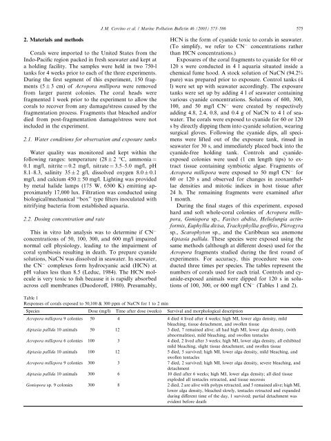
Table 1<br />
Responses of corals exposed to 50,100 & 300 ppm of NaCN for 1 to 2 m<strong>in</strong><br />
Species Dose (mg/l) Time after dose (weeks) Survival <strong>and</strong> morphological description<br />
Acropora millepora 9 colonies 50 4 4 died 4 lived after 4 weeks; high MI, lower alga <strong>density</strong>, mild<br />
bleach<strong>in</strong>g, tissue detachment, <strong>and</strong> swollen tissue<br />
Aiptasia pallida 10 animals 50 12 3 died, 7 rema<strong>in</strong>ed alive; all had high MI, lower alga <strong>density</strong>, (with<br />
abnormalities), mild bleach<strong>in</strong>g, <strong>and</strong> swollen tentacles<br />
Acropora millepora 6 colonies 100 3 4 died, 2 lived after 3 weeks; high MI, lower alga <strong>density</strong>, all exhibited<br />
mild bleach<strong>in</strong>g, slight tissue detachment, <strong>and</strong> swollen tissue<br />
Aiptasia pallida 10 animals 100 12 5 died, 5 survived; high MI, lower alga <strong>density</strong>, mild bleach<strong>in</strong>g, <strong>and</strong><br />
swollen tentacles<br />
Acropora millepora 9 colonies 300 3 7 died, 2 survived; high MI, lower alga <strong>density</strong>, severe bleach<strong>in</strong>g, <strong>and</strong><br />
detachment<br />
Aiptasia pallida 10 animals 300 6 10 died after 6 weeks; high MI, lower alga <strong>density</strong>; all died tissue<br />
exploded all tentacles retracted, <strong>and</strong> tissue necrosis<br />
Goniopora sp. 9 colonies 300 8 2 died, 2 are alive with polyps retracted, <strong>and</strong> 5 rema<strong>in</strong>ed alive; high MI,<br />
lower alga <strong>density</strong>, bleached slowly, tentacles retracted <strong>and</strong> exp<strong>and</strong>ed<br />
dur<strong>in</strong>g different time of the day, 1 survived; partial detachment was<br />
evident before death


