altering vertical dimension in the perio-restorative patient - Kokich ...
altering vertical dimension in the perio-restorative patient - Kokich ...
altering vertical dimension in the perio-restorative patient - Kokich ...

You also want an ePaper? Increase the reach of your titles
YUMPU automatically turns print PDFs into web optimized ePapers that Google loves.
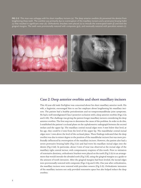
FIG 2-2 This man was unhappy with his short maxillary <strong>in</strong>cisors (a). The deep anterior overbite (b) prevented <strong>the</strong> dentist from<br />
leng<strong>the</strong>n<strong>in</strong>g <strong>the</strong>se teeth. The overbite was primarily due to overeruption of <strong>the</strong> maxillary <strong>in</strong>cisors and a protrusive brux<strong>in</strong>g habit<br />
(c) that resulted <strong>in</strong> significant wear (d). Orthodontic brackets were placed (e) to <strong>in</strong>trude <strong>the</strong> overerupted teeth (f) and level <strong>the</strong><br />
g<strong>in</strong>gival marg<strong>in</strong>s. The teeth were provisionally restored with composite (g,h), and eventually restored with porcela<strong>in</strong> veneers (i).<br />
a<br />
b<br />
Case 2: Deep anterior overbite and short maxillary <strong>in</strong>cisors<br />
This 42-year-old male firefighter was concerned about his short maxillary anterior teeth. His<br />
wife, a hygienist, encouraged him to see her employer about leng<strong>the</strong>n<strong>in</strong>g his maxillary <strong>in</strong>cisors.<br />
The <strong>patient</strong> had a healthy <strong>perio</strong>dontium and no temporomandibular jo<strong>in</strong>t symptoms.<br />
He had a well <strong>in</strong>terdigitated Class I posterior occlusion with a deep anterior overbite (Figs 2-2a<br />
and 2-2b). The challenge was giv<strong>in</strong>g <strong>the</strong> <strong>patient</strong> longer maxillary <strong>in</strong>cisors consider<strong>in</strong>g his deep<br />
anterior overbite. The first step was to determ<strong>in</strong>e <strong>the</strong> cause of <strong>the</strong> problem. In order to do this,<br />
I established <strong>the</strong> <strong>patient</strong>’s occlusal plane on <strong>the</strong> cephalometric radiograph between <strong>the</strong> second<br />
molars and <strong>the</strong> upper lip. The maxillary central <strong>in</strong>cisal edges were 3 mm below that level; at<br />
his age, <strong>the</strong>y could be 2 mm from <strong>the</strong> level of <strong>the</strong> upper lip. The mandibular central <strong>in</strong>cisal<br />
edges were 1 mm above <strong>the</strong> level of <strong>the</strong> occlusal plane. These f<strong>in</strong>d<strong>in</strong>gs <strong>in</strong>dicated that <strong>the</strong> deep<br />
overbite was due <strong>in</strong> m<strong>in</strong>or degree to <strong>the</strong> position of <strong>the</strong> mandibular <strong>in</strong>cisors but was more profoundly<br />
<strong>in</strong>fluenced by overeruption of <strong>the</strong> maxillary <strong>in</strong>cisors. However, <strong>the</strong> <strong>patient</strong> also had a<br />
severe protrusive brux<strong>in</strong>g habit (Fig 2-2c) and had worn his maxillary <strong>in</strong>cisal edges <strong>in</strong>to <strong>the</strong><br />
dent<strong>in</strong> (Fig 2-2d). In particular, about 3 mm of wear was observed at <strong>the</strong> <strong>in</strong>cisal edge of <strong>the</strong><br />
maxillary right central <strong>in</strong>cisor, with compensatory eruption of this tooth. Prior to <strong>in</strong>itiation<br />
of <strong>restorative</strong> dentistry, orthodontic brackets were placed on <strong>the</strong> teeth (Fig 2-2e) <strong>in</strong> an arrangement<br />
that would <strong>in</strong>trude <strong>the</strong> abraded teeth (Fig 2-2f), us<strong>in</strong>g <strong>the</strong> g<strong>in</strong>gival marg<strong>in</strong>s as a guide to<br />
<strong>the</strong> amount of tooth <strong>in</strong>trusion. After <strong>the</strong> g<strong>in</strong>gival marg<strong>in</strong>s had been leveled, <strong>the</strong> <strong>in</strong>cisal edges<br />
were provisionally restored with composite (Figs 2-2g and 2-2h). One year after orthodontics,<br />
<strong>the</strong> maxillary <strong>in</strong>cisors were restored with porcela<strong>in</strong> veneers (Fig 2-2i). Orthodontic <strong>in</strong>trusion<br />
of <strong>the</strong> maxillary <strong>in</strong>cisors not only provided <strong>restorative</strong> space but also helped reduce <strong>the</strong> deep<br />
overbite.<br />
8