BSE - Curriculum Based Assessement Tool for Basic Training in ...
BSE - Curriculum Based Assessement Tool for Basic Training in ...
BSE - Curriculum Based Assessement Tool for Basic Training in ...
You also want an ePaper? Increase the reach of your titles
YUMPU automatically turns print PDFs into web optimized ePapers that Google loves.
British<br />
Society of<br />
Echocardiography<br />
1<br />
Affiliated to the British Cardiovascular Society<br />
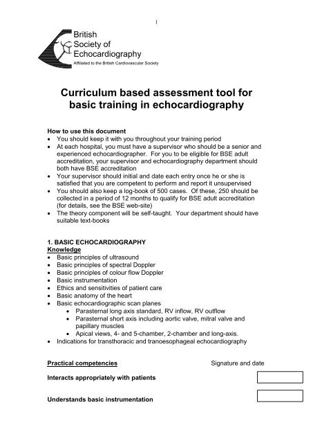
<strong>Curriculum</strong> based assessment tool <strong>for</strong><br />
basic tra<strong>in</strong><strong>in</strong>g <strong>in</strong> echocardiography<br />
How to use this document<br />
You should keep it with you throughout your tra<strong>in</strong><strong>in</strong>g period<br />
At each hospital, you must have a supervisor who should be a senior and<br />
experienced echocardiographer. For you to be eligible <strong>for</strong> <strong>BSE</strong> adult<br />
accreditation, your supervisor and echocardiography department should<br />
both have <strong>BSE</strong> accreditation<br />
Your supervisor should <strong>in</strong>itial and date each entry once he or she is<br />
satisfied that you are competent to per<strong>for</strong>m and report it unsupervised<br />
You should also keep a log-book of 500 cases. Of these, 250 should be<br />
collected <strong>in</strong> a period of 12 months to qualify <strong>for</strong> <strong>BSE</strong> adult accreditation<br />
(<strong>for</strong> details, see the <strong>BSE</strong> web-site)<br />
The theory component will be self-taught. Your department should have<br />
suitable text-books<br />
1. BASIC ECHOCARDIOGRAPHY<br />
Knowledge<br />
<strong>Basic</strong> pr<strong>in</strong>ciples of ultrasound<br />
<strong>Basic</strong> pr<strong>in</strong>ciples of spectral Doppler<br />
<strong>Basic</strong> pr<strong>in</strong>ciples of colour flow Doppler<br />
<strong>Basic</strong> <strong>in</strong>strumentation<br />
Ethics and sensitivities of patient care<br />
<strong>Basic</strong> anatomy of the heart<br />
<strong>Basic</strong> echocardiographic scan planes<br />
Parasternal long axis standard, RV <strong>in</strong>flow, RV outflow<br />
Parasternal short axis <strong>in</strong>clud<strong>in</strong>g aortic valve, mitral valve and<br />
papillary muscles<br />
Apical views, 4- and 5-chamber, 2-chamber and long-axis.<br />
Indications <strong>for</strong> transthoracic and tranoesophageal echocardiography<br />
Practical competencies<br />
Signature and date<br />
Interacts appropriately with patients<br />
Understands basic <strong>in</strong>strumentation
2<br />
Cares <strong>for</strong> mach<strong>in</strong>e appropriately<br />
Can obta<strong>in</strong> standard views<br />
Can obta<strong>in</strong> standard measurements us<strong>in</strong>g 2D or M-mode<br />
Can recognise normal variants<br />
Eustachian valve, chiari net, LV tendon<br />
Can use colour exam<strong>in</strong>ation <strong>in</strong> at least two planes <strong>for</strong> all valves<br />
optimis<strong>in</strong>g ga<strong>in</strong> and box-size<br />
Can obta<strong>in</strong> pulsed Doppler at<br />
a) left ventricular <strong>in</strong>flow (mitral valve)<br />
b) left ventricular outflow tract ( LVOT )<br />
c) right ventricular <strong>in</strong>flow ( tricuspid valve)<br />
d) right ventricular outflow tract, pulmonary valve & ma<strong>in</strong> pulmonary<br />
artery<br />
2. LEFT VENTRICLE<br />
Knowledge<br />
Coronary anatomy and correlation with 2D views of left ventricle.<br />
Segmentation of the left ventricle<br />
Wall motion<br />
Measurements of global systolic function. (LVOT VTI, stroke volume,<br />
fractional shorten<strong>in</strong>g<br />
Doppler mitral valve fill<strong>in</strong>g patterns & normal range<br />
Appearance of complications after myocardial <strong>in</strong>farction<br />
Aneurysm, pseudoaneurysm,<br />
Ventricular septal and papillary muscle rupture<br />
Ischaemic mitral regurgitation<br />
Features of dilated, and hypertrophic cardiomyopathy<br />
Common differential diagnosis<br />
Athletic heart, hypertensive disease<br />
Practical competencies<br />
Can differentiate normal from abnormal LV systolic function
3<br />
Can recognise large wall motion abnormalities<br />
Can describe wall motion abnormalities and myocardial segments<br />
Can obta<strong>in</strong> basic measures of systolic function<br />
VTI, FS, LVEF<br />
Understands & can differentiate diastolic fill<strong>in</strong>g patterns<br />
Can detect and recognise complications after myocardial <strong>in</strong>farction<br />
Understands causes of a hypok<strong>in</strong>etic left ventricle<br />
Can recognise features associated with hypertrophic cardiomyopathy<br />
3. MITRAL VALVE DISEASE<br />
Knowledge<br />
Normal anatomy of the mitral valve, and the subvalvar apparatus and their<br />
relationship with LV function<br />
Causes of mitral stenosis and regurgitation<br />
Ischaemic, functional, prolapse, rheumatic, endocarditis<br />
Practical competencies<br />
Can recognise rheumatic disease<br />
Can recognise mitral prolapse<br />
Can recognise functional mitral regurgitation<br />
Can assess mitral stenosis<br />
2D planimetry, pressure half-time, gradient<br />
Can assess severity of regurgitation,<br />
chamber size, signal density, proximal flow acceleration & vena<br />
contracta,
4<br />
4. AORTIC VALVE DISEASE and AORTA<br />
Knowledge<br />
Causes of aortic valve disease<br />
Causes of aortic disease<br />
Methods of assessment of aortic stenosis and regurgitation<br />
<strong>Basic</strong> criteria <strong>for</strong> surgery to understand reasons <strong>for</strong> mak<strong>in</strong>g measurements<br />
Practical competencies<br />
Can recognise bicuspid, rheumatic, and degenerative disease<br />
Can recognise a significantly stenotic aortic valve<br />
Can derive peak & mean gradients us<strong>in</strong>g cont<strong>in</strong>uous wave Doppler<br />
Can recognise severe aortic regurgitation<br />
Can recognise dilatation of the ascend<strong>in</strong>g aorta<br />
Knows the echocardiographic signs of dissection<br />
5. RIGHT HEART<br />
Knowledge<br />
Causes of tricuspid and pulmonary valve disease<br />
Causes of right ventricular dysfunction<br />
Causes of pulmonary hypertension<br />
The imag<strong>in</strong>g features of pulmonary hypertension<br />
The estimation of pulmonary pressures<br />
Practical competencies<br />
Recognises right ventricular dilatation
5<br />
Can estimate PA systolic pressure<br />
6. REPLACEMENT HEART VALVES<br />
Knowledge<br />
Types of valve replacement<br />
Criteria of normality<br />
Signs of failure<br />
Indications <strong>for</strong> TOE<br />
Practical competencies<br />
Can recognise broad types of replacement valve<br />
Can recognise severe paraprosthetic regurgitation<br />
Can recognise prosthetic obstruction<br />
7. INFECTIVE ENDOCARDITIS<br />
Knowledge<br />
Duke criteria <strong>for</strong> diagnos<strong>in</strong>g endocarditis<br />
Echocardiographic features of endocarditis<br />
Criteria <strong>for</strong> TOE<br />
Practical competencies<br />
Can recognise typical vegetations<br />
Can recognise an abscess<br />
8. INTRACARDIAC MASSES<br />
Knowledge<br />
Types of mass found <strong>in</strong> the heart<br />
Features of a mxyoma
6<br />
<br />
<br />
Differentiation of atrial mass<br />
Normal variants and artifacts<br />
Practical competencies<br />
Can recognise a LA myxoma<br />
9. PERICARDIAL DISEASE<br />
Knowledge<br />
<br />
Features of tamponade<br />
RV collapse, effect on IVC, A-V valve flow velocities<br />
Practical competencies<br />
Can differentiate a pleural and pericardial effusion<br />
Can recognise the features of tamponade<br />
Can judge the route <strong>for</strong> pericardiocentesis<br />
Annex<br />
List of supervisors<br />
Name Date Specimen signature


