Download
Download
Download

You also want an ePaper? Increase the reach of your titles
YUMPU automatically turns print PDFs into web optimized ePapers that Google loves.
March 2004<br />
Nursing Best Practice Guideline<br />
Shaping the future of Nursing<br />
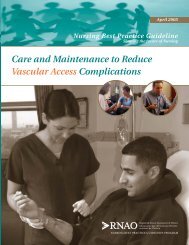
Assessment and Management<br />
of Venous Leg Ulcers
Greetings from Doris Grinspun<br />
Executive Director<br />
Registered Nurses Association of Ontario<br />
It is with great excitement that the Registered Nurses Association of Ontario (RNAO)<br />
disseminates this nursing best practice guideline to you. Evidence-based practice supports<br />
the excellence in service that nurses are committed to deliver in our day-to-day practice.<br />
We offer our endless thanks to the many institutions and individuals that are making<br />
RNAO’s vision for Nursing Best Practice Guidelines (NBPGs) a reality. The Ontario Ministry<br />
of Health and Long-Term Care recognized RNAO’s ability to lead this project and is providing multi-year<br />
funding. Tazim Virani – NBPG project director – with her fearless determination and skills, is moving the<br />
project forward faster and stronger than ever imagined. The nursing community, with its commitment and<br />
passion for excellence in nursing care, is providing the knowledge and countless hours essential to the creation<br />
and evaluation of each guideline. Employers have responded enthusiastically to the request for proposals<br />
(RFP), and are opening their organizations to pilot test the NBPGs.<br />
Now comes the true test in this phenomenal journey: Will nurses utilize the guidelines in their day-to-day practice?<br />
Successful uptake of these NBPGs requires a concerted effort of four groups: nurses themselves, other<br />
healthcare colleagues, nurse educators in academic and practice settings, and employers. After lodging<br />
these guidelines into their minds and hearts, knowledgeable and skillful nurses and nursing students need<br />
healthy and supportive work environments to help bring these guidelines to life.<br />
We ask that you share this NBPG, and others, with members of the interdisciplinary team. There is much to<br />
learn from one another. Together, we can ensure that Ontarians receive the best possible care every time they<br />
come in contact with us. Let’s make them the real winners of this important effort!<br />
RNAO will continue to work hard at developing and evaluating future guidelines. We wish you the<br />
best for a successful implementation!<br />
Doris Grinspun, RN, MScN, PhD (candidate)<br />
Executive Director<br />
Registered Nurses Association of Ontario
Nursing Best Practice Guideline<br />
How to Use this Document<br />
This nursing best practice guideline is a comprehensive document providing<br />
resources necessary for the support of evidence-based nursing practice. The document<br />
needs to be reviewed and applied based on the specific needs of the organization or practice<br />
setting/environment, as well as the needs and wishes of the client. Guidelines should not be<br />
applied in a “cookbook” fashion but used as a tool to assist in decision making for individualized<br />
client care, as well as ensuring that appropriate structures and supports are in place to<br />
provide the best possible care.<br />
Nurses, other healthcare professionals and administrators who are leading and facilitating<br />
practice changes will find this document valuable for the development of policies, procedures,<br />
protocols, educational programs, assessment and documentation tools. It is recommended<br />
that the nursing best practice guidelines be used as a resource tool. It is not necessary, nor<br />
practical that every nurse have a copy of the entire guideline. Nurses providing direct client<br />
care will benefit from reviewing the recommendations, the evidence in support of the<br />
recommendations and the process that was used to develop the guidelines. However, it is<br />
highly recommended that practice settings/environments adapt these guidelines in formats<br />
that would be user-friendly for daily use. This guideline has some suggested formats for such<br />
local adaptation and tailoring.<br />
1<br />
Organizations wishing to use the guideline may decide to do so in a number of ways:<br />
Assess current nursing and healthcare practices using the recommendations in the<br />
guideline.<br />
Identify recommendations that will address identified needs or gaps in services.<br />
Systematically develop a plan to implement the recommendations using associated<br />
tools and resources.<br />
RNAO is interested in hearing how you have implemented this guideline. Please contact<br />
us to share your story. Implementation resources will be made available through the<br />
RNAO website at www.rnao.org/bestpractices to assist individuals and organizations to<br />
implement best practice guidelines.
Assessment and Management of Venous Leg Ulcers<br />
Guideline Development Panel Members<br />
2<br />
Kathryn Kozell, RN, BA, BScN, MScN,<br />
ACNP/CNS, ET<br />
(Co-Team Leader)<br />
GI Surgery Ostomy/Wound<br />
St. Joseph’s Healthcare London<br />
St. Joseph’s Site<br />
London, Ontario<br />
Susan Mills-Zorzes, RN, BScN, CWOCN<br />
(Co-Team Leader)<br />
Enterostomal Therapy Nurse<br />
St. Joseph’s Care Group<br />
Thunder Bay, Ontario<br />
Patti Barton, RN, PHN, ET<br />
Ostomy, Wound and Skin Consultant<br />
Specialty ET Services<br />
Toronto, Ontario<br />
Marion Chipman, RN<br />
ONA Representative<br />
Staff Nurse<br />
Shaver Rehabilitation Hospital<br />
St. Catharines, Ontario<br />
Patricia Coutts, RN<br />
Wound Care & Clinical Trials Coordinator<br />
The Mississauga Dermatology Centre<br />
Office of Dr. R. Gary Sibbald<br />
Mississauga, Ontario<br />
Diane Gregoire, RN, ET, MScN<br />
Spina Bifida Service Coordinator<br />
Coordinatrice des Services de Spina Bifida<br />
Ottawa, Ontario<br />
Margaret Harrison, RN, PhD<br />
Associate Professor<br />
School of Nursing<br />
Queen’s University<br />
Kingston, Ontario<br />
Nurse Scientist<br />
Clinical Epidemiology Program<br />
Ottawa Health Research Institute<br />
Ottawa, Ontario<br />
Terri Labate, RN, CRRN, GNC(C), BScN (candidate)<br />
Staff Nurse<br />
St. Joseph’s Healthcare London<br />
Parkwood Site<br />
London, Ontario<br />
Karen Lorimer, RN, MScN (candidate)<br />
Clinical Leader<br />
Ottawa-Carleton Regional Leg Ulcer Project<br />
Ottawa, Ontario<br />
Sheri Oliver, RPN<br />
Project Coordinator<br />
Nursing Education Initiative<br />
Registered Practical Nurses Association<br />
of Ontario<br />
Mississauga, Ontario<br />
Nancy Parslow, RN, ET<br />
Enterostomal/Wound Care Consultant<br />
Calea<br />
Toronto, Ontario<br />
Josephine Santos, RN, MN<br />
Facilitator, Project Coordinator<br />
Nursing Best Practice Guidelines Project<br />
Registered Nurses Association of Ontario<br />
Toronto, Ontario
Nursing Best Practice Guideline<br />
Assessment & Management<br />
of Venous Leg Ulcers<br />
Project team:<br />
Tazim Virani, RN, MScN<br />
Project Director<br />
Josephine Santos, RN, MN<br />
Project Coordinator<br />
3<br />
Heather McConnell, RN, BScN, MA(Ed)<br />
Project Manager<br />
Jane Schouten, RN, BScN, MBA<br />
Project Coordinator<br />
Stephanie Lappan-Gracon, RN, MN<br />
Coordinator – Best Practice Champions Network<br />
Carrie Scott<br />
Project Assistant<br />
Elaine Gergolas, BA<br />
Project Coordinator – Advanced Clinical/Practice<br />
Fellowships<br />
Melissa Kennedy, BA<br />
Project Assistant<br />
Keith Powell, BA, AIT<br />
Web Editor<br />
Registered Nurses Association of Ontario<br />
Nursing Best Practice Guidelines Project<br />
111 Richmond Street West, Suite 1100<br />
Toronto, Ontario M5H 2G4<br />
www.rnao.org/bestpractices
Assessment and Management of Venous Leg Ulcers<br />
Acknowledgement<br />
Stakeholders representing diverse perspectives were solicited for their feedback<br />
and the Registered Nurses Association of Ontario wishes to acknowledge the<br />
following for their contribution in reviewing this Nursing Best Practice Guideline.<br />
Marlene Allen<br />
Physiotherapist<br />
Oshawa, Ontario<br />
Connie Harris<br />
Enterostomal Therapist/Consultant<br />
Kitchener, Ontario<br />
4<br />
Lucy Cabico<br />
Nurse Practitioner/Clinical Nurse Specialist<br />
Baycrest Centre for Geriatric Care<br />
Toronto, Ontario<br />
Karen Campbell<br />
Nurse Practitioner/Clinical Nurse Specialist<br />
Parkwood Hospital<br />
London, Ontario<br />
Dawn-Marie Clarke<br />
Chiropodist<br />
Shaver Rehabilitation Hospital<br />
St. Catharines, Ontario<br />
Debra Clutterbuck<br />
Registered Practical Nurse<br />
Cambridge, Ontario<br />
Nicole Denis<br />
Enterostomal Therapy Nurse<br />
The Ottawa Hospital<br />
Ottawa, Ontario<br />
Cheri Hernandez<br />
Associate Professor<br />
Faculty of Nursing<br />
University of Windsor<br />
Windsor, Ontario<br />
Dr. Pamela Houghton<br />
Associate Professor<br />
School of Physiotherapy<br />
University of Western Ontario<br />
London, Ontario<br />
Madge Legrace<br />
Registered Nurse<br />
Unionville, Ontario<br />
Dr. Ronald Mahler<br />
Dermatologist<br />
Thunder Bay Medical Centre<br />
Thunder Bay, Ontario<br />
Stephanie McIntosh<br />
Consumer<br />
Elaine Diebold<br />
Enterostomal Therapy Nurse<br />
Durham, Ontario<br />
Marie-Andre Meloche<br />
Victorian Order of Nurses – Peel<br />
Mississauga, Ontario<br />
Geneviève Grégoire<br />
Dietetic Intern<br />
Moncton, New Brunswick<br />
Beverly Monette<br />
Clinical Nurse Consultant<br />
Dell Pharmacy, Home Health Care Centre<br />
Hamilton, Ontario
Nursing Best Practice Guideline<br />
Sue Morrell-DeVries<br />
Nurse Coordinator, Vascular Surgery<br />
Toronto General Hospital<br />
Toronto, Ontario<br />
Dr. Gary Sibbald<br />
Director of Dermatology Day Care and<br />
Wound Healing Clinic<br />
Sunnybrook & Women’s College Health<br />
Sciences Centre<br />
Associate Professor & Director<br />
Continuing Education<br />
Department of Medicine<br />
University of Toronto<br />
Toronto, Ontario<br />
The Mississauga Dermatology Centre<br />
Mississauga, Ontario<br />
Hélène Villeneuve<br />
Dietitian<br />
Sarsfield, Ontario<br />
Claire Westendorp<br />
Enterostomal Therapist<br />
Kingston General Hospital<br />
Kingston, Ontario<br />
Meta Wilson<br />
Consumer<br />
A special acknowledgement also goes<br />
to Barbara Willson, RN, MSc, and<br />
Anne Tait, RN, BScN, who served as<br />
Project Coordinators at the onset of the<br />
guideline development.<br />
5<br />
Jennifer Skelly<br />
Associate Professor<br />
McMaster University<br />
Hamilton, Ontario<br />
Louise Spence<br />
Hamilton-Wentworth Community Care<br />
Access Centre<br />
Hamilton, Ontario<br />
Dr. Terry Trusdale<br />
Varicose & Spider Vein Treatment<br />
Kakabeka Falls, Ontario
Assessment and Management of Venous Leg Ulcers<br />
RNAO also wishes to acknowledge the<br />
following organizations for their role in<br />
pilot testing this guideline:<br />
Pilot Project Sites<br />
Saint Elizabeth Health Care<br />
Toronto, Ontario<br />
St. Peter’s Hospital<br />
Hamilton, Ontario<br />
As well, RNAO sincerely acknowledges the leadership and dedication of the<br />
researchers who have directed the evaluation phase of the Nursing Best Practice<br />
Guidelines Project. The Evaluation Team is comprised of:<br />
6<br />
Principal Investigators:<br />
Nancy Edwards, RN, PhD<br />
Barbara Davies, RN, PhD<br />
University of Ottawa<br />
Evaluation Team:<br />
Maureen Dobbins, RN, PhD<br />
Jenny Ploeg, RN, PhD<br />
Jennifer Skelly, RN, PhD<br />
McMaster University<br />
Patricia Griffin, RN, PhD<br />
University of Ottawa<br />
Project Staff:<br />
University of Ottawa<br />
Barbara Helliwell, BA(Hons); Marilynn Kuhn, MHA; Diana Ehlers, MA(SW), MA(Dem);<br />
Lian Kitts, RN; Elana Ptack, BA; Isabelle St-Pierre, BScN, MScN(cand.)<br />
Contact Information<br />
Registered Nurses Association<br />
of Ontario<br />
Nursing Best Practice Guidelines Project<br />
111 Richmond Street West, Suite 1100<br />
Toronto, Ontario<br />
M5H 2G4<br />
Registered Nurses Association<br />
of Ontario<br />
Head Office<br />
438 University Avenue, Suite 1600<br />
Toronto, Ontario<br />
M5G 2K8
Nursing Best Practice Guideline<br />
Assessment and Management<br />
of Venous Leg Ulcers<br />
Disclaimer<br />
These best practice guidelines are related only to nursing practice and not intended to take into<br />
account fiscal efficiencies. These guidelines are not binding for nurses and their use should be<br />
flexible to accommodate client/family wishes and local circumstances. They neither constitute<br />
a liability or discharge from liability. While every effort has been made to ensure the accuracy<br />
of the contents at the time of publication, neither the authors nor RNAO give any guarantee as<br />
to the accuracy of the information contained in them, nor accept any liability, with respect to<br />
loss, damage, injury or expense arising from any such errors or omissions in the contents of this<br />
work. Any reference throughout the document to specific pharmaceutical products as examples<br />
does not imply endorsement of any of these products.<br />
7<br />
Copyright<br />
With the exception of those portions of this document for which a specific prohibition or<br />
limitation against copying appears, the balance of this document may be produced, reproduced<br />
and published in its entirety, in any form, including in electronic form, for educational or<br />
non-commercial purposes only, without requiring the consent or permission of the Registered<br />
Nurses Association of Ontario, provided that an appropriate credit or citation appears in the<br />
copied work as follows:<br />
Registered Nurses Association of Ontario (2004). Assessment and Management of Venous<br />
Leg Ulcers. Toronto, Canada: Registered Nurses Association of Ontario.
Assessment and Management of Venous Leg Ulcers<br />
table of contents<br />
Summary of Recommendations . . . . . . . . . . . . . . . . . . . . . . . . . . . . . . . . . . . . . . . . . . .10<br />
Interpretation of Evidence . . . . . . . . . . . . . . . . . . . . . . . . . . . . . . . . . . . . . . . . . . . . . . .18<br />
Responsibility for Development . . . . . . . . . . . . . . . . . . . . . . . . . . . . . . . . . . . . . . . . . . .19<br />
8<br />
Purpose and Scope . . . . . . . . . . . . . . . . . . . . . . . . . . . . . . . . . . . . . . . . . . . . . . . . . . . .19<br />
Guideline Development Process . . . . . . . . . . . . . . . . . . . . . . . . . . . . . . . . . . . . . . . . . . .20<br />
Definition of Terms . . . . . . . . . . . . . . . . . . . . . . . . . . . . . . . . . . . . . . . . . . . . . . . . . . . .24<br />
Background Context . . . . . . . . . . . . . . . . . . . . . . . . . . . . . . . . . . . . . . . . . . . . . . . . . . .26<br />
Guiding Principles/Assumptions in Venous Leg Ulcers Care . . . . . . . . . . . . . . . . . . . . . . .27<br />
Interactive Guiding Principles of Venous Leg Ulcers Care . . . . . . . . . . . . . . . . . . . . . . . .28<br />
Practice Recommendations . . . . . . . . . . . . . . . . . . . . . . . . . . . . . . . . . . . . . . . . . . . . . .29<br />
Education Recommendations . . . . . . . . . . . . . . . . . . . . . . . . . . . . . . . . . . . . . . . . . . . . .53<br />
Organization & Policy Recommendations . . . . . . . . . . . . . . . . . . . . . . . . . . . . . . . . . . . .55<br />
Evaluation & Monitoring . . . . . . . . . . . . . . . . . . . . . . . . . . . . . . . . . . . . . . . . . . . . . . . .56<br />
Implementation Tips . . . . . . . . . . . . . . . . . . . . . . . . . . . . . . . . . . . . . . . . . . . . . . . . . . .58<br />
Process for Update/Review of Guideline . . . . . . . . . . . . . . . . . . . . . . . . . . . . . . . . . . . . .60<br />
References . . . . . . . . . . . . . . . . . . . . . . . . . . . . . . . . . . . . . . . . . . . . . . . . . . . . . . . . . .61<br />
Bibliography . . . . . . . . . . . . . . . . . . . . . . . . . . . . . . . . . . . . . . . . . . . . . . . . . . . . . . . . .64
Nursing Best Practice Guideline<br />
Appendix A – Search Strategy for Existing Evidence . . . . . . . . . . . . . . . . . . . . . . . . . . .70<br />
Appendix B – Glossary of Terms . . . . . . . . . . . . . . . . . . . . . . . . . . . . . . . . . . . . . . . . . .74<br />
Appendix C – Different Types of Leg Ulcers and Their Causes . . . . . . . . . . . . . . . . . . . .88<br />
Appendix D – Leg Ulcer Assessment Form . . . . . . . . . . . . . . . . . . . . . . . . . . . . . . . . . .89<br />
Appendix E – Leg Ulcer Measurement Tool . . . . . . . . . . . . . . . . . . . . . . . . . . . . . . . . . .95<br />
Appendix F – Quality of Life Assessment Tool . . . . . . . . . . . . . . . . . . . . . . . . . . . . . . .101<br />
Appendix G – Pain Assessment Tools . . . . . . . . . . . . . . . . . . . . . . . . . . . . . . . . . . . . .103<br />
Appendix H – Cleansing Agents and Their Associated Toxicities . . . . . . . . . . . . . . . . . .106<br />
Appendix I – Potential Allergens . . . . . . . . . . . . . . . . . . . . . . . . . . . . . . . . . . . . . . . .107<br />
Appendix J – Topical Antimicrobial Agents . . . . . . . . . . . . . . . . . . . . . . . . . . . . . . . . .108<br />
Appendix K – Classes of Compression Bandages . . . . . . . . . . . . . . . . . . . . . . . . . . . .110<br />
Appendix L – Description of the Toolkit . . . . . . . . . . . . . . . . . . . . . . . . . . . . . . . . . . .111<br />
9
Assessment and Management of Venous Leg Ulcers<br />
Summary of Recommendations<br />
RECOMMENDATION<br />
*LEVEL OF EVIDENCE<br />
Practice A. ASSESSMENT<br />
Recommendations 1. Assessment and clinical investigations should be undertaken by healthcare C<br />
professional(s) trained and experienced in leg ulcer management.<br />
10<br />
2. A comprehensive clinical history and physical examination including blood C<br />
pressure measurement, weight, urinalysis, blood glucose level and Doppler<br />
measurement of Ankle Brachial Pressure Index (ABPI) should be recorded<br />
for a client presenting with either their first or recurrent leg ulcer and<br />
should be ongoing thereafter.<br />
3. Information relating to ulcer history should be documented in a C<br />
structured format.<br />
4. Examine both legs and record the presence/absence of the following C<br />
to aid in the assessment of underlying etiology.<br />
Venous Disease:<br />
usually shallow moist ulcers<br />
situated on the gaiter area of the leg<br />
edema<br />
eczema<br />
ankle flare<br />
lipodermatosclerosis<br />
varicose veins<br />
hyperpigmentation<br />
atrophie blanche<br />
Arterial Disease:<br />
ulcers with a “punched out” appearance<br />
base of wound poorly perfused, pale, dry<br />
cold legs/feet (in a warm environment)<br />
shiny, taut skin<br />
dependent rubor<br />
pale or blue feet<br />
gangrenous toes<br />
* See page 18 for details regarding Interpretation of Evidence
Nursing Best Practice Guideline<br />
RECOMMENDATION<br />
*LEVEL OF EVIDENCE<br />
Practice 5. Measure the surface areas of ulcers, at regular intervals, to monitor B<br />
Recommendations progress. Maximum length and width, or tracings onto a transparency<br />
are useful methods.<br />
6. The client’s estimate of the quality of life should be included in the initial C<br />
discussion of the treatment plan, throughout the course of treatment,<br />
and when the ulcer has healed.<br />
7. Assess the functional, cognitive and emotional status of the client and C<br />
family to manage self-care.<br />
11<br />
8. Regular ulcer assessment is essential to monitor treatment effectiveness C<br />
and healing goals.<br />
B. DIAGNOSTIC EVALUATION<br />
9. Venous disease of the leg is most commonly detected by a combination A<br />
of clinical examination and measurement of a reliably taken Ankle Brachial<br />
Pressure Index (ABPI).<br />
10. Doppler ultrasound measurement of Ankle Brachial Pressure Index (ABPI) B<br />
should be done by practitioners trained to undertake this measure.<br />
11. If there are no signs of chronic venous insufficiency and the Ankle Brachial C<br />
Pressure Index (ABPI) is abnormal (greater than 1.2 or less than 0.8),<br />
arterial etiology should be assumed and a vascular opinion sought.<br />
12. Vascular assessment, such as Ankle Brachial Pressure Index (ABPI) is C<br />
recommended for ulcers in lower extremities, prior to debridement,<br />
to rule out vascular compromise.<br />
C. PAIN<br />
13. Assess Pain. C<br />
14. Pain may be a feature of both venous and arterial disease, and should B<br />
be addressed.
Assessment and Management of Venous Leg Ulcers<br />
RECOMMENDATION<br />
*LEVEL OF EVIDENCE<br />
Practice 15. Prevent or manage pain associated with debridement. Consult with a C<br />
Recommendations physician and pharmacist as needed.<br />
D. VENOUS ULCER CARE<br />
16. Choose the technique of debridement, considering the type, quantity C<br />
and location of non-viable tissue, the depth of the wound, the amount of<br />
wound fluid and the general condition and goals of the client.<br />
17. Cleansing of the ulcer should be kept simple; warm tap water or saline C<br />
is usually sufficient.<br />
12<br />
18. Dressings must be simple, low adherent, acceptable to the client and A<br />
should be low cost.<br />
19. Avoid products that commonly cause skin sensitivity, such as those C<br />
containing lanolin, phenol alcohol, or topical antibiotics.<br />
20. Choose a type of dressing depending on the amount of exudate and C<br />
the phase of healing.<br />
21. No specific dressing has been demonstrated to encourage ulcer healing. A<br />
22. In contrast to drying out, moist wound conditions allow optimal A<br />
cell migration, proliferation, differentiation and neovascularization.<br />
23. Refer clients with suspected sensitivity reactions to a dermatologist for B<br />
patch testing. Following patch testing, identified allergens must be<br />
avoided, and medical advice on treatment should be sought.<br />
24. Venous surgery followed by graduated compression hosiery is an option C<br />
for consideration in clients with superficial venous insufficiency.<br />
25. Biological wound coverings and growth factor treatments should not be C<br />
applied in cases of wound infection.
Nursing Best Practice Guideline<br />
RECOMMENDATION<br />
*LEVEL OF EVIDENCE<br />
Practice 26. Optimal nutrition facilitates wound healing, maintains immune B<br />
Recommendations competence, and decreases the risk of infection.<br />
E. INFECTION<br />
27. Assess for infection. A<br />
28. An infection is indicated when > 10 5 bacteria/gram tissue is present. B<br />
29. The treatment of infection is managed by debridement, wound cleansing A<br />
and systemic antibiotics.<br />
30. Antibiotics should only be considered if the ulcer is clinically cellulitic C<br />
(presence of some of the following signs and symptoms: pyrexia;<br />
increasing pain; increasing erythema of surrounding skin; purulent exudate;<br />
rapid increase in ulcer size).<br />
13<br />
31. Do not use topical antiseptics to reduce bacteria in wound tissue, B<br />
e.g., povidone iodine, iodophor, sodium hypochlorite, hydrogen peroxide,<br />
or acetic acid.<br />
32. Topical antibiotics and antibacterial agents are frequent sensitizers and B<br />
should be avoided.<br />
F. COMPRESSION<br />
33. The treatment of choice for clinical venous ulceration uncomplicated A<br />
by other factors, is graduated compression bandaging, properly applied,<br />
and combined with exercise. Graduated compression is the main<br />
treatment for venous eczema.<br />
34. High compression increases venous ulcer healing and is more effective A<br />
than low compression, but should only be used where ABPI ≥ 0.8 and<br />
ulcer is clinically venous.<br />
35. Compression bandages should only be applied by a suitably trained and B<br />
experienced practitioner.
Assessment and Management of Venous Leg Ulcers<br />
RECOMMENDATION<br />
*LEVEL OF EVIDENCE<br />
Practice 36. Venous ulceration should be treated with high compression bandaging C<br />
Recommendations to achieve a pressure between 35-40 mm Hg. at the ankle, graduating<br />
to half at calf in the normally shaped limb, as per La Place’s Law.<br />
37. Use protective padding over bony prominences when applying C<br />
high compression.<br />
38. Arterial insufficiency is a contraindication to the use of high compression. C<br />
A modified form of compression may be used under specialist supervision.<br />
14<br />
39. Use compression with caution in clients with diabetes, those with C<br />
connective tissue disease and the elderly.<br />
40. Compression therapy should be modified until clinical infection is treated. C<br />
41. Bandages should be applied according to manufacturer’s recommendations. C<br />
42. When using elastic systems such as “high compression” bandages, the C<br />
ankle circumference must be more than or padded to equal 18 cms.<br />
43. Ankle circumference should be measured at a distance of 2.5 cm (one inch) C<br />
above the medial malleolus.<br />
44. The concepts, practice, and hazards of graduated compression should A<br />
be fully understood by those prescribing and fitting compression stockings.<br />
45. Graduated compression hosiery should be measured and fitted by a C<br />
certified fitter.<br />
46. To maintain a therapeutic level of compression, stockings should be C<br />
cared for as per manufacturer’s instructions, and replaced every six months.<br />
47. Graduated compression hosiery should be prescribed for life. B<br />
48. External compression applied using various forms of pneumatic compression A<br />
pumps is indicated for individuals with chronic venous insufficiency.
Nursing Best Practice Guideline<br />
RECOMMENDATION<br />
*LEVEL OF EVIDENCE<br />
Practice 49. The client should be prescribed regular vascular exercise by means A<br />
Recommendations of intensive controlled walking and exercises to improve the function of<br />
the upper ankle joint and calf muscle pump.<br />
G. COMPLEMENTARY THERAPIES<br />
50. Consider electrical stimulation in the treatment of venous leg ulcers. B<br />
51. Hyperbaric oxygen may reduce ulcer size in non-diabetic, A<br />
non-atherosclerotic leg ulcers.<br />
52. Therapeutic ultrasound may be used to reduce the size of chronic A<br />
venous ulcers.<br />
15<br />
H. REASSESSMENT<br />
53. With no evidence of healing, a comprehensive assessment should be carried C<br />
out at three-month intervals, or sooner if clinical condition deteriorates.<br />
54. For resolving and healing venous leg ulcers, routine assessment at six-month C<br />
intervals should include: physical assessment; Ankle Brachial Pressure Index<br />
(ABPI); replacement of compression stockings; and reinforcement of teaching.<br />
I. SECONDARY PREVENTION<br />
55. Measures to prevent recurrence of a venous leg ulcer include: wearing C<br />
compression stockings, regular follow-up to monitor Ankle Brachial<br />
Pressure Index (ABPI), discouragement of self-treatment with over-the-counter<br />
preparations, and avoidance of accidents or trauma to legs.<br />
56. Inform the client after the ulcer has healed regarding: wearing and C<br />
maintenance of compression stockings; elevation of affected limb above<br />
level of heart when at rest; early referral at first sign of skin breakdown or<br />
trauma to limb; need for exercise and ankle-joint mobility; appropriate skin<br />
care; avoidance of products likely to be sensitizers; and life-long use<br />
of compression.
Assessment and Management of Venous Leg Ulcers<br />
RECOMMENDATION<br />
*LEVEL OF EVIDENCE<br />
Education 57. Guidelines are more likely to be effective if they take into account local C<br />
Recommendations circumstances and are disseminated by an ongoing education and<br />
training program.<br />
58. Develop educational programs that target appropriate healthcare providers, C<br />
clients, family members, and caregivers. Develop programs that maximize<br />
retention, ensure carryover into practice, and support lifestyle changes.<br />
Present information at an appropriate level for the target audience using<br />
principles of adult learning.<br />
16<br />
59. Design, develop, and implement educational programs that reflect a C<br />
continuum of care. The program should begin with a structured,<br />
comprehensive, and organized approach to prevention and should<br />
culminate in effective treatment protocols that promote healing as well<br />
as prevent recurrence.<br />
60. All healthcare professionals should be trained in leg ulcer assessment C<br />
and management.<br />
61. Education programs for healthcare professionals should include: C<br />
pathophysiology of leg ulceration<br />
leg ulcer assessment<br />
need for Doppler ultrasound to measure Ankle Brachial Pressure Index (ABPI)<br />
normal and abnormal wound healing<br />
compression therapy theory, management, and application<br />
dressing selection<br />
principles of debridement<br />
principles of cleansing and infection control<br />
skin care of the lower leg<br />
peri-wound skin care and management<br />
psychological impact of venous stasis disease<br />
quality of life<br />
pain management<br />
teaching and support for care provider<br />
health education<br />
preventing recurrence<br />
principles of nutritional support with regard to tissue integrity<br />
mechanisms for accurate documentation and monitoring of pertinent<br />
data, including treatment interventions and healing progress<br />
criteria for referral for specialized assesment
Nursing Best Practice Guideline<br />
RECOMMENDATION<br />
*LEVEL OF EVIDENCE<br />
Education 62. Healthcare professionals with recognized training in leg ulcer care should C<br />
Recommendations cascade their knowledge and skills to local healthcare teams.<br />
63. The knowledge and understanding of the healthcare professional is a C<br />
major factor in adherence to treatment regimens.<br />
Organization 64. Successful implementation of a venous ulcer treatment C<br />
& Policy<br />
policy/strategy requires:<br />
Recommendations dedicated funding<br />
integration of healthcare services<br />
support from all levels of government<br />
management support<br />
human resources<br />
financial resources<br />
functional space<br />
commitment<br />
collection of baseline information about vulnerable populations<br />
resources and existing knowledge<br />
interpretation of above data and identification of<br />
organizational problems<br />
17<br />
65. Nursing best practice guidelines can be successfully implemented only C<br />
where there are adequate planning, resources, organizational and<br />
administrative support, as well as appropriate facilitation. Organizations<br />
may wish to develop a plan for implementation that includes:<br />
An assessment of organizational readiness and barriers to education.<br />
Involvement of all members (whether in a direct or indirect supportive<br />
function) who will contribute to the implementation process.<br />
Dedication of a qualified individual to provide the support needed for<br />
the education and implementation process.<br />
Ongoing opportunities for discussion and education to reinforce the<br />
importance of best practices.<br />
Opportunities for reflection on personal and organizational experience<br />
in implementing guidelines.<br />
In this regard, RNAO (through a panel of nurses, researchers and<br />
administrators) has developed the Toolkit: Implementation of Clinical<br />
Practice Guidelines, based on available evidence, theoretical perspectives<br />
and consensus. The RNAO strongly recommends the use of this Toolkit for<br />
guiding the implementation of the best practice guideline on Assessment<br />
and Management of Venous Leg Ulcers.
Assessment and Management of Venous Leg Ulcers<br />
Interpretation of Evidence<br />
This RNAO guideline is a synthesis of a number of source guidelines. In order to fully<br />
inform the reader, every effort has been made to maintain the original level of evidence<br />
cited in the source document. No alterations have been made to the wording of the<br />
source documents involving recommendations based on randomized controlled trials or<br />
research studies. Where a source document has demonstrated an “expert opinion” level<br />
of evidence, wording may have been altered and the notation of RNAO Consensus Panel<br />
2004 has been added.<br />
18<br />
In the guidelines reviewed, the panel assigned each recommendation a rating of A, B, or<br />
C level of evidence (LOE), to indicate the strength of the evidence supporting the<br />
recommendation. It is important to clarify that these ratings represent the strength of<br />
the supporting research evidence to date.<br />
LEVEL OF EVIDENCE A: Evidence obtained from at least one randomized controlled trial or<br />
meta-analysis of randomized controlled trials.<br />
LEVEL OF EVIDENCE B: Evidence from well designed clinical studies but no randomized<br />
controlled trials.<br />
LEVEL OF EVIDENCE C: Evidence from expert committee reports or opinion and/or clinical<br />
experience or respected authorities. Indicates absence of directly applicable studies of<br />
good quality.
Nursing Best Practice Guideline<br />
Responsibility for Development<br />
The Registered Nurses Association of Ontario, with funding from the Ontario Ministry of<br />
Health and Long-Term Care, has embarked on a multi-year project of nursing best practice<br />
guideline development, pilot implementation, evaluation and dissemination. Assessment<br />
and management of venous leg ulcers is one of six nursing best practice guidelines developed<br />
in the third cycle of the project. The RNAO convened a panel to develop this guideline,<br />
conducting its work independent of any bias or influence from the Ministry of Health and<br />
Long-Term Care.<br />
Purpose and Scope<br />
19<br />
The purpose of this guideline is to:<br />
improve outcomes for venous leg ulcer clients;<br />
assist practitioners to apply the best available research evidence to clinical decisions;<br />
and<br />
promote the responsible use of healthcare resources.<br />
Best practice guidelines are systematically developed statements to assist practitioners and<br />
clients’ decisions about appropriate healthcare (Field & Lohr, 1990; McKibbon, Eady & Marks, 1999).<br />
This best practice guideline is intended to provide direction to practicing nurses in all care<br />
settings, both institutional and community, in the assessment and management of venous<br />
leg ulcers, including prevention of recurrence wherever possible.<br />
The guideline focuses on:<br />
1. Practice Recommendations: directed at the nurse to guide practice regarding assessment,<br />
planning and interventions.<br />
2. Education Recommendations: directed at educational institutions and organizations in<br />
which nurses work to support its implementation.<br />
3. Organization and Policy Recommendations: directed at practice settings and environment<br />
to facilitate nurses’ practice.<br />
4. Evaluation and monitoring indicators.
Assessment and Management of Venous Leg Ulcers<br />
This nursing best practice guideline contains recommendations for Registered Nurses (RNs)<br />
and Registered Practical Nurses (RPNs). Although these guidelines are written for the nurse,<br />
venous leg ulcer care is an interdisciplinary endeavour. Many settings have formalized<br />
interdisciplinary teams and the panel strongly supports this structure. Collaborative assessment<br />
and treatment planning with the client is essential. The recommendations made are not<br />
binding for nurses and should accommodate client/family wishes and local circumstances.<br />
20<br />
It is the intention of this guideline to identify best nursing practices in the treatment of<br />
venous leg ulcers. It is acknowledged that the individual competency of nurses varies<br />
between nurses and across categories of nursing professionals (RNs and RPNs) and is based<br />
on the knowledge, skills, attitudes and judgment enhanced over time by experience<br />
and education.<br />
It is expected that individual nurses will perform only those aspects of venous leg ulcer<br />
assessment and management for which they have appropriate education and experience.<br />
Further, it is expected that nurses, both RNs and RPNs, will seek consultation in instances<br />
where the client’s care needs surpass the individual nurse’s ability to act independently. It is<br />
acknowledged that effective client care depends on a coordinated interdisciplinary approach<br />
incorporating ongoing communication between health professionals and clients, ever<br />
mindful of the personal preferences and unique needs of each individual client.<br />
Guideline Development Process<br />
In February of 2001, a panel of nurses with expertise in the practice and research related to<br />
venous leg ulcers, from community and academic settings, was convened under the auspices<br />
of the RNAO. At the onset the panel discussed and came to consensus on the scope of the<br />
best practice guideline.<br />
A search of the literature for systematic reviews, clinical practice guidelines, relevant articles<br />
and websites was conducted. See Appendix A for a detailed outline of the search strategy<br />
employed.
Nursing Best Practice Guideline<br />
The panel identified a total of eleven clinical practice guidelines related to venous leg<br />
ulcers. An initial screening was conducted with the following criteria:<br />
Guideline was in English.<br />
Guideline was dated no earlier than 1998 as significant changes in venous leg ulcer<br />
management occurred in that year.<br />
Guideline was strictly about the topic area.<br />
Guideline was evidence-based (e.g., contained references, description of evidence,<br />
sources of evidence).<br />
Complete guideline was available and accessible for retrieval.<br />
Eight guidelines were short-listed for critical appraisal using the “Appraisal Instrument for<br />
Clinical Practice Guidelines” (Cluzeau et al., 1997). This appraisal tool allowed for evaluation<br />
in three key dimensions: rigour, content and context, and application.<br />
21<br />
The panel, following the appraisal process, identified the following guidelines, and related<br />
updates, to adapt and modify recommendations:<br />
Clement, D. L. (1999). Venous ulcer reappraisal: Insights from an international task force.<br />
Journal of Vascular Research, 36(Suppl.1), 42-47.<br />
Clinical Resource Efficiency Support Team (CREST) (1998a). Guidelines for the assessment<br />
and management of leg ulceration. CREST, Belfast, Northern Ireland [On-line].<br />
Available: http://www.ni-nhs.uk/crest/index.htm<br />
Compliance Network Physicians/Health Force Initiative, Inc. (1999). Guideline for the outpatient<br />
treatment – venous and venous-arterial mixed leg ulcer. Compliance Network<br />
Physicians/Health Force Initiative, Inc., Berlin, Germany [On-line]. Available:<br />
http://www.cnhfi.de/index-engl.html<br />
Kunimoto, B., Cooling, M., Gulliver, W., Houghton, P., Orsted, H., & Sibbald, R. G. (2001).<br />
Best practices for the prevention and treatment of venous leg ulcers. Ostomy/Wound<br />
Management, 47(2), 34-50.<br />
New Zealand Guidelines Group (NZGG) (1999). Care of people with chronic leg ulcers:<br />
An evidence based guideline. New Zealand Guidelines Group [On-line]. Available:<br />
http://www.nzgg.org.nz/library.cfm
Assessment and Management of Venous Leg Ulcers<br />
Ottawa-Carleton Community Care Access Centre Leg Ulcer Care Protocol Task Force (2000).<br />
Ottawa-Carleton Community Care Access Centre (CCAC) venous leg ulcer care protocol:<br />
Development, methods, and clinical recommendations. Ottawa, Ontario: Ottawa-Carleton<br />
CCAC Leg Ulcer Protocol Task Force.<br />
Royal College of Nursing (RCN) (1998). Clinical practice guideline: The management of<br />
patients with venous leg ulcers. RCN Institute, Centre for Evidence-Based Nursing, University<br />
of York and the School of Nursing, Midwifery and Health Visiting, University of Manchester<br />
[On-line]. Available: http://www.rcn.org.uk<br />
22<br />
Scottish Intercollegiate Guidelines Network (SIGN) (1998). The care of patients with chronic<br />
leg ulcers: A national clinical guideline. Scottish Intercollegiate Guidelines Network<br />
[On-line]. Available: http://www.show.scot.nhs.u.k/sign/home.htm<br />
The Ottawa-Carleton Community Care Access Centre Venous Leg Ulcer Care Protocol (2000)<br />
is a synthesis guideline that was based on all of the above noted guidelines with the exception<br />
of the Care of People with Chronic Leg Ulcers: An Evidence Based Guideline which was<br />
developed by the New Zealand Guidelines Group (1999).<br />
A critique of systematic review articles and pertinent literature was conducted to update the<br />
existing guidelines. Through a process of evidence gathering, synthesis and consensus, a<br />
draft set of recommendations was established. This draft document was submitted to a set<br />
of external stakeholders for review and feedback – an acknowledgement of these reviewers<br />
is provided at the front of this document. Stakeholders represented various healthcare<br />
professional groups, clients and families, as well as professional associations. External<br />
stakeholders were provided with specific questions for comment, as well as the opportunity<br />
to give overall feedback and general impressions. The results were compiled and reviewed by<br />
the development panel – discussions and consensus resulted in revisions to the draft document<br />
prior to pilot testing.
Nursing Best Practice Guideline<br />
A pilot implementation practice setting was identified through a “Request for Proposal” (RFP)<br />
process. Practice settings in Ontario were asked to submit a proposal if they were interested in<br />
pilot testing the recommendations of the guideline. These proposals were then subjected to a<br />
review process, from which a successful practice setting was identified. A nine-month pilot<br />
implementation was undertaken to test and evaluate the recommendations. The evaluation<br />
took place in a chronic care hospital and community care organization in Southern Ontario.<br />
An acknowledgement of these organizations is included at the front of this document. The<br />
development panel reconvened after the pilot implementation in order to review the experiences<br />
of the pilot site, consider the evaluation results, and review any new literature published since<br />
the initial development phase. All these sources of information were used to update/revise<br />
the document prior to publication.<br />
23
Assessment and Management of Venous Leg Ulcers<br />
Definition of Terms<br />
An additional Glossary of Terms related to clinical aspects of the document is located in<br />
Appendix B.<br />
Clinical Practice Guidelines or Best Practice Guidelines: Systematically<br />
developed statements (based on best available evidence) to assist practitioner and client<br />
decisions about appropriate healthcare for specific clinical (practice) circumstances<br />
(Field & Lohr, 1990).<br />
24<br />
Consensus: A process for making policy decisions, not a scientific method for creating<br />
new knowledge. At its best, consensus development merely makes the best use of available<br />
information, be that of scientific data or the collective wisdom of the participants (Black et al., 1999).<br />
Education Recommendations: Statements of educational requirements and<br />
educational approaches/strategies for the introduction, implementation and sustainability<br />
of the best practice guideline.<br />
Evidence: “An observation, fact or organized body of information offered to support or<br />
justify inferences or beliefs in the demonstration of some proposition or matter at issue”<br />
(Madjar & Walton, 2001, p.28).<br />
Meta-analysis: The use of statistical methods to summarize the results of independent<br />
studies, thus providing more precise estimates of the effects of healthcare than those derived<br />
from the individual studies included in a review (Clarke & Oxman, 1999).
Nursing Best Practice Guideline<br />
Organization & Policy Recommendations: Statements of conditions required for<br />
a practice setting that enable the successful implementation of the best practice guideline.<br />
The conditions for success are largely the responsibility of the organization, although they<br />
may have implications for policy at a broader government or societal level.<br />
Practice Recommendations: Statements of best practice directed at the practice of<br />
healthcare professionals that are ideally evidence-based.<br />
25<br />
Randomized Controlled Trial: For the purposes of this guideline, a study in which<br />
subjects are assigned to conditions on the basis of chance, and where at least one of the<br />
conditions is a control or comparison condition.<br />
Stakeholder: A stakeholder is an individual, group or organization with a vested interest<br />
in the decisions and actions of organizations who may attempt to influence decisions and<br />
actions (Baker et al., 1999). Stakeholders include all individuals or groups who will be directly or<br />
indirectly affected by the change or solution to the problem. Stakeholders can be of various<br />
types, and can be divided into opponents, supporters, and neutrals (Ontario Public Health<br />
Association, 1996).<br />
Systematic Review: Application of a rigorous scientific approach to the preparation of<br />
a review article (National Health and Medical Research Council, 1998). Systematic reviews establish<br />
where the effects of healthcare are consistent and research results can be applied across<br />
populations, settings, and differences in treatment (e.g., dose); and where effects may vary<br />
significantly. The use of explicit, systematic methods in reviews limits bias (systematic errors)<br />
and reduces chance effects, thus providing more reliable results upon which to draw conclusions<br />
and make decisions (Clarke & Oxman, 1999).
Assessment and Management of Venous Leg Ulcers<br />
Background Context<br />
Leg ulcer disease is typically cyclical and chronic, with periods of healing followed by recurrence.<br />
It is not uncommon for leg ulcers to persist for years, with recurrence rates as high as 76 percent<br />
within one year (Nelzen, Bergquist & Lindhagen, 1995). Leg ulcers are a major cause of morbidity,<br />
suffering and high health service costs. The negative impact on the sufferer’s quality of life is<br />
significant, as individuals may experience mobility loss, chronic pain, fear, anger, depression,<br />
and social isolation (Phillips, Stanton, Provan & Lew, 1994; Pieper, Szczepaniak & Templin, 2000; Price & Harding, 1996).<br />
26<br />
International studies on leg ulcer prevalence from all etiologies have demonstrated rates of<br />
between 1 and 6 per 1, 000 population in Western countries (Baker, Stacy, Jopp-McKay & Thompson,<br />
1991; Callam, Ruckley, Harper & Dale, 1985; Cornwall, Dore & Lewis, 1986; Nelzen et al., 1995). A one-month<br />
prevalence study in one large Canadian region found a prevalence rate of 1.8 per 1,000 for<br />
the population over the age of 25 (Harrison, Graham, Friedberg, Lorimer & Vandervelde-Coke, 2001). The<br />
care of this population is compounded by the fact that the condition is highly associated with<br />
age, with the prevalence rate reported in the 2 percent range for those over age 65 (Callam et<br />
al., 1985; Cornwall et al., 1986). Reports on the percentage of lower limb ulcerations that result<br />
predominantly from a venous etiology range from 37 to 62 percent (Baker et al., 1991; Callam et<br />
al., 1985; Cornwall et al., 1986; Nelzen, Bergquist, Lindhagen & Halbrook, 1991; Nelzen et al., 1995). Some<br />
studies found venous leg ulcers had a longer duration and a higher recurrence rate than those<br />
of a non-venous etiology (Baker et al., 1991; Nelzen et al., 1995).<br />
Surveys have shown wide variation in the clinical management of leg ulcers. Numerous types<br />
of wound dressings, bandages and stocking are used in the treatment and prevention of<br />
recurrence (Lees & Lambert, 1992; Stevens, Franks & Harrington, 1997). In leg ulcer care, using treatments<br />
with known efficacy leads to improvements in both healing rates and quality of life for the leg ulcer<br />
sufferer (Cullum, Nelson, Fletcher & Sheldon, 2000; Franks et al., 1995a). Despite the evidence supporting<br />
effective leg ulcer management, many clients are not receiving this care (Harrison et al., 2001; Hickie,<br />
Ross & Bond, 1998).<br />
The cost of caring for individuals with leg ulcers is significant. Reports from the United<br />
Kingdom and France indicate that the cost of venous diseases of the leg accounts for 2 percent<br />
of their total national health budgets (Laing, 1992). One study in the UK estimated that district<br />
nurses spend as much as 30 to 50 percent of their time with clients in leg ulcer care (Lees &<br />
Lambert, 1992). Over 80 percent of the ongoing management of chronic wounds such as leg<br />
ulcers occurs mainly in the community (Callam et al., 1985; Lees & Lambert, 1992; Lindholm, Bjellerup,<br />
Christensen & Zederfeldt, 1992). As the prevalence of leg ulcers increases with age, the swell in the<br />
elderly population with the advance of the “boomer” generation, and an anticipated increment<br />
in longevity will result in higher resource demand for community leg ulcer care.
Nursing Best Practice Guideline<br />
Guiding Principles/Assumptions in<br />
Venous Leg Ulcers Care<br />
1. Venous leg ulcers can significantly compromise quality of life.<br />
2. Interdisciplinary, collaborative assessment, and treatment planning with the client<br />
is essential.<br />
3 Early prevention strategies decrease the potential for ulcer development.<br />
4. Therapy involves client acceptance and participation.<br />
27<br />
5. Clinicians must be knowledgeable of the features and management of venous disease.<br />
6. Venous leg ulcers are managed with effective compression and wound management therapy.<br />
7. An Ankle Brachial Pressure Index (ABPI) measurement must be done prior to commencement<br />
of compression therapy.<br />
8. Clinicians must have sound practical knowledge and experience in the use of Ankle<br />
Brachial Pressure Index (ABPI).<br />
9. Clinicians must have sound practical knowledge and expertise in the use of therapeutic<br />
compression.<br />
10.Maintaining therapeutic measures reduces the risk of re-occurrence.<br />
11.Proactive care supports rehabilitation and return of client independence.
Assessment and Management of Venous Leg Ulcers<br />
Interactive Guiding Principles of<br />
Venous Leg Ulcers Care<br />
A graphic depiction of the previously listed guiding principle statements can be visualized in<br />
the following diagram:<br />
Early<br />
Prevention<br />
28<br />
Proactive<br />
Care<br />
Interdisciplinary<br />
Collaboration<br />
Maintenance of<br />
therapeutic<br />
measures<br />
Client with<br />
Venous Leg Ulcers<br />
Client<br />
acceptance and<br />
participation<br />
Quality of Life<br />
Therapeutic<br />
Compression<br />
Knowledgeable<br />
Clinicians<br />
ABPI prior to<br />
compression
Nursing Best Practice Guideline<br />
Practice Recommendations<br />
A. ASSESSMENT OF VENOUS LEG ULCERS<br />
Recommendation • 1<br />
Assessment and clinical investigations should be undertaken by healthcare professional(s)<br />
trained and experienced in leg ulcer management.<br />
(Level of Evidence = C – RNAO Consensus Panel, 2004)<br />
A complete client assessment precedes evaluation of the limb and ulcer characteristics. A<br />
comprehensive assessment is essential to determine the underlying ulcer etiology and<br />
appropriate treatment approaches.<br />
29<br />
Discussion of Evidence:<br />
Although little guidance is given, the literature strongly supports the importance of assessment<br />
and clinical investigations for venous leg ulcers. Recognizing significant arterial insufficiency<br />
is important, as no healing will occur in the presence of severe occlusive arterial disease of the<br />
affected limb. Kunimoto et al. (2001) caution that the high levels of compression necessary to<br />
correct venous hypertension will be potentially dangerous in this situation. Keast & Orsted<br />
(1998) add that a chronic wound should prompt a search for underlying causes.<br />
According to Zink, Rousseau & Holloway (2000), twenty-one percent of individuals with venous<br />
ulcers experience concomitant arterial disease, with the risk of co-existing arterial dysfunction<br />
increasing with age, which again supports the importance of a thorough assessment.<br />
Research repeatedly confirms the necessity of trained healthcare professionals in leg ulcer<br />
management. Surveys of reported practice by nurses demonstrate that knowledge of nurses<br />
in wound care often falls short of what is ideal (RCN, 1998). Providers of healthcare recognize<br />
that the mismanagement of wounds is both costly and unnecessary. Kerstein, van Rijswijk &<br />
Betiz (1998), among others, maintain that providing optimal cost-effective wound care<br />
requires extensive skills, as well as knowledge, and that classroom teaching alone will not<br />
meet the needs of our aging population.<br />
While findings as to what constitutes adequate training levels for nurses involved in leg ulcer<br />
care are inconclusive, the essential point is that the person conducting the assessment must
Assessment and Management of Venous Leg Ulcers<br />
be trained and experienced. The RNAO guideline development panel found no trials assessing<br />
and comparing reliability and accuracy based on levels of training.<br />
Recommendation • 2<br />
A comprehensive clinical history and physical examination including blood pressure<br />
measurement, weight, urinalysis, blood glucose level and Doppler measurement of Ankle<br />
Brachial Pressure Index (ABPI) should be recorded for a client presenting with either their<br />
first or recurrent leg ulcer and should be ongoing thereafter.<br />
(Level of Evidence = C – RNAO Consensus Panel, 2004)<br />
30<br />
An assessment for a history of venous insufficiency also includes:<br />
Family history of venous disease.<br />
Client history of deep vein thrombosis (DVT).<br />
Lower leg fracture or other major leg injury, previous vein surgery, varicose veins, or prior<br />
history of ulceration with/without use of compression stockings.<br />
History of episodes of chest pain, hemoptysis, or history of a pulmonary embolus.<br />
Lifestyle factors (e.g., sedentary lifestyle, chair-bound), obesity, poor nutrition.<br />
An assessment for signs indicative of Non-Venous Disease also includes:<br />
Family history of non-venous etiology.<br />
Heart disease, stroke, transient ischemic attack.<br />
Diabetes mellitus.<br />
Peripheral vascular disease (PVD)/intermittent claudication.<br />
Smoking.<br />
Rheumatoid arthritis.<br />
Ischemic rest pain.<br />
A combination of the features described above may be indicative of mixed arterial/venous<br />
disease (RCN, 1998).<br />
Discussion of Evidence:<br />
Several clinical studies show strong support for the need for thorough history taking for<br />
assessment of venous insufficiency (NZGG, 1999; RCN, 1998). The New Zealand Guidelines<br />
Group (1999) further suggests assessing the history of the ulcer, the mechanism of injury, and<br />
previous methods of treatment.
Nursing Best Practice Guideline<br />
Zink et al. (2000) recommend a guided interview to obtain the history most pertinent to the<br />
cause of the ulcer, explaining that while the client may be able to relate important symptoms,<br />
living with a chronic disease often blunts the ability to be discriminatory. Zink et al. (2000)<br />
further adds that the initial encounter with the client is critical to establishing a positive,<br />
therapeutic relationship. Establishing trust is instrumental for successful client outcomes,<br />
particularly as venous leg ulcers often involve a lengthy healing time.<br />
Misdiagnosis of ulcers can cause harm or lead to long periods of inappropriate treatment. It<br />
is therefore important to have an accurate diagnosis of ulcer etiology (NZGG, 1999). Despite this,<br />
there is only one population study that has systematically investigated and published data<br />
on the etiology of identified ulcers.<br />
31<br />
Recommendation • 3<br />
Information relating to ulcer history should be documented in a structured format.<br />
(Level of Evidence = C – RNAO Consensus Panel, 2004)<br />
An ulcer history should include:<br />
The year first ulcer occurred.<br />
Site of ulcer and of any previous ulcers.<br />
Number of previous episodes of ulceration.<br />
Length of time taken to heal in previous episodes.<br />
Length of time with no recurrence of ulcers.<br />
Past treatment methods (both successful and unsuccessful).<br />
Previous operations on venous system.<br />
Previous and current use of compression hosiery.<br />
Discussion of Evidence:<br />
Although no specific evidence was cited, the Royal College of Nursing (1998) supports the<br />
theory that collection of data in a structured format will enable consideration of clinical factors<br />
that may have an impact on treatment and healing progress, as well as provide baseline<br />
information on ulcer history. They cautioned, however, that a diagnosis of ulcer type should<br />
not be made solely on this information.
Assessment and Management of Venous Leg Ulcers<br />
The literature also stresses the importance of clear and comprehensive documentation of<br />
information during history taking, and suggests several examples of leg ulcer assessment<br />
forms. The RNAO guideline development panel does not consider one assessment form to<br />
be superior to another. (For examples of leg ulcer assessment forms, see Appendices D and E).<br />
Recommendation • 4<br />
Examine both legs and record the presence/absence of the following to aid in the<br />
assessment of underlying etiology.<br />
32<br />
Venous Disease<br />
usually shallow moist ulcers<br />
situated on the gaiter area of the leg<br />
edema<br />
eczema<br />
ankle flare<br />
lipodermatosclerosis<br />
varicose veins<br />
hyperpigmentation<br />
atrophie blanche<br />
Arterial Disease<br />
ulcers with a “punched out” appearance<br />
base of wound poorly perfused, pale, dry<br />
cold legs/feet (in a warm environment)<br />
shiny, taut skin<br />
dependent rubor<br />
pale or blue feet<br />
gangrenous toes<br />
(Level of Evidence = C – RNAO Consensus Panel, 2004)<br />
Discussion of Evidence:<br />
Research strongly recommends that the person conducting the assessment should be aware<br />
that ulcers may result from many different causes, such as arterial insufficiencies, diabetes,<br />
rheumatoid arthritis, or malignancy. Where there is mixed venous/arterial etiology, this<br />
condition will have the features of venous ulcer in combination with signs of arterial<br />
impairment (RCN, 1998).<br />
Several studies confirm that malignancy can cause and may be a sequel of leg ulceration<br />
(NZGG, 1999). The RNAO guideline development panel supports the practice of checking for a
Nursing Best Practice Guideline<br />
history of skin cancers, although little guidance is offered in the literature. Signs suggestive<br />
of malignancy include:<br />
irregular nodular appearance of the surface ulcer<br />
raised or rolled edge<br />
rapid increase in ulcer size<br />
failure to respond to treatment.<br />
Any unusual appearance or signs of malignancy should be documented, and if present, refer<br />
to a physician or to a dermatologist for a biopsy.<br />
For characteristics specific to ulcer types, see:<br />
Different Types of Leg Ulcers and Their Causes (Appendix C)<br />
33<br />
Recommendation • 5<br />
Measure the surface areas of ulcers, at regular intervals, to monitor progress. Maximum<br />
length and width, or tracings onto a transparency are useful methods.<br />
(Level of Evidence = B)<br />
Discussion of Evidence:<br />
The New Zealand Guidelines Group (1999) confirms that surface area and volume measurement<br />
are indicators of ulcer healing. Common reproducible techniques, such as those described in this<br />
recommendation, closely correlate to wound area determined by computerized planimetry of<br />
photographs (a reliable and valid objective measure, but not widely available).<br />
During an assessment, the following characteristics should be observed and recorded:<br />
location<br />
pain<br />
depth<br />
infection<br />
size (mm, cm)<br />
appearance of the wound bed<br />
odour<br />
(eschar, slough, fibrin, granulation<br />
sinus tracts<br />
tissue, epithelial tissue)<br />
undermining<br />
condition of the surrounding skin<br />
tunneling<br />
(peri-wound) and wound edges<br />
exudate
Assessment and Management of Venous Leg Ulcers<br />
Recommendation • 6<br />
The client’s estimate of the quality of life should be included in the initial discussion of the<br />
treatment plan, throughout the course of treatment, and when the ulcer has healed.<br />
(Level of Evidence = C – RNAO Consensus Panel, 2004)<br />
34<br />
Discussion of Evidence:<br />
Issues relating to quality of life for leg ulcer clients have been well documented in the literature,<br />
with several studies confirming that the negative impact of venous ulcers on quality of life<br />
is significant (Phillips et al.,1994; Pieper et al., 2000; Price & Harding, 1996). Healing the ulcer and<br />
normalizing the clients’ lives can and should form the basis of care (Husband, 2001a).<br />
Increased sensitivity to, and understanding of the impact of painful venous ulcers on quality of<br />
life may lead to more effective intervention strategies and improved outcomes for these clients<br />
(Krasner, Sibbald & Coutts, 2001). Although it is widely accepted among healthcare professionals that<br />
the individual needs of the client should be considered, and that a positive management<br />
outcome will likely be influenced by client insight into the severity of the venous disorder, there<br />
has been little conclusive research done in this area. One qualitative study cited by Krasner<br />
(1998), focused on understanding and interpreting the meaning of living with a painful venous<br />
leg ulcer, and the resulting quality of life.<br />
From the point of view of the client, quality of life is crucial in assessing the effectiveness of<br />
medical treatments (Phillips et al., 1994). Compliance Network Physicians (1999) add that<br />
compliance in clients may be enhanced as a result of regular communication in the<br />
physician-client and nurse-client interaction.<br />
In one study conducted in Sweden, where standard questionnaires were distributed to clients,<br />
results showed that chronic leg ulcers had a marked impact on the client’s subjectively<br />
perceived health. Males exhibited elevated scores, while for women the impact of leg ulcer<br />
disease, although obvious, seemed much less marked (Lindholm et al., 1992). Lindholm et al.<br />
(1992) further added that the impact of chronic disease on health is closely related to<br />
personal, social, and environmental factors.<br />
Research also indicates that quality of life is impacted if clients attend a leg ulcer clinic. Liew,<br />
Law & Sinha (2000) found an improvement in three quality of life indicators – pain, sleep and
Nursing Best Practice Guideline<br />
mobility, over an average of one to three visits to the clinic, and home visits by primary care<br />
nurses. There is also some evidence demonstrating an improvement in the quality of life<br />
resulting from healing of leg ulcers, but again, results are inconclusive.<br />
See Appendix F for a Quality of Life Assessment Tool.<br />
Recommendation • 7<br />
Assess the functional, cognitive and emotional status of the client and family to manage<br />
self-care. (Level of Evidence = C – RNAO Consensus Panel, 2004)<br />
35<br />
Communicate with the client, family and caregivers to establish realistic expectations for the<br />
healability of the venous leg ulcers. The basis for a treatment plan begins with the client when<br />
the individual aims of the overall treatment are defined and agreed upon.<br />
The RNAO guideline development panel believes that the presence or absence of a social<br />
support system is important for the treatment and prevention of venous leg ulcers.<br />
Discussion of Evidence:<br />
Pieper, Rossi & Templin (1998) describe how persons with leg ulcers describe interferences<br />
in their functional status and psychological well-being. They experience more pain, less vitality,<br />
more restrictions in physical and social functioning, and poorer general health and limitations<br />
in their physical and emotional roles compared with age-matched cohorts.<br />
Pain and increased sensitivity can serve as a constant reminder of the presence of an ulcer,<br />
and contribute to sleep disturbances and decreased mobility (Liew et al., 2000). In a study where<br />
62 individuals with chronic leg ulcers were interviewed, Phillips et al.<br />
(1994) found the leg ulcer was associated with altered mobility<br />
(81 percent of cases), burdensome care (58 percent), negative<br />
emotional impact on life such as fear, isolation, anger,<br />
depression, and negative depression (60 percent). Pieper et al.<br />
(2000) documented similar findings.
Assessment and Management of Venous Leg Ulcers<br />
Recommendation • 8<br />
Regular ulcer assessment is essential to monitor treatment effectiveness and healing<br />
goals. (Level of Evidence = C – RNAO Consensus Panel, 2004)<br />
36<br />
Common features of venous ulcers include:<br />
Irregular borders that are flat and slope into a shallow crater.<br />
Loss of epidermis with a dermal base.<br />
Base may be covered with yellow fibrin, or ruddy granulation.<br />
Ulcer located often over medial malleolus where long saphenous vein is most superficial<br />
and has the greatest curvature. In severe cases, ulcers may extend over the circumference<br />
of the ankle.<br />
Exudate evident and may be minimal to copious.<br />
Periwound skin that may be dry, scaly, irritated (stasis dermatitis) or macerated.<br />
Edema that may be pitting or firm.<br />
B. DIAGNOSTIC EVALUATION<br />
Recommendation • 9<br />
Venous disease of the leg is most commonly detected by a combination of clinical examination<br />
and measurement of a reliably taken Ankle Brachial Pressure Index (ABPI).<br />
(Level of Evidence = A)<br />
Recommendation • 10<br />
Doppler ultrasound measurement of Ankle Brachial Pressure Index (ABPI) should be done<br />
by practitioners trained to undertake this measure. (Level of Evidence = B)<br />
Recommendation • 11<br />
If there are no signs of chronic venous insufficiency and the Ankle Brachial Pressure Index<br />
(ABPI) is abnormal (greater than 1.2 or less than 0.8), arterial etiology should be assumed<br />
and a vascular opinion sought.<br />
(Level of Evidence = C – RNAO Consensus Panel, 2004)<br />
Recommendation • 12<br />
Vascular assessment, such as Ankle Brachial Pressure Index (ABPI) is recommended for<br />
ulcers in lower extremities, prior to debridement, to rule out vascular compromise.<br />
(Level of Evidence = C – RNAO Consensus Panel, 2004)
Nursing Best Practice Guideline<br />
Discussion of Evidence:<br />
The importance of making an objective etiological diagnosis by measuring the Ankle Brachial<br />
Pressure Index (ABPI), in addition to visual inspection of the ulcer, pedal pulse palpation and<br />
a thorough clinical and physical assessment, is highlighted in a number of studies (CREST, 1998a;<br />
Moffatt, Oldroyd, Greenhalgh & Franks, 1994).<br />
Expert opinion recommends that the ABPI is used to rule out arterial disease and to determine<br />
the safe use of therapeutic compression therapy (RNAO Consensus Panel, 2004). The Royal College<br />
of Nursing (1998) also notes that all clients should be given the benefit of Doppler ultrasound<br />
management to ensure detection of arterial insufficiency, which could result in commencement<br />
of inappropriate or even dangerous therapy.<br />
37<br />
According to Zink et al. (2000), the Trendelenburg test also assists in the physical evaluation<br />
of venous valve competence in the perforators and saphenous system.<br />
Research evidence cautions that Doppler ultrasound measurements of ABPI can be unreliable<br />
if operators have not undergone training, adding that reliability can be considerably<br />
improved if operators have received instruction and training to undertake this measure<br />
(Cornwall et al.,1986).<br />
Based on available research from the New Zealand Guidelines Group (1999), Doppler<br />
ultrasound measurement of ABPI should be repeated when:<br />
a leg ulcer deteriorates<br />
an ulcer is not fully healed within three months<br />
clients present with recurrence (of whichever leg)<br />
there is a sudden increase in pain<br />
colour and/or temperature of foot changes (RCN, 1998).<br />
In addition, the New Zealand Guidelines Group (1999) recommended that:<br />
The presence of palpable foot pulses alone are insufficient to rule out arterial disease.<br />
All ulcers should be screened for arterial disease using Doppler ultrasound to determine<br />
the Ankle Brachial Pressure Index (ABPI). A single measure of ABPI < 0.8 makes the presence<br />
of peripheral arterial occlusive disease (PAOD) highly likely.<br />
Further tests should be considered prior to initiating compression bandaging if a client has<br />
an ABPI > 0.8 in the presence of signs and symptoms of PAOD, rheumatoid arthritis,<br />
diabetes mellitus or systemic vasculitis.<br />
Clients with ABPI < 0.6 should be considered for referral to a vascular surgeon.
Assessment and Management of Venous Leg Ulcers<br />
38<br />
A Specialist medical referral may be appropriate for:<br />
treatment of underlying medical problems<br />
ulcers of non-venous etiology (rheumatoid; diabetic; arterial; mixed etiology)<br />
suspected malignancy<br />
diagnostic uncertainty<br />
reduced ABPI (e.g., 1.2 as in calcification of vessels)<br />
rapid deterioration of ulcers<br />
newly diagnosed diabetes mellitus<br />
signs of contact dermatitis (spreading eczema; increased itch)<br />
cellulitis<br />
consideration for venous surgery<br />
ulcers which have received adequate treatment, and have not improved for three months<br />
recurring ulceration<br />
ischemic foot<br />
infected foot<br />
pain management (LOE = C – RCN, 1998; RNAO Consensus Panel, 2004)<br />
clients with suspected sensitivity reactions (should be referred to a dermatologist for patch<br />
testing). Following patch testing, identified allergens must be avoided and medical advice<br />
on treatment should be sought (RCN, 1998)<br />
a non-healing or atypical leg ulcer which should be considered for biopsy (CREST, 1998a)<br />
In the case of clients with diabetes, some studies note that there may be a higher risk of<br />
peripheral vascular disease, and as a result ABPI readings may be unreliable (greater than 1.2)<br />
due to arterial calcification. As results are inconclusive, further investigation is required.
Nursing Best Practice Guideline<br />
C. PAIN<br />
Recommendation • 13<br />
Assess Pain (Level of Evidence = C – RNAO Consensus Panel, 2004 )<br />
Recommendation • 14<br />
Pain may be a feature of both venous and arterial disease, and should be addressed.<br />
(Level of Evidence = B)<br />
Recommendation • 15<br />
Prevent or manage pain associated with debridement. Consult with a physician and<br />
pharmacist as needed. (Level of Evidence = C – RNAO Consensus Panel, 2004)<br />
39<br />
Discussion of Evidence:<br />
Research results consistently indicate that clients with venous leg ulcers can experience<br />
considerable pain (RCN, 1998), and that a significant proportion of clients with venous leg<br />
ulcers report moderate to severe pain. Sibbald (1998a) reports that 76 percent of severe<br />
venous ulcers are painful. In a study cited by Kunimoto et al. (2001), pain in three distinct<br />
locations was reported by clients – within ulcers, around ulcers, and elsewhere in the leg. Pain<br />
often increases when the limb is in a dependent position.<br />
Assessment of pain is complex, but a structured discussion and frequent re-assessment are<br />
important (CREST, 1998a; SIGN, 1998). The importance of pain management in venous leg ulcer<br />
clients is often cited in the literature, yet in one particular study, 55 percent of district nurses<br />
did not assess the clients’ pain.<br />
Pieper et al. (1998) identified a need for better control of venous leg ulcer pain so people felt<br />
more confident and positive about treatment and could reduce activity restrictions,<br />
complementing the observation by Liew et al. (2000), that pain can significantly reduce<br />
clients’ quality of life (see Recommendation 6).<br />
Although utilization of a pain assessment tool is strongly recommended in the literature, no<br />
research evidence could be identified that examined the use of a pain assessment method<br />
specifically designed for clients with venous leg ulcers, or compared different methods of
Assessment and Management of Venous Leg Ulcers<br />
relief. There are several samples of pain assessment tools currently available being used;<br />
the RNAO guideline development panel does not consider one tool superior to others.<br />
(See Appendix G for examples of Pain Assessment Tools).<br />
Although other pain relief strategies may be considered, there is little conclusive research on<br />
interventions such as exercise or leg elevation (RCN, 1998). However, Johnson (1995) observed<br />
that increased pain on mobility may be associated with poorer healing rates.<br />
40<br />
The presence of severe pain does not necessarily indicate arterial disease or infection, and<br />
Krasner (1998) observes that pain is often inadequately controlled in these clients. According<br />
to Scottish Intercollegiate Guidelines Network (1998), “the pain associated with a dressing<br />
change can be reduced by adequate soakage before the dressing is removed. In two trials, one<br />
of a hydrocolloid and the other of a foam dressing, the ulcer pain was less when compared<br />
with a non-adherent dressing” (p.8).<br />
The RNAO guideline development panel has found that there is very limited guidance in the<br />
literature on how best to manage pain associated with debridement.<br />
D. VENOUS ULCER CARE<br />
Recommendation • 16<br />
Choose the technique of debridement, considering the type, quantity and location of nonviable<br />
tissue, the depth of the wound, the amount of wound fluid and the general condition<br />
and goals of the client. (Level of Evidence = C – RNAO Consensus Panel, 2004)<br />
Recommendation • 17<br />
Cleansing of the ulcer should be kept simple; warm tap water or saline is usually sufficient.<br />
(Level of Evidence = C – RNAO Consensus Panel, 2004)<br />
Recommendation • 18<br />
Dressings must be simple, low adherent, acceptable to the client and should be low cost.<br />
(Level of Evidence = A)
Nursing Best Practice Guideline<br />
Recommendation • 19<br />
Avoid products that commonly cause skin sensitivity, such as those containing lanolin,<br />
phenol alcohol, or topical antibiotics. (Level of Evidence = C – RNAO Consensus Panel, 2004)<br />
Recommendation • 20<br />
Choose a type of dressing depending on the amount of exudate and the phase of healing.<br />
(Level of Evidence = C – RNAO Consensus Panel, 2004)<br />
Recommendation • 21<br />
No specific dressing has been demonstrated to encourage ulcer healing.<br />
(Level of Evidence = A)<br />
41<br />
Recommendation • 22<br />
In contrast to drying out, moist wound conditions allow optimal cell migration, proliferation,<br />
differentiation and neovascularization. (Level of Evidence = A)<br />
Recommendation • 23<br />
Refer clients with suspected sensitivity reactions to a dermatologist for patch testing.<br />
Following patch testing, identified allergens must be avoided, and medical advice on<br />
treatment should be sought. (Level of Evidence = B)<br />
Recommendation • 24<br />
Venous surgery followed by graduated compression hosiery is an option for consideration<br />
in clients with superficial venous insufficiency.<br />
(Level of Evidence = C – RNAO Consensus Panel, 2004)<br />
Recommendation • 25<br />
Biological wound coverings and growth factor treatments should not be applied in cases<br />
of wound infection. (Level of Evidence = C – RNAO Consensus Panel, 2004)<br />
Recommendation • 26<br />
Optimal nutrition facilitates wound healing, maintains immune competence and<br />
decreases the risk of infection. (Level of Evidence = B)
Assessment and Management of Venous Leg Ulcers<br />
Debridement is necessary to remove devitalized tissue and exudate, reduce the risk of infection,<br />
prepare the wound bed and promote healing. Debridement can be:<br />
Autolytic, the natural self-clearance of debris in the wound bed by phagocytosis and<br />
proteolytic enzymes<br />
Mechanical, the use of wet-to-dry dressings, hydrotherapy or irrigation with saline solution<br />
Enzymatic<br />
Sharp, using a scalpel or scissors (Fowler, 1992)<br />
Select the method of debridement most appropriate to the client’s condition and goals.<br />
42<br />
Sharp debridement is a high-risk procedure. Debridement with a scalpel should be undertaken<br />
with caution and performed by specially trained and experienced healthcare professionals.<br />
Subcutaneous debridement with a scalpel is a controlled act that must be carried out by a<br />
physician or the delegate.<br />
Discussion of Evidence:<br />
There is no evidence to favour any one method of debridement, whether mechanical, autolytic,<br />
enzymatic/chemical or sharp (NZGG, 1999). Fowler (1992) states that debridement of non-viable<br />
tissue in open wounds is clearly an overlapping function of medicine and nursing, and nurses<br />
who are trained to perform this function are practicing within the scope of nursing.<br />
There is a body of research showing a wide variation in the clinical management of venous<br />
leg ulcers through the use of dressings, however it is unlikely that a single type of dressing will<br />
be appropriate for all types of wounds (Bryant, 2001). Bryant (2001)also explains that if the<br />
dressing material transmits less moisture than the wound loses, then the wound will remain<br />
moist. Several articles confirm that there are numerous types of wound dressings, bandages,<br />
and stockings used in the treatment and prevention of recurrence (Lees & Lambert, 1992; Stevens,<br />
Franks & Harrington, 1997). The RNAO guideline development panel, however, found insufficient<br />
evidence to determine whether any particular dressing increases healing or reduces the pain<br />
of venous leg ulcers. The most important factor, according to the Royal College of Nursing<br />
(1998), is that the dressings be simple, low adherent, acceptable to the client, and low in cost.<br />
The New Zealand Guidelines Group (1999) cautions that a number of cleansing agents<br />
currently on the market may commonly cause skin sensitivity, and that some antiseptic and
Nursing Best Practice Guideline<br />
chemical agents have been shown to damage cells. (See Appendix H for a list of Cleansing<br />
Agents and Their Associated Toxicities). Health professionals should also be aware that clients<br />
can become sensitized to elements of their treatment at any time (RCN, 1998). (See Appendix I<br />
for a list of Potential Allergens).<br />
Wound cleansing can be accomplished by showering and ensuring peri-wound skin is dried<br />
carefully.<br />
The nutritional status of clients appears to be a key factor in the management of venous leg<br />
ulcers. Several studies show a strong link between deteriorating nutritional status and the<br />
development and healing of chronic, non-healing wounds (Himes, 1999; Whitney & Heirkemper,<br />
1999; Wissing, Unosson, Lennernas & Ek, 1997).<br />
43<br />
Himes (1999) observed that clients with chronic wounds require higher intake of protein and<br />
calories, suggesting that aggressive intervention may be required to prevent malnutrition.<br />
While the importance of specific nutrients to wound healing, including ascorbic acid,<br />
Vitamin A and zinc is well documented, the actual understanding of the precise nutritional<br />
requirements needed for tissue repair is still developing (Whitney & Heirkemper, 1999).<br />
Kunimoto et al. (2001) state that a nutritionist or dietitian should be consulted if nutritional<br />
deficiency is thought to be significant enough to possibly impair wound healing. Wipke-Tevis<br />
& Stotts (1998) note that there are many factors that can contribute to inadequate dietary<br />
intake:<br />
financial constraints<br />
mobility limitations<br />
social isolation<br />
coexistent medical problems<br />
inadequate cooking facilities<br />
poor eating habits<br />
lack of nutritional knowledge.<br />
In clinical practice, Wipke-Tevis & Stotts (1998) recommend that a multidisciplinary<br />
approach is desirable, as the factors listed above are quite different in nature.
Assessment and Management of Venous Leg Ulcers<br />
E. INFECTION<br />
Recommendation • 27<br />
Assess for infection. (Level of Evidence = A)<br />
Recommendation • 28<br />
An infection is indicated when > 10 5 bacteria/gram tissue is present.<br />
(Level of Evidence = B)<br />
44<br />
Recommendation • 29<br />
The treatment of infection is managed by debridement, wound cleansing and systemic<br />
antibiotics. (Level of Evidence = A)<br />
Recommendation • 30<br />
Antibiotics should only be considered if the ulcer is clinically cellulitic (presence of some<br />
of the following signs and symptoms: pyrexia; increasing pain; increasing erythema of<br />
surrounding skin; purulent exudate; rapid increase in ulcer size).<br />
(Level of Evidence = C – RNAO Consensus Panel, 2004)<br />
Recommendation • 31<br />
Do not use topical antiseptics to reduce bacteria in wound tissue, e.g., povidone iodine,<br />
iodophor, sodium hypochlorite, hydrogen peroxide, or acetic acid. (Level of Evidence = B)<br />
Recommendation • 32<br />
Topical antibiotics and antibacterial agents are frequent sensitizers and should be avoided.<br />
(Level of Evidence = B)<br />
Discussion of Evidence:<br />
Avoidance of wound infection is a primary concern in the healing of acute injuries and<br />
wounds (Whitney & Heirkemper, 1999), and wound care regimens should be aimed at preventing<br />
infection, which delays healing (Mureebe & Kerstein, 1998). All wounds are colonized by a colony<br />
of bacteria, but most do not become infected. In chronic wounds, an infection should be<br />
suspected if the wound does not begin to show signs of healing.
Nursing Best Practice Guideline<br />
There are numerous strategies cited in the literature regarding assessment of infection, but<br />
the Royal College of Nursing (1998) recommends that routine bacteriological swabbing is<br />
unnecessary unless there is evidence of clinical infection such as:<br />
inflammation/redness/cellulitis<br />
increased pain<br />
purulent exudate<br />
rapid deterioration of the ulcer<br />
pyrexia<br />
When a client with an ulcer develops sudden pain, a bacterial infection may be present<br />
(Kunimoto et al., 2001). An infection may be indicated based on the presence of bacteria/gram<br />
tissue (Compliance Network Physicians, 1999). The presence of bacteria in a leg ulcer, however, does<br />
not mean that it is infected as all chronic ulcers can be colonized by microorganisms which<br />
are not producing any inflammatory reaction. A diagnosis of infection should therefore be<br />
made on clinical evidence, e.g., cellulitis. Odour or increased exudate also does not necessarily<br />
indicate infection and can be managed with selective dressings. Again, a clinical assessment<br />
is recommended if infection is suspected.<br />
45<br />
There is strong support in the literature, citing clinical studies, stressing the avoidance of<br />
topical antibiotics and antiseptics to treat infections, as they are frequent sensitizers or<br />
cytotoxics (CREST, 1998a; Compliance Network Physicians, 1999; NZGG, 1999). (See Appendix H for<br />
Cleansing Agents and Their Associated Toxicities; and Appendix J for a list of Topical<br />
Antimicrobial Agents).<br />
F. COMPRESSION<br />
Recommendation • 33<br />
The treatment of choice for clinical venous ulceration uncomplicated by other factors, is<br />
graduated compression bandaging, properly applied, and combined with exercise.<br />
Graduated compression is the main treatment for venous eczema.<br />
(Level of Evidence = A)
Assessment and Management of Venous Leg Ulcers<br />
Recommendation • 34<br />
High compression increases venous ulcer healing and is more effective than low<br />
compression, but should only be used where ABPI ≥ 0.8 and ulcer is clinically venous.<br />
(Level of Evidence = A)<br />
Recommendation • 35<br />
Compression bandages should only be applied by a suitably trained and experienced<br />
practitioner. (Level of Evidence = B)<br />
46<br />
Recommendation • 36<br />
Venous ulceration should be treated with high compression bandaging to achieve a<br />
pressure between 35-40 mm Hg. at the ankle, graduating to half at calf in the normally<br />
shaped limb, as per La Place’s Law. (Level of Evidence = C – RNAO Consensus Panel, 2004)<br />
Recommendation • 37<br />
Use protective padding over bony prominences when applying high compression.<br />
(Level of Evidence = C – RNAO Consensus Panel, 2004)<br />
Recommendation • 38<br />
Arterial insufficiency is a contraindication to the use of high compression. A modified<br />
form of compression may be used under specialist supervision.<br />
(Level of Evidence = C- RNAO Consensus Panel, 2004)<br />
Recommendation • 39<br />
Use compression with caution in clients with diabetes, those with connective tissue<br />
disease and the elderly. (Level of Evidence = C – RNAO Consensus Panel, 2004)<br />
Recommendation • 40<br />
Compression therapy should be modified until clinical infection is treated.<br />
(Level of Evidence = C – RNAO Consensus Panel, 2004)<br />
Recommendation • 41<br />
Bandages should be applied according to manufacturer’s recommendations.<br />
(Level of Evidence = C – RNAO Consensus Panel, 2004)
Nursing Best Practice Guideline<br />
Recommendation • 42<br />
When using elastic systems such as “high compression” bandages, the ankle circumference<br />
must be more than or padded to equal 18 cms.<br />
(Level of Evidence = C – RNAO Consensus Panel, 2004)<br />
Recommendation • 43<br />
Ankle circumference should be measured at a distance of 2.5 cm (one inch) above the<br />
medial malleolus. (Level of Evidence = C – RNAO Consensus Panel, 2004)<br />
Recommendation • 44<br />
The concepts, practice, and hazards of graduated compression should be fully understood<br />
by those prescribing and fitting compression stockings. (Level of Evidence = A)<br />
47<br />
Recommendation • 45<br />
Graduated compression hosiery should be measured and fitted by a certified fitter.<br />
(Level of Evidence = C – RNAO Consensus Panel, 2004)<br />
Recommendation • 46<br />
To maintain a therapeutic level of compression, stockings should be cared for as per<br />
manufacturer’s instructions, and replaced every six months.<br />
(Level of Evidence = C – RNAO Consensus Panel, 2004)<br />
Recommendation • 47<br />
Graduated compression hosiery should be prescribed for life. (Level of Evidence = B)<br />
Recommendation • 48<br />
External compression applied using various forms of pneumatic compression pumps is<br />
indicated for individuals with chronic venous insufficency. (Level of Evidence = A)<br />
Recommendation • 49<br />
The client should be prescribed regular vascular exercise by means of intensive controlled<br />
walking and exercises to improve the function of the upper ankle joint and calf muscle<br />
pump. (Level of Evidence = A)
Assessment and Management of Venous Leg Ulcers<br />
Discussion of Evidence:<br />
Results of at least one randomized controlled trial recommends the use of graduated compression<br />
bandaging as the treatment of choice for clinical venous ulceration, uncomplicated by other<br />
factors (CREST, 1998a). Cotton (1996) notes that compression therapy increases skin perfusion<br />
pressure and decreases interstitial fluid volume, thereby increasing tissue oxygenation, thus<br />
promoting tissue healing.<br />
Graduated compression may be achieved by prescribed bandages or by compression stockings.<br />
48<br />
Tensor bandages and post-operative anti-embolic stockings, e.g., TEDS, do not provide<br />
therapeutic compression for treatment and management of venous stasis disease. Thomas<br />
(1999) explains that these materials have limited elasticity and tend to “lock out” at relatively<br />
low levels of extension, further adding that they are not suitable for significant levels of pressure.<br />
Compression systems must be applied correctly so that sufficient (but not excessive)<br />
pressure is applied. Incorrectly applied systems may be harmful or ineffective. CREST (1998a)<br />
in recommending the use of graduated compression bandaging, cautions that compression<br />
bandages should only be applied by a suitably trained and experienced practitioner.<br />
Practitioners without training in compression bandages apply bandages at inappropriate<br />
and wide varying pressures (NZGG, 1999; RCN, 1998). More research is needed to see what training<br />
strategies improve compression techniques, and if the effects of training are maintained over<br />
time (RCN, 1998).<br />
It is important to note, however, that no controlled studies have compared recurrence rates<br />
of venous ulceration achieved with or without compression hosiery, nor are there conclusive<br />
studies to indicate which high compression system (3 layer, 4 layer, or short stretch) is most<br />
effective (NZGG, 1999). The systematic review by Nelson, Bell-Syer and Cullum (2003) has found<br />
that there is no evidence to indicate that high compression hosiery is more effective than<br />
moderate compression in the prevention of ulcer recurrence. Compliance is lower in people<br />
wearing high compression stockings. From the same systematic review, it was suggested<br />
that clients should be prescribed the highest grade stocking they are able to wear.<br />
Kunimoto et al. (2001) note that recognition of significant arterial insufficiency is important,<br />
and that no healing will occur in the presence of severe occlusive arterial disease of the affected<br />
limb. Kunimoto et al. (2001) also add that the high levels of compression necessary to correct venous<br />
hypertension will be potentially dangerous in this situation.
Nursing Best Practice Guideline<br />
External compression using various forms of pneumatic compression pumps (PCPs) are<br />
indicated for individuals with chronic venous insufficiency. However, there is no strong evidence<br />
about the effects of intermittent pneumatic compression (IPC) on venous leg ulcer. In the<br />
review by Mani, Vowden and Nelson (2003), they found that there is conflicting evidence<br />
whether or not IPC can help heal venous leg ulcers.<br />
Note: At time of publishing, this recommendation is not inclusive of elastic and non-elastic<br />
bandages (RNAO Consensus Panel, 2004).<br />
Findings from several studies indicate that high compression bandaging should be used for<br />
venous leg ulceration, as per La Place’s Law (NZGG, 1999):<br />
49<br />
La Place’s Law:<br />
The theoretical pressure produced beneath a bandage can be calculated as follows:<br />
P = 4630 x N x T<br />
C x W<br />
Where<br />
P = sub-bandage pressure (mmHg)<br />
N = number of layers<br />
T = tension within bandage (Kgforce)<br />
C = limb circumference (cm)<br />
W = width of bandage (cm)<br />
It can be seen that sub-bandage pressure is directly proportional to the tension in the bandage<br />
during application and the number of layers applied, but inversely proportional to the radius<br />
of curvature of the limb (Logan, Thomas, Harding & Collyer, 1992).<br />
The treatment of venous stasis disease demands the life-long use of therapeutic compression.<br />
The concepts, practice, and hazards of graduated compression should be fully understood by<br />
those prescribing and fitting (SIGN, 1998). Graduated compression hosiery should be prescribed<br />
for life (CREST, 1998a).<br />
Provided the client has been measured by a certified stocking fitter for the correct size, elasticized<br />
stockings are a safe alternative to bandages. They can be full-length, but generally
Assessment and Management of Venous Leg Ulcers<br />
below-knee stockings are the most frequently used, and are more acceptable to the client.<br />
There are three classes of compression stockings. The stockings are unsuitable in clients with<br />
a high level of exudate, and are prescribed for use after compression therapy with bandages has<br />
reduced the edema. When edema and exudate are controlled, the use of therapeutic compression<br />
stockings may be considered. (See Appendix K for Classes of Compression Bandages).<br />
The client should be prescribed regular vascular exercise by means of intensive controlled<br />
walking and exercises to improve the function of the upper ankle joint and calf muscle pump<br />
(Compliance Network Physicians, 1999; Kan & Delis, 2001).<br />
50<br />
Loss of ankle joint movement can accompany venous ulceration. Good calf muscle pump<br />
function is an important aspect of ulcer healing. Walking and passive ankle exercises should<br />
be encouraged. The immobility of the ankle joint is thought to influence ambulatory venous<br />
hypertension and may be a factor in causing venous ulceration. Exercise is necessary to<br />
enhance compression therapy. These can be modified to accommodate the needs of<br />
non-ambulatory and obese individuals. A physical and/or occupational therapist should be<br />
consulted. When resting, elevation of the limb above chest level is beneficial.<br />
G. COMPLEMENTARY THERAPIES<br />
Recommendation • 50<br />
Consider electrical stimulation in the treatment of venous leg ulcers. (Level of Evidence = B)<br />
Recommendation • 51<br />
Hyperbaric oxygen may reduce ulcer size in non-diabetic, non-atherosclerotic leg ulcers.<br />
(Level of Evidence = A)<br />
Recommendation • 52<br />
Therapeutic ultrasound may be used to reduce the size of chronic venous ulcers.<br />
(Level of Evidence = A)<br />
Discussion of Evidence:<br />
The clinical research evidence for complementary therapies to treat chronic venous leg<br />
ulcers is the subject of an article by Kunimoto et al. (2001).
Nursing Best Practice Guideline<br />
The New Zealand Guidelines Group (1999) reports that there is sufficient evidence to conclude<br />
that hyperbaric oxygen may reduce ulcer size in non-diabetic, non-atherosclerotic leg ulcers,<br />
and should be considered as a complementary therapy for venous leg ulcer clients.<br />
There have been several randomized controlled trials examining the effect of ultrasound on<br />
chronic venous leg ulcers. In addition, a meta-analysis published by Johannsen, Gam &<br />
Karlsmark (1998) found a significant effect of ultrasound on wound size of chronic venous<br />
leg ulcers.<br />
In a search of the literature, the RNAO guideline development panel also found that there is<br />
insufficient evidence to give clear direction on the use of laser therapy, maggot therapy, sugar,<br />
honey, vitamins, hormones, Vacuum Assisted Closure (VAC) Therapy , growth factors,<br />
mineral elements, and normalthermic therapies in the treatment of venous ulcers.<br />
51<br />
H. REASSESSMENT<br />
Recommendation • 53<br />
With no evidence of healing, a comprehensive assessment should be carried out at threemonth<br />
intervals, or sooner if clinical condition deteriorates.<br />
(Level of Evidence = C – RNAO Consensus Panel, 2004)<br />
Recommendation • 54<br />
For resolving and healing venous leg ulcers, routine assessment at six-month intervals<br />
should include:<br />
physical assessment<br />
Ankle Brachial Pressure Index (ABPI)<br />
replacement of compression stockings<br />
reinforcement of teaching<br />
(Level of Evidence = C – RNAO Consensus Panel, 2004)<br />
Discussion of Evidence:<br />
Refer to a specialist (Dermatologist or Vascular physician) if there is a deterioration of the<br />
ulcer status, client status or if non-venous etiology is identified or suspected (e.g., rheumatoid<br />
disease; suspected malignancy; acute congestive heart failure (CHF); renal failure; diagnostic<br />
uncertainty; rapid deterioration of ulcers; new diagnosis of diabetes; lack of healing; recurrent<br />
ulceration; ischemic limb or foot infection; pain management; or for potential surgery).
Assessment and Management of Venous Leg Ulcers<br />
The active management of leg ulcers may be required over many months or years and may<br />
be carried out by several different healthcare professionals. It is important to reassess<br />
progress 12 weeks after the institution of treatment. This should involve a comprehensive<br />
review, similar to the initial assessment. Likewise, when an ulcer recurs, a full assessment<br />
should be carried out even though the client may be well known to the nurse or doctor.<br />
I. SECONDARY PREVENTION<br />
52<br />
Recommendation • 55<br />
Measures to prevent recurrence of a venous leg ulcer include:<br />
wearing compression stockings<br />
regular follow-up to monitor Ankle Brachial Pressure Index (ABPI)<br />
discouragement of self-treatment with over-the-counter preparations<br />
avoidance of accidents or trauma to legs.<br />
(Level of Evidence = C – RNAO Consensus Panel, 2004)<br />
Recommendation • 56<br />
Inform the client after the ulcer has healed regarding:<br />
wearing and maintenance of compression stockings<br />
elevation of affected limb above level of heart when at rest<br />
early referral at first sign of skin breakdown or trauma to limb<br />
need for exercise and ankle-joint mobility<br />
appropriate skin care<br />
avoidance of products likely to be sensitizers<br />
life-long use of compression.<br />
(Level of Evidence = C – RNAO Consensus Panel, 2004)<br />
Discussion of Evidence:<br />
The majority of venous leg ulcers recur. The rates of recurrence vary, but several large<br />
population studies found 59 to 76 percent of all identified ulcers were recurrent (NZGG, 1999).<br />
Secondary prevention must be put into place and maintained in order to prevent this recurrence.<br />
Secondary prevention presently takes the form of graduated compression, surgery, or drugs.
Nursing Best Practice Guideline<br />
With graduated compression, it is important to take note that not all stockings produce an<br />
adequate pressure. Stockings are more effective when client adherence is taken into account.<br />
The hazards of incorrectly fitting stockings are the same as those of improperly applied<br />
compression bandages.<br />
In order to maintain a proper level of compression, stockings should be replaced every 3 to<br />
6 months as per manufacturer’s instructions. If a stocking is uncomfortable a change of the<br />
brand of stocking within the same class of stocking may be beneficial to the comfort and<br />
compliance of the client.<br />
Education Recommendations<br />
53<br />
Recommendation • 57<br />
Guidelines are more likely to be effective if they take into account local circumstances and<br />
are disseminated by an ongoing education and training program.<br />
(Level of Evidence = C – RNAO Consensus Panel, 2004)<br />
Recommendation • 58<br />
Develop educational programs that target appropriate healthcare providers, clients, family<br />
members, and caregivers. Develop programs that maximize retention, ensure carryover<br />
into practice, and support lifestyle changes. Present information at an appropriate level<br />
for the target audience using principles of adult learning.<br />
(Level of Evidence = C – RNAO Consensus Panel, 2004)<br />
Recommendation • 59<br />
Design, develop, and implement educational programs that reflect a continuum of care.<br />
The program should begin with a structured, comprehensive, and organized approach to<br />
prevention and should culminate in effective treatment protocols that promote healing as<br />
well as prevent recurrence. (Level of Evidence = C – RNAO Consensus Panel, 2004)<br />
Recommendation • 60<br />
All healthcare professionals should be trained in leg ulcer assessment and management.<br />
(Level of Evidence = C – RNAO Consensus Panel, 2004)
Assessment and Management of Venous Leg Ulcers<br />
54<br />
Recommendation • 61<br />
Education programs for healthcare professionals should include:<br />
pathophysiology of leg ulceration<br />
leg ulcer assessment<br />
need for Doppler ultrasound to measure Ankle Brachial Pressure Index (ABPI)<br />
normal and abnormal wound healing<br />
compression therapy theory, management, and application<br />
dressing selection<br />
principles of debridement<br />
principles of cleansing and infection control<br />
skin care of the lower leg<br />
peri-wound skin care and management<br />
psychological impact of venous stasis disease<br />
quality of life<br />
pain management<br />
teaching and support for care provider<br />
health education<br />
preventing recurrence<br />
principles of nutritional support with regard to tissue integrity<br />
mechanisms for accurate documentation and monitoring of pertinent data, including<br />
treatment interventions and healing progress<br />
criteria for referral for specialized assessment.<br />
(Level of Evidence = C – RNAO Consensus Panel, 2004)<br />
Recommendation • 62<br />
Healthcare professionals with recognized training in leg ulcer care should cascade their<br />
knowledge and skills to local healthcare teams.<br />
(Level of Evidence = C – RNAO Consensus Panel, 2004)<br />
Recommendation • 63<br />
The knowledge and understanding of the healthcare professional is a major factor in<br />
adherence to treatment regimens.<br />
(Level of Evidence = C – RNAO Consensus Panel, 2004)
Nursing Best Practice Guideline<br />
Discussion of Evidence:<br />
Research using non-randomized comparison groups on pre- and post-test designs have<br />
shown that community nurses’ knowledge of leg ulcer management is often inadequate, but<br />
that knowledge can be improved by provision of training (RCN, 1998).<br />
In an article by Ruane-Morris (1995), programs of continuing support and education offer an<br />
alternative to discharge and loss of contact for clients in the United Kingdom. All clients with<br />
a healed ulcer and an ABPI of 0.8 or greater, are invited to participate in Healed Ulcer Groups<br />
(HUGS). The program focuses on daily life activities, exercise and movement, skin care, and the<br />
reasons for developing ulcers, with a goal to shifting clients from nurse-led to self-directed care.<br />
Organization & Policy<br />
Recommendations<br />
Recommendation • 64<br />
Successful implementation of a venous ulcer treatment policy/strategy requires:<br />
dedicated funding<br />
integration of healthcare services<br />
support from all levels of government<br />
management support<br />
human resources<br />
financial resources<br />
functional space<br />
commitment<br />
collection of baseline information about vulnerable populations<br />
resources and existing knowledge<br />
interpretation of above data and identification of organizational problems.<br />
(Level of Evidence = C – RNAO Consensus Panel, 2004)<br />
55
Assessment and Management of Venous Leg Ulcers<br />
56<br />
Recommendation • 65<br />
Nursing best practice guidelines can be successfully implemented only where there are<br />
adequate planning, resources, organizational and administrative support, as well as<br />
appropriate facilitation. Organizations may wish to develop a plan for implementation<br />
that includes:<br />
An assessment of organizational readiness and barriers to education.<br />
Involvement of all members (whether in a direct or indirect supportive function) who<br />
will contribute to the implementation process.<br />
Dedication of a qualified individual to provide the support needed for the education<br />
and implementation process.<br />
Ongoing opportunities for discussion and education to reinforce the importance of best<br />
practices.<br />
Opportunities for reflection on personal and organizational experience in implementing<br />
guidelines.<br />
In this regard, RNAO (through a panel of nurses, researchers and administrators) has<br />
developed the Toolkit: Implementation of Clinical Practice Guidelines, based on available<br />
evidence, theoretical perspectives and consensus. The RNAO strongly recommends the<br />
use of this Toolkit for guiding the implementation of the best practice guideline on<br />
Assessment and Management of Venous Leg Ulcers.<br />
(Level of Evidence = C – RNAO Consensus Panel, 2004)<br />
Evaluation & Monitoring<br />
Organizations implementing the recommendations in this nursing best practice guideline<br />
are advised to consider how the implementation and its impact will be monitored and<br />
evaluated. The following table, based on the framework outlined in the RNAO Toolkit:<br />
Implementation of Clinical Practice Guidelines (2002), illustrates some suggested indicators<br />
for monitoring and evaluation.
Nursing Best Practice Guideline<br />
Indicator<br />
Structure<br />
Process<br />
Outcome<br />
Objectives<br />
• To evaluate the supports<br />
available in the organization<br />
that allow for nurses to integrate<br />
into their practice the<br />
assessment and management<br />
of venous leg ulcers.<br />
• To evaluate the changes in<br />
practice that lead towards<br />
assessment and management<br />
of venous leg ulcers.<br />
• To evaluate the impact<br />
of implementing the<br />
recommendations.<br />
Organization/Unit<br />
• Review of best practice<br />
recommendations by<br />
organizational committee(s)<br />
responsible for policies and<br />
procedures.<br />
• Availability of client education<br />
resources that are consistent<br />
with best practice<br />
recommendations.<br />
• Provision of accessible<br />
resource people for nurses to<br />
consult for ongoing support<br />
during and after initial<br />
implementation period.<br />
• Development of forms or<br />
documentation systems that<br />
encourage documentation of<br />
assessment and management<br />
of venous leg ulcers.<br />
• Concrete procedures for<br />
making referrals to internal<br />
and external resources and<br />
services.<br />
• Incorporation of assessment<br />
and management of venous<br />
leg ulcers in staff orientation<br />
program.<br />
• Referrals internally and<br />
externally.<br />
57<br />
Provider<br />
• Percentage of nurses attending<br />
the best practice guideline<br />
education sessions on<br />
assessment and management<br />
of venous leg ulcers.<br />
• Nurses’ self-assessed knowledge<br />
of assessment and management<br />
of venous leg ulcers.<br />
• Nurses’ average self-reported<br />
awareness levels of community<br />
referral sources for clients<br />
with venous leg ulcers.<br />
• Evidence of documentation<br />
in the client’s record<br />
consistent with the guideline<br />
recommendations:<br />
a) Referral to the following<br />
services or resources within<br />
the community or within<br />
the organization as necessary<br />
– Wound Care Clinic,<br />
Wound Care Specialist or<br />
Enterostomal Nurse,<br />
Dermatologist, Infectious<br />
Disease Specialist, Vascular<br />
Surgeon, Plastic Surgeon,<br />
Family Physician,<br />
Dietitian, Occupational<br />
Therapist, Physiotherapist,<br />
Chiropodist/Podiatrist,<br />
Certified Compression<br />
Stocking Fitter.<br />
b) Provision of education and<br />
support to client and family<br />
members.<br />
c) Client/family satisfaction.<br />
Client<br />
Eligibility criteria:<br />
• new ulcer or<br />
• recurrent venous<br />
leg ulcer<br />
Exclusion:<br />
• arterial<br />
• diabetic<br />
• mixed<br />
• primary<br />
lymphadema<br />
• vasculitis<br />
• Percentage of clients admitted<br />
to unit/facility or seen at the<br />
clinic with venous leg ulcers.<br />
• Percentage of clients with<br />
venous leg ulcers who have<br />
a Doppler assessment<br />
completed and recorded by<br />
a trained professional.<br />
• Percentage of clients with<br />
venous leg ulcers where<br />
compression is appropriately<br />
used.<br />
• Percentage of clients adhering<br />
to treatment plan at three<br />
months post discharge.<br />
• Percentage of clients reporting<br />
reduced leg ulcer pain at three<br />
months post discharge.<br />
• Percentage of clients with<br />
ulcers partially or fully healed<br />
at three months post discharge.<br />
• Percentage of clients accessing<br />
referral sources in community.<br />
• Percentage of clients seen or<br />
to be seen for referral.<br />
Financial Costs<br />
• Cost related to equipment<br />
and products (e.g., Doppler,<br />
bandages).<br />
• Cost related to implementing<br />
guideline:<br />
• Education and access to on<br />
the job supports.<br />
• New documentation systems.<br />
• Support systems.<br />
• Cost for treatments.
Assessment and Management of Venous Leg Ulcers<br />
Implementation Tips<br />
This best practice guideline was pilot tested in a chronic care hospital and community care<br />
setting. There were many strategies that the pilot sites found helpful during the implementation,<br />
and those who are interested in implementing this guideline may consider these strategies<br />
or implementation tips. A summary of these strategies follows:<br />
Have a dedicated person such as a clinical resource nurse who will provide support, clinical<br />
expertise and leadership. The individual should also have good interpersonal, facilitation<br />
and project management skills.<br />
58<br />
Establishment of a steering committee comprised of key stakeholders and members<br />
committed to leading the initiative. A work plan can assist as a means of keeping track of<br />
activities, responsibilities and timelines.<br />
Provide educational sessions and ongoing support for implementation. At the pilot sites,<br />
training was set up for maximum flexibility to respond to the various levels of experience<br />
of the nurses and accommodate their work schedule. The key resource for training nurses<br />
was a manual produced by the pilot sites, which is based on this RNAO best practice guideline.<br />
It contained two modules available in both print and electronic format: (1) basic wound<br />
care; and (2) care of venous leg ulcers. The training consisted of four phases. In the first phase,<br />
nurses were given the training manual, both in hard copy and on a CD, as a self-directed<br />
learning package. The nurses were given four to six weeks to review the material. The second<br />
phase was a two-hour, face-to-face session where participants had a short quiz on the<br />
training manual, an opportunity to ask questions, and a demonstration and practice<br />
session on bandaging. The third phase involved training a group of consultants who were<br />
available to support the further learning of staff. The final phase was support through a<br />
series of monthly newsletters. Each newsletter included an update of the project and<br />
focused on a specific group of recommendations from the guideline, such as product<br />
assessment, exercise, or nutrition.<br />
Samples of these implementation tools developed by the pilot sites can be found at the<br />
RNAO website, www.rnao.org/bespractices.<br />
Organizational support, such as having the structures in place to facilitate the implementation.<br />
For examples, hiring of replacement staff so participants would not be distracted by concerns<br />
about work, and having an organizational philosophy that reflects the values of best practices<br />
through policies and procedures and documentation tools.
Nursing Best Practice Guideline<br />
Organizations implementing this guideline should look at a range of self-learning, group<br />
learning, mentorship and reinforcement strategies that will, over time, build the knowledge<br />
and confidence of nurses in implementing this guideline.<br />
Beyond skilled nurses, the infrastructure required to implement this guideline includes<br />
access to specialized equipment and treatment materials. A vigilant monitoring of the<br />
most effective pressure bandage products in the market needs to be established. A formal<br />
allocation of this monitoring activity to an appropriate staff or team is required.<br />
Timely access to Doppler ultrasound measurements is essential to proper assessment.<br />
Staff using the Doppler must have appropriate training and make frequent use of their<br />
skills in order to maintain a high standard of quality.<br />
59<br />
Orientation of the staff to the use of specific products must be provided and regular<br />
refresher training planned.<br />
Teamwork, collaborative assessment and treatment planning with the client and family<br />
and through interdisciplinary work are beneficial in implementing guidelines successfully.<br />
Referral should be made as necessary to the following services or resources in the<br />
community or within the organization: Wound Care Clinic, Wound Care Specialist or<br />
Enterostomal Nurse, Dermatologist, Infectious Disease Specialist, Vascular Surgeon,<br />
Plastic Surgeon, Other healthcare professionals who provide care to clients with venous<br />
leg ulcers such as family physician, dietitian, occupational therapist, physiotherapist,<br />
chiropodist/podiatrist, and Certified Compression Stocking fitter.<br />
The RNAO’s Advanced/Clinical Practice Fellowships (ACPF) Project is another way that<br />
registered nurses in Ontario may apply for a fellowship and have an opportunity to work<br />
with a mentor who has expertise in venous leg ulcer management. With the ACPF, the<br />
nurse fellow will have the opportunity to hone their skills in assessing and managing<br />
venous leg ulcers.<br />
In addition to the tips mentioned above, the RNAO has developed resources that are available<br />
on the website. A toolkit for implementing guidelines can be helpful if used appropriately.<br />
A brief description of this Toolkit can be found in Appendix L. A full version of the document, in<br />
pdf file, is also available at the RNAO website, www.rnao.org/bestpractices.
Assessment and Management of Venous Leg Ulcers<br />
Process For Update/ Review<br />
of Guideline<br />
The Registered Nurses Association of Ontario proposes to update the Best<br />
Practice Guidelines as follows:<br />
1. Following dissemination, each nursing best practice guideline will be reviewed by a team of<br />
specialists (Review Team) in the topic area every three years following the last set of revisions.<br />
60<br />
2. During the three-year period between development and revision, RNAO Nursing Best<br />
Practice Guideline project staff will regularly monitor for new systematic reviews, metaanalysis<br />
and randomized controlled trials (RCT) in the field.<br />
3. Based on the results of the monitor, project staff may recommend an earlier revision period.<br />
Appropriate consultation with a team of members, comprised of original panel members<br />
and other specialists in the field, will help inform the decision to review and revise the best<br />
practice guideline earlier than the three year milestone.<br />
4. Three months prior to the three year milestone, the project staff will commence the planning<br />
of the review process as follows:<br />
a) Invite specialists in the field to participate in the Review Team. The Review Team will be<br />
comprised of members from the original panel, as well as other recommended specialists.<br />
b) Compilation of feedback received, questions encountered during the dissemination<br />
phase, as well as other comments and experiences of implementation sites.<br />
c) Compilation of new clinical practice guidelines in the field, systematic reviews,<br />
meta-analysis papers, technical reviews and randomized controlled trial research.<br />
d) Detailed work plan with target dates for deliverables will be established.<br />
The revised guideline will undergo dissemination based on established structures and processes.
Nursing Best Practice Guideline<br />
References<br />
Baker, S., Stacy, M., Jopp-McKay, A., & Thompson,<br />
P. (1991). Epidemiology of chronic venous ulcers.<br />
British Journal of Surgery, 78(7), 864-867.<br />
Baker, C., Ogden, S., Prapaipanich, W., Keith,<br />
C. K., Beattie, L.C., Nickleson, L. (1999). Hospital<br />
consolidation: Applying stakeholder analysis to<br />
merger life-cycle. Journal of Nursing<br />
Administration, 29(3), 11-20.<br />
Black, N., Murphy, M., Lamping, D., McKee,<br />
M., Sanderson, C., Ashkam, J. et. al. (1999).<br />
Consensus development methods: Review of best<br />
practice in creating clinical guidelines. Journal of<br />
Health Services Research & Policy, 4(4), 236-248.<br />
Blair, S. D., Wright, D. D. I., Backhouse, C. M.,<br />
Riddle, E., & McCollum, C. (1988). Sustained<br />
compression and healing of chronic venous ulcers.<br />
British Medical Journal, 297, 1159-1161.<br />
Brignell, A. (2000). Guidelines for developing a<br />
pain management program – A resource guide for<br />
long term facilities. [On-line]. Available:<br />
http://www.lambtoncounty.com/hospice/manualmain.htm<br />
Bryant, R. A. (2001). Acute and chronic wounds:<br />
Nursing management. (2nd ed.) St. Louis: Mosby.<br />
Callam, M. J., Ruckley, C. V., Harper, D. R., & Dale,<br />
J. J. (1985). Chronic ulceration of the leg: Extent of<br />
the problem and provision of care. British Medical<br />
Journal, 290(6485), 1855-1857.<br />
Clinical Resource Efficiency Support Team (CREST)<br />
(1998a). Guidelines for the assessment and<br />
management of leg ulceration. CREST, Belfast,<br />
Northern Ireland [On-line].<br />
Available: http://www.ni-nhs.uk/crest/index.htm<br />
Clarke M. & Oxman, A. D. (1999). Cochrane<br />
Reviewers’ Handbook 4.0 (updated July 1999)<br />
(Version 4.0) [Computer software]. Oxford: Review<br />
Manager (RevMan).<br />
Clement, D. L. (1999). Venous ulcer reappraisal:<br />
Insights from an international task force. Journal of<br />
Vascular Research, 36(Suppl. 1), 42-47.<br />
Cluzeau, F., Littlejohns, P., Grimshaw, J., et al.<br />
(1997). Appraisal instrument for clinical guidelines.<br />
St.Georges’s Hospital Medical School, England<br />
[On-line]. Available: http://sghms.ac.uk/phs/hceu/<br />
Compliance Network Physicians/Health Force<br />
Initiative, Inc. (1999). Guideline for the outpatient<br />
treatment – venous and venous-arterial mixed leg<br />
ulcer. Compliance Network Physicians/Health Force<br />
Initiative, Inc., Berlin, Germany [On-line].<br />
Available: http:www.cnhfi.de/index-engl.html<br />
Cornwall, J., Dore, C., & Lewis, J. (1986). Leg<br />
ulcers: Epidemiology and aetiology.<br />
British Journal of Surgery, 73(9), 693-696.<br />
Cotton, C. (1996). Compression therapy in the<br />
management of leg ulcers. Canadian Association<br />
for Enterostomal Therapy (CAET) Journal, 15(1), 8-9.<br />
Cullum, N., Nelson, E. A., Fletcher, A., & Sheldon,<br />
T. (2000). Compression bandaging and stockings<br />
for venous leg ulcers. (Cochrane Review). In The<br />
Cochrane Library, Issue 3. Oxford: Update Software.<br />
Field, M. J. & Lohr, K. N. (1990). Guidelines for<br />
Clinical Practice: Directions for a new program.<br />
Washington, DC: Institute of Medicine, National<br />
Academy Press.<br />
Fowler, E. (1992). Instrument/sharp debridement<br />
of non-viable tissue in wounds. Ostomy/Wound<br />
Management, 38(8),26, 28-33.<br />
Franks, P.J., Moffatt, C.J., Connolly, M., Bosanquet,<br />
N., Oldroyd, M.I. & Greenhalgh, R.M. (1995a).<br />
Factors associated with healing leg ulceration with<br />
high compression. Age and Ageing, 24(5), 407-410.<br />
Gilchrist, B. (1997). Shoud iodine be reconsidered<br />
in wound management? Journal of Wound Care,<br />
6(3), 148-150<br />
Harrison, M. B., Graham, I. D., Friedberg, E.,<br />
Lorimer, K., & Vandervelde-Coke, S. (2001).<br />
Assessing the population with leg and foot ulcers.<br />
Canadian Nurse, 97(2), 18-23.<br />
61
Assessment and Management of Venous Leg Ulcers<br />
62<br />
Hickie, S., Ross, S., & Bond, C. (1998). A survey of<br />
the management of leg ulcers in primary care<br />
settings in Scotland. Journal of Clinical Nursing, 7(1),<br />
45-50.<br />
Himes, D. (1999). Protein-calorie malnutrition and<br />
involuntary weight loss: The role of aggressive<br />
nutritional intervention in wound healing.<br />
Ostomy/Wound Management, 45(3), 46-55.<br />
Husband, L. L. (2001a). Shaping the trajectory of<br />
patients with venous ulceration in primary care.<br />
Health Expectations, 4, 189-198.<br />
Johannsen, F., Gam, A. N., & Karlsmark, T. (1998).<br />
Ultrasound therapy in chronic leg ulceration: A<br />
meta-analysis. Wound Repair & Regeneration, 6,<br />
121-126.<br />
Johnson, M. (1995). Patient characteristics and<br />
environmental factors in leg ulcer healing. Journal<br />
of Wound Care, 4(6), 277-282.<br />
Kan, Y. M. & Delis, K. T. (2001). Hemodynamic<br />
effects of supervised calf muscle exercise in<br />
patients with venous leg ulceration: A prospective<br />
controlled study. Archives of Surgery, 136, 1364-1369.<br />
Keast, D. H. & Orsted, H. (1998). The basic principles<br />
of wound care. Ostomy/Wound Management, 44(8),<br />
24-31.<br />
Kerstein, M. D., van Rijswijk, L., & Betiz, J. M.<br />
(1998). Improved coordination: The wound care<br />
specialist. Ostomy/Wound Management, 44(5), 42-53.<br />
Krasner, D. (1998). Painful venous ulcers: Themes<br />
and stories about their impact on quality of life.<br />
Ostomy/Wound Management, 44(9), 38-49.<br />
Krasner, D., Sibbald, G., & Coutts, P. (2001).<br />
Best practices for a new millennium: Options for<br />
compression therapy. Toronto, Ontario, Dumex<br />
Medical 2001. Ref Type: Video Recording<br />
Kunimoto, B., Cooling, M., Gulliver, W., Houghton,<br />
P., Orsted, H., & Sibbald, R. G. (2001). Best<br />
practices for the prevention and treatment of venous<br />
leg ulcers. Ostomy/Wound Management, 47(2),<br />
34-50.<br />
Laing, W. (1992). Chronic venous diseases of the<br />
leg. London, England: Office of Health Economics,<br />
United Kingdom.<br />
Lees, T. & Lambert, D. (1992). Prevalence of lower<br />
limb ulceration in an urban district. British Journal<br />
of Surgery, 79(10), 269-274.<br />
Liew, I. H., Law, A. K., & Sinha, S. N. (2000). Do<br />
leg ulcer clinics improve patients’ quality of life?<br />
Journal of Wound Care, 9(9), 423-426.<br />
Lindholm, C., Bjellerup, M., Christensen, O. B., &<br />
Zederfeldt, B. (1992). Leg and foot ulcers. Nursing<br />
care in Malmo, Sweden. Acta Derm Venereol, 72(3),<br />
224-226.<br />
Logan, R. A., Thomas, S., Harding, E. F., & Collyer,<br />
G. J. (1992). A comparison of sub-bandage pressures<br />
produced by experienced and inexperienced<br />
bandagers. Journal of Wound Care, 1(3), 23-26.<br />
Madjar, I. & Walton, J. A. (2001). What is problematic<br />
about evidence? Thousand Oaks: Sage.<br />
Mani, R., Vowden, K., Nelson, E. A. (2003).<br />
Intermittent pneumatic compression for treating<br />
venous leg ulcers. (Cochrane Review). In The<br />
Cochrane Library, Volume 3. Oxford: Update Software.<br />
McKibbon, A., Eady, A., & Marks, S. (1999).<br />
Secondary publications: Clinical practice guidelines.<br />
In PDQ Evidence-Based Principles And Practice<br />
(pp. 153-172). Hamilton, Ontario: B. C. Decker Inc.<br />
Moffatt, C. J., Oldroyd, M. I., Greenhalgh, R. M., &<br />
Franks, P. J. (1994). Palpating ankle pulses is insufficient<br />
in detecting arterial insufficiency in patients with<br />
leg ulceration. Phlebology, 9, 170-172.<br />
Mureebe, L. & Kerstein, M. D. (1998). Wound<br />
infection: A physician’s perspective. Ostomy/Wound<br />
Management, 44(8), 56-64.<br />
National Health and Medical Research<br />
Council (1998). A guide to the development,<br />
implementation and evaluation of clinical<br />
practice guidelines. National Health and Medical<br />
Research Council [On-line]. Available:<br />
www.ausinfo.gov.au/general/gen_hottobuy.htm
Nursing Best Practice Guideline<br />
Nelson, E. A., Bell-Syer, S. E. M., & Cullum, N. A.<br />
(2003). Compression for preventing recurrence of<br />
venous ulcers. (Cochrane Review). In The Cochrane<br />
Library, Issue 4. Oxford: Update Software.<br />
Nelzen, O., Bergquist, D., Lindhagen, A., &<br />
Halbrook, T. (1991). Chronic leg ulcers:<br />
An underestimated problem in primary health care<br />
among elderly patients. Journal of Epidemiology<br />
and Community Health, 45, 184-187.<br />
Nelzen, O., Bergquist, D., & Lindhagen, A. (1995).<br />
The prevalence of chronic lower-limb ulceration<br />
has been underestimated: Results of a validated<br />
population study. British Journal of Surgery, 83,<br />
255-258.<br />
New Zealand Guidelines Group (NZGG) (1999).<br />
Care of people with chronic leg ulcers:<br />
An evidence based guideline. New Zealand<br />
Guidelines Group [On-line].<br />
Available: http://www.nzgg.org.nz/library.cfm.<br />
Ontario Public Health Association (1996). Making a<br />
difference! A workshop on the basics of policy<br />
change. Toronto, Ontario: Government of Ontario.<br />
Ottawa-Carleton Community Care Access Centre<br />
Leg Ulcer Protocol Task Force (2000). Ottawa-<br />
Carleton Community Care Access Centre (CCAC)<br />
venous leg ulcer care protocol: Development,<br />
methods, and clinical recommendations.<br />
Ottawa, Ontario: Ottawa-Carleton CCAC Leg Ulcer<br />
Protocol Task Force.<br />
Phillips, T., Stanton, B., Provan., A. & Lew, R. (1994).<br />
A study of the impact of leg ulcers on quality of<br />
life: Financial, social, and psychological implications.<br />
Journal of the Academy of Dermatology, 31, 49-53.<br />
Pieper, B., Szczepaniak, K., & Templin, T. (2000).<br />
Psychosocial adjustment, coping, and quality of life<br />
in persons with venous ulcers and a history of<br />
intravenous drug use. Journal of Wound, Ostomy,<br />
and Continence, 27(4), 227-237.<br />
Pieper, B., Rossi, R., & Templin, T. (1998). Pain<br />
associated with venous ulcers in injecting drug<br />
users. Ostomy/Wound Management, 44(11), 54-67.<br />
Price, P. & Harding, K. (1996). Measuring<br />
health-related quality of life in patients with<br />
chronic leg ulcers. Wounds: A Compendium of<br />
Clincial Research and Practice, 8(3), 91-94.<br />
Registered Nurses Association of Ontario (2002).<br />
Toolkit: Implementation of Clinical Practice<br />
Guidelines. Toronto, Canada: Registered Nurses<br />
Association of Ontario.<br />
Rodeheaver, G. T. (2001). Wound cleansing, wound<br />
irrigation, wound disinfection. In D. L. Krasner,<br />
G. T. Rodeheaver & R. G. Sibbald (Eds.), Chronic<br />
wound care: A clinical source book for healthcare<br />
professionals, Third Edition. (pp. 369-383). Wayne,<br />
PA: HMP Communications.<br />
Royal College of Nursing (RCN) (1998). Clinical<br />
practice guideline: The management of patients<br />
with venous leg ulcers. RCN Institute, Centre for<br />
Evidence-Based Nursing, University of York and the<br />
School of Nursing, Midwifery and Health Visiting,<br />
University of Manchester [On-line].<br />
Available: http://www.rcn.org.uk<br />
Ruane-Morris, M. (1995). Supporting patients with<br />
healed leg ulcers. Professional Nurse,10(12), 765-770.<br />
Scottish Intercollegiate Guidelines Network (SIGN).<br />
(1998). The care of patients with chronic leg ulcer:<br />
A national clinical guideline. Scottish Intercollegiate<br />
Guidelines Network [On-line]. Available:<br />
http://www.show.scot.nhs.u.k/sign/home.htm<br />
Sibbald, R. G. (1998a). An approach to leg and<br />
foot ulcers: A brief overview. Ostomy/Wound<br />
Management, 44(9), 28-35.<br />
Stevens, J., Franks, P., & Harrington, M. (1997). A<br />
community/hospital leg ulcer service. Journal of<br />
Wound Care, 6(2), 62-68.<br />
Thomas, S. (1999). Bandages and bandaging: The<br />
science behind the art. Care, Science and Practice,<br />
8(2), 56-60.<br />
Whitney, J. D. & Heirkemper, M. M. (1999). Issues<br />
in wound healing–Modifying perfusion, nutrition,<br />
and stress to promote wound healing in patients<br />
with acute wounds. Heart & Lung, 28, 124-133.<br />
Wipke-Tevis, D. D. & Stotts, N. A. (1998). Nutrition,<br />
tissue oxygenation, and healing of venous leg<br />
ulcers. Journal of Vascular Nursing, 14(3), 48-56.<br />
Wissing, U., Unosson, M., Lennernas, M.A.-C. &<br />
Ek, A.-C. (1997). Nutritional intake and physical<br />
activity in leg ulcer patients. Journal of Advanced<br />
Nursing, 25(3), 57-8.<br />
63
Assessment and Management of Venous Leg Ulcers<br />
64<br />
Zink, M., Rousseau, P., & Holloway, G. A. Jr. (2000).<br />
Lower extremity ulcers. In R. Bryant (Ed.), Chronic<br />
wounds: Nursing management. (pp. 64-212).<br />
St. Louis: Mosby.<br />
Bibliography<br />
Adams, C., Munasinghe, I., & Bryar, R. (1999).<br />
Clinical effectiveness information bulletin. CPHVA:<br />
Clinical Effectiveness Information Bulletin 1, 1-4.<br />
Adams, C., Munasinghe, I., & Bryar, R. (2000).<br />
Clinical effectiveness information bulletin. CPHVA:<br />
Clinical Effectiveness Information Bulletin 2, 1-4.<br />
American Society of Plastic and Reconstructive<br />
Surgeons. (1998). Lower extremity ulceration.<br />
National Guideline Clearinghouse[On-line].<br />
Available: www.guidelines.gov/index.asp<br />
Anderson, E. & Ippen, H. (1998). Report on<br />
experience with a new hydroactive wound dressing<br />
for treating leg ulcers. Compression Aided Wound<br />
Care (CAWC), 1(3), 1-4.<br />
Baharestani, M. M. (1999). International wound<br />
care ambassadorship: Exploring healthcare system<br />
paradigms and wound care practices in France.<br />
Ostomy/Wound Management, 45(2), 46-54.<br />
Balaji, P. & Mosley, G. J. (1995). Evaluation of<br />
vascular and metabolic deficiency in patients with<br />
large leg ulcers. Annals of the Royal College of<br />
Surgeons of England, 77, 270-272.<br />
Bauer, N. (2000). Limitations of the ankle: Brachial<br />
index (ABI). World Council Enterostomal Therapist<br />
(WCET) Journal, 20(4), 33-35.<br />
Bergan, J. J. & Sparks, S. R. (2000). Non-elastic<br />
compression: An alternative in management of chronic<br />
venous insufficiency. Journal of Wound, Ostomy &<br />
Continence Nursing (JWOCN), 27(2), 83-89.<br />
Bolton, L. L., Monte, K., & Pirone, L. A. (2000).<br />
Moisture and healing: Beyond the jargon.<br />
Ostomy/Wound Management, 46(Suppl. 1A),<br />
51S-64S.<br />
Bosanquet, N. (1992). Costs of venous ulcers: From<br />
maintenance therapy to investment programmes.<br />
Phlebology, (Suppl. 1), 44-46.<br />
Bosanquet, N., Franks, P., Moffatt, C., Connolly,<br />
M., Oldroyd, M., Brown, P., et al. (1993).<br />
Community leg ulcer clinics: Cost-effectiveness.<br />
Health Trends, 25(4), 146-148.<br />
Bradbury, A. W. (2003). Modern management of<br />
chronic venous insufficiency. Asian Journal of<br />
Surgery, 26(3), 129-132.<br />
Briggs, M. and Nelson, E. A. (2003). Topical agents<br />
or dressings for pain in venous leg ulcers (Cochrane<br />
Review). In The Cochrane Library, Issue 3. Oxford:<br />
Update Software.<br />
Bucknole, M. W. (1996). Treating venous leg ulcers<br />
in the community. Journal of Wound Care, 5(6),<br />
258-260.<br />
Callam, M. (1992). Prevalence of chronic leg<br />
ulceration and severe chronic venous disease in<br />
Western Countries. Phlebology, Suppl. 1(6), 2-8.<br />
Callam, M. J., Harper, D. R., Dale, J. J., Brown, D.,<br />
Gibson, B., Prescott, J. R., & Ruckley, C. V. (1992).<br />
Lothian and forth valley leg ulcer healing trial, Part 1:<br />
Elastic versus non-elastic bandaging in the treatment<br />
of chronic leg ulceration. Phlebology, 7, 136-141.<br />
Callam, M. J., Dale, J. J., Harper, D. R. & Ruckley, C. V.<br />
(1987). A controlled trial of weekly ultrasound therapy<br />
in chronic leg ulceration. The Lancet, 204-205.
Nursing Best Practice Guideline<br />
Callam, M. J., Ruckley, C. V., Dale, J. J., & Harper,<br />
D. R. (1987). Hazard of compression treatment of<br />
the leg: An estimate from Scottish surgeons. British<br />
Medical Journal, 295, 1392.<br />
Campbell, L. V., Graham, A. R., Kidd, R. M., Molloy,<br />
H. F., O’Rourke, S. R., & Colagiuri, S. (2000).<br />
The lower limb in people with diabetes: Position<br />
statement of the Australian Diabetes Society.<br />
EMJA [On-line]. Available:<br />
http://www.mja.com.au/public/issues/173_07_0210<br />
00/campbell/campbell.html<br />
Charles, H., Callicott, C., Mathuri, D., Hart, J., &<br />
Ballard, K. (1998). Short stretch compression: An<br />
effective and cost-saving system for venous ulcer<br />
management. Compression Aided Wound Care<br />
(CAWC), 1(1), 1-4.<br />
Cherry, G. W. & Wilson, J. (1999). The treatment of<br />
ambulatory venous ulcer patients with warming<br />
therapy. Ostomy/Wound Management, 45(9),<br />
65-70.<br />
Clinical Resource Efficiency Support Team (CREST).<br />
(1998b). Wound management guidelines:<br />
Summary of conclusions and key recommendations.<br />
CREST, Belfast, Northern Ireland [On-line]. Available:<br />
http://www.n-i.nhs.uk.crest/Wound/summary<br />
Cluzeau, F., Littlejohns, P., Grimshaw, J., Feder, G.,<br />
& Moran, S. (1999). Development and application<br />
of a generic methodology to assess the quality of<br />
clinical guidelines. International Journal for Quality<br />
in Health Care, 11, 21-28.<br />
Connell-Gifford, O. E. (2000). Contact dermatitis<br />
complicated by environment. Ostomy/Wound<br />
Management, 46(9), 16-17.<br />
Cullum, N., Nelson, E. A., Fletcher, A. W., &<br />
Sheldon, T. A. (1999). Compression bandages and<br />
stockings for venous leg ulcers. (Cochrane Review).<br />
In The Cochrane Library, Issue 4. Oxford: Update<br />
Software.<br />
Declair, V. (1999). The importance of growth factors<br />
in wound healing. Ostomy/Wound Management, 45(4),<br />
64-80.<br />
Devlin, H. R. (1998). Bacteria for the nineties.<br />
Ostomy/Wound Management, 44(8), 32-40.<br />
Dolynchuk, K., Hull, P., Gunether, L., Sibbald, R. G.,<br />
Brassard, A., Cooling, M., et al. (1999). The role of<br />
apligraf in the treatment of venous leg ulcers.<br />
Ostomy/Wound Management, 45(1), 34-43.<br />
Douglas, V. (2001). Living with a chronic leg ulcer:<br />
An insight into patients’ experiences and feelings.<br />
Journal of Wound Care, 10(9), 355-360.<br />
Dow, G., Browne, A., & Sibbald, R. G. (1999).<br />
Infection in chronic wounds: Controversies in diagnosis<br />
and treatment. Ostomy/Wound Management, 45(8),<br />
23-40.<br />
Duby, T., Hoffman, D., Cameron, J., Doblhoff-Brown,<br />
D., Cherry, G., & Ryan, T. (1993). A randomized trial<br />
in the treatment of venous leg ulcers comparing<br />
short stretch bandages, four layer bandage system,<br />
and a long stretch-paste bandage system. Wounds: A<br />
Compendium of Clincial Research and Practice,<br />
5(6), 276-278.<br />
Ennis, W. J. & Meneses, P. (2000). Wound healing<br />
at the local level: The stunned wound.<br />
Ostomy/Wound Management, 46(Suppl 1A), 39S-48S.<br />
Eriksson, S. V., Lundeberg, T., & Malm, M. (1991).<br />
A placebo controlled trial of ultrasound therapy in<br />
chronic leg ulceration. Scandinavian Journal of<br />
Rehabilitative Medicine, 23(4), 211-213.<br />
Falanga, V. (1999). Care of venous leg ulcers.<br />
Ostomy/Wound Management, 45(Suppl 1A), 33S-43S.<br />
Faller, N. A. (1999). Clean versus sterile: A review<br />
of the literature. Ostomy/Wound Management, 45(5),<br />
56-68.<br />
Federman, D. G., Trent, J. T., Forelich, C. W.,<br />
Demirovic, J., & Kirsner, R. S. (1998). Epidemiology<br />
of peripheral vascular disease: A predictor of<br />
systemic vascular disease. Ostomy/Wound<br />
Management, 44(5), 58-69.<br />
65
Assessment and Management of Venous Leg Ulcers<br />
66<br />
Ferris, F., Kenshole, A., Krasner, D., & Sibbald, G.<br />
(2001). Understanding Venous Ulcers. Toronto,<br />
Ontario, Dumex Medical 2001. Ref Type: Video<br />
Recording<br />
Fleming, K. & Cullum, N. (2003). Electromagnetic<br />
therapy for treating venous leg ulcers. (Cochrane<br />
Review). In The Cochrane Library, Volume 3.<br />
Oxford: Update Software.<br />
Fleming, K. & Cullum, N. (2001). Laser therapy for<br />
venous leg ulcers. (Cochrane Review). In The<br />
Cochrane Library, Issue 4. Oxford: Update Software.<br />
Fleming, K. & Cullum, N. (2000). Therapeutic<br />
ultrasound for venous leg ulcers. (Cochrane Review).<br />
In The Cochrane Library, Issue 4. Oxford: Update<br />
Software.<br />
Franks, P. J., Oldroyd, M. I., Dickson, D., Sharp, E. J.,<br />
& Moffatt, C. J. (1995b). Risk factors for leg ulcer<br />
recurrence: A randomized trial of two types of<br />
compression stocking. Age and Ageing, 24(6),<br />
490-494.<br />
Furlong, W. (2001). Venous disease treatment and<br />
compliance: The nursing role. British Journal of<br />
Nursing, 10(11), S18-S20, S25-S35.<br />
Gaber, Y., Siemens, H.-J., & Schmeller, W. (2001).<br />
Resistance to activated protein C due to factor V<br />
Leiden mutation: High prevalence in patients with<br />
post-thrombotic leg ulcers. British Journal of<br />
Dermatology, 144, 546-548.<br />
Gallagher, S. M. (1999). The beneficent nature of<br />
chronic wound care. Ostomy/Wound Management,<br />
45(5), 16-17.<br />
Gardner, S. E., Frantz, R. A., Troia, C., Eastman,<br />
S., MacDonald, M., Buresh, K., et al. (2001). A tool<br />
to assess clinical signs and symptoms of localized<br />
infection in chronic wounds: Development and<br />
reliability. Ostomy/Wound Management, 47(1), 40-47.<br />
Gardon-Mollard, C. (2000). Tubular compression in<br />
the treatment of venous ulcers of the leg: A new<br />
graduated tubular device. Phlebology, 15, 169-174.<br />
Gaylarde, P. M., Sarkany, I. & Dodd, H. J. (1993).<br />
The effect of compression on venous stasis. British<br />
Journal of Dermatology, 128(3), 255-8.<br />
Gilchrist, B. (1998). Innovations in leg ulcer care.<br />
Journal of Wound Care, 7(3), 151-152.<br />
Graham, I. D., Lorimer, K., Harrison, M. B., &<br />
Pierscianowski, T. (2001). Evaluating the quality and<br />
content of international clinical practice guidelines for<br />
leg ulcers: Preparing for Canadian adaptation.<br />
Canadian Association for Enterostomal Therapy<br />
(CAET) Journal, 19(3), 15-31.<br />
Groeneveld, A., Allen, S., Goodhelpsen, S., &<br />
Bressmer, S. (2000). A wound decision tree for<br />
pressure ulcer management. Abstracts 2000, 122.<br />
Hafner, J., Schadd, I., Schneider, E., Seifert, B.,<br />
Burg, G., & Cassina, P. C. (2000). Leg ulcers in<br />
peripheral arterial disease (arterial leg ulcers):<br />
Impaired healing above the threshold of chronic<br />
critical limb ischemia. Journal of the American<br />
Academy of Dermatolgy, 43(6), 1001-1008.<br />
Heit, J. A., Rooke, T. W., Silverstein, M. C., Mohr,<br />
D. N., Lohse, M., Petterson, T. M. et al. (2001).<br />
Trends in the incidence of venous stasis syndrome<br />
and venous ulcer: A 25-year population-based<br />
study. Journal of Vascular Surgery, 33(5), 1022-1027.<br />
Hendricks, W. M. & Swallow, R. T. (1985).<br />
Management of statis leg ulcers with Unna’s boots<br />
versus elastic support stockings. Journal of the<br />
American Academy of Dermatology, 12(1), 90-98.<br />
Hoffman, D. (1995). Intermittent compression<br />
treatment for venous leg ulcers. Journal of Wound<br />
Care, 4(4), 163-165.<br />
Houghton, P. E., Kincaid, C. B., Campbell, K. E.,<br />
Woodbury, M. G., & Keast, D. H. (2000).<br />
Photographic assessment of the appearance of<br />
chronic pressure and leg ulcers. Ostomy/Wound<br />
Management, 46(4), 20-30.<br />
Husband, L. L. (2001b). Venous ulceration: The<br />
pattern of pain and the paradox. Clinical<br />
Effectiveness in Nursing, 5, 35-40.<br />
Jennings, B. M. & Loan, L. A. (2001).<br />
Misconceptions among nurses about evidence-based<br />
practice. Journal of Nursing Scholarship, 33(2),<br />
121-126.<br />
Jones, L. & Carroll, M. (1999a). The art of bandaging:<br />
Unna’s boot – cut and smooth technique.<br />
Compression Aided Wound Care (CAWC), 2(5), 1-4.
Nursing Best Practice Guideline<br />
Jones, L. & Carroll, M. (1999b). The art of bandaging:<br />
Unna’s boot. Compression Aided Wound Care<br />
(CAWC), 2(4), 1-4.<br />
Kerstein, M. D. & Gahtan, V. (2000). Outcomes of<br />
venous ulcer care: Results of a longitudinal study.<br />
Ostomy/Wound Management, 46(6), 22-29.<br />
Kohr, R. (2001). Moist healing versus wet-to-dry.<br />
Canadian Nurse, 97(1), 17-19.<br />
Kolari, P. I. & Pekanmaki, K. (1986). Intermittent<br />
pneumatic compression in healing of venous<br />
ulcers. Lancet, 2(8515),1108.<br />
Krasner, D. (2000). Painful venous ulcers: Themes<br />
and stories about living with the pain and suffering.<br />
Journal of Wound, Ostomy, and Continence<br />
Nursing (JWOCN), 25(3), 158-168.<br />
Kunimoto, B. T. (2001). Assessment of venous leg<br />
ulcers: An in-depth discussion of a literature-guided<br />
approach. Ostomy/Wound Management, 47(5),<br />
38-49.<br />
Lagan, K. M., McKenna, T., Witherow, A., Johns,<br />
J., McDonough, S. M., & Baxter, G. D. (2002).<br />
Low-intensity laser therapy/combined phototherapy<br />
in the management of chronic venous ulceration:<br />
A placebo-controlled study. Journal of Clinical Laser<br />
Medicine & Surgery, 20(3), 109-116.<br />
Lait, M. E. & Smith, L. N. (1998). Wound management:<br />
A literature review. Journal of Clinical Nursing, 7,<br />
11-17.<br />
Lauder, W. (1999). A survey of self-neglect in<br />
patients living in the community. Journal of Clinical<br />
Nursing 8(1), 95-102.<br />
Limova, M. & Toyer-Caudle, J. (2002). Controlled,<br />
randomized clinical trial of 2 hydrocolloid dressings<br />
in the management of venous insufficiency ulcers.<br />
Journal of Vascular Nursing, 20(1), 22-34.<br />
Lindholm, C., Bjellerup, M., Christensen, O. B., &<br />
Zederfeldt, B. (1993). Quality of life in chronic leg<br />
ulcer patients. Acta Derm Venereol (Stockh), 73,<br />
440-443.<br />
Lundeberg, T., Nordstrom, F., Brodda-Jansen, G.,<br />
Eriksson, S. V., Kjartansson, J., & Samuelson, U. E.<br />
(1990). Pulsed ultrasound does not improve healing<br />
of venous ulcers. Scandinavian Journal of<br />
Rehabilitation Medicine, 22, 195-197.<br />
Mahan, L. K. & Escott-Stump, S. (2001). Krause’s<br />
food nutrition and diet therapy. (10 th ed.) Toronto:<br />
W. B. Saunders Co.<br />
Marshall, J. L., Mead, P., Jones, K., Kaba, E., &<br />
Roberts, A. P. (2001). The implementation of<br />
venous leg ulcer guidelines: Process analysis of the<br />
intervention used in a multi-centre, pragmatic,<br />
randomized, controlled trial. Journal of Clinical<br />
Nursing, 10, 758-766.<br />
McCulloch, J. (1991). Intermittent compression in<br />
the treatment of a chronic stasis ulceration.<br />
Physical Therapy, 61(10),1452-1455.<br />
McGuckin, M., Kerstein, M. D., McGann, L., &<br />
Turnbull, G. (1997). Venous leg ulcer video.<br />
Pittsburgh, PA, University of Pennsylvania School of<br />
Medicine. Ref Type: Video Recording.<br />
McGuckin, M., Stineman, M. G., Goin, J. E., &<br />
Williams, S. V. (1997). Venous leg ulcer guideline.<br />
Pittsburgh, PA: University of Pennsylvania School<br />
of Medicine.<br />
McGuckin, M., Williams, L., Brooks, J., & Cherry, G.<br />
(2001). Guidelines in practice: The effect on healing<br />
of venous ulcers. Advances in Skin and Wound<br />
Care, 14(1), 33-36.<br />
Moffatt, C., Harper, P., & Marks-Maren, D. (1997).<br />
Leg ulcers. New York: Churchill-Livingstone.<br />
Neil, J. A. & Munjas, B. A. (2000). Living with a<br />
chronic wound: The voices of sufferers.<br />
Ostomy/Wound Management, 46(5), 28-38.<br />
Nelson, E., Cullum, N., & Jones, J. (2000). Venous<br />
leg ulcers. In BMJ Publishing Group (Ed.), Clinical<br />
Evidence (pp. 1167-1178). London, England: BMJ<br />
Publishing Group.<br />
Nelson, E. A. (1998). The evidence in support of<br />
compression bandaging. Journal of Wound Care,<br />
7(3), 148-150.<br />
67
Assessment and Management of Venous Leg Ulcers<br />
68<br />
O’Brien, S. P., Mureebe, L., Lossing, A., & Kerstein,<br />
M. D. (1998). Epidemiology, risk factors, and<br />
management of peripheral vascular disease.<br />
Ostomy/Wound Management, 44(9), 68-75.<br />
O’Meara, S. & Ovington, L. (2003). Antibiotics<br />
and antiseptics for venous leg ulcers. (Cochrane<br />
Review). In The Cochrane Library, Volume 3.<br />
Oxford: Updated Software.<br />
Orsted, H. (1996). Venous disease in the aged<br />
related to decreased mobility. Canadian Association<br />
for Enterostomal Therapy (CAET) Journal, 15(3), 7-10.<br />
Ostomy Wound Management (1999). Wounds: A<br />
compendium of clinical research and practice – Leg<br />
compression & wrapping systems. Ostomy/Wound<br />
Management, 11(1), 8, 114-116.<br />
Palfreyman, S. J., Michaels, J. A., Lochiel, R. &<br />
Nelson, E. A. (2003). Dressings for venous leg<br />
ulcers. (Cochrane Review). In The Cochrane Library,<br />
Volume 3. Oxford: Updated Software.<br />
Partsch, H. & Horakova, M. A. (1994).<br />
Compression stockings for the treatment of venous<br />
leg ulcers. Wiener Medizinische Wochenschrift,<br />
144, 242-249.<br />
Pediani, R. (2001). What has pain relief to do with<br />
acute surgical wound healing? World Wide<br />
Wounds [On-line]. Available:<br />
http://www.worldwidewounds.com/2001/march/Pe<br />
diani/Pain-relief-surgical-wounds.html<br />
Penn, E. (2002). Nurses’ education and skills in<br />
bandaging the lower limb. British Journal of<br />
Nursing, 11(3), 164-170.<br />
Peschen, M., Weichenthal, M., Schopf, E. &<br />
Vanscheidt, W. (1997). Low-frequency ultrasound<br />
treatment of chronic venous leg ulcers in an<br />
outpatient therapy. Acta Derm Venereol (Stockh), 77,<br />
311-314.<br />
Peters, J. (1998). A review of the factors influencing<br />
nonrecurrence of venous leg ulcers. Journal of<br />
Clinical Nursing, 7(1), 3-9.<br />
Phillips, T. J., Machado, F., Trout, R., Porter, J., Olin,<br />
J., Falanga, V., et al. (2001). Prognostic indicators<br />
in venous ulcers. Journal of the American Academy<br />
of Dermatology, 43(4), 627-630.<br />
Reynolds, S. (1999). The impact of a bandage<br />
training programme. Journal of Wound Care, 8(2),<br />
55-60.<br />
Roche C. & West J. A. (1984). Controlled trial<br />
investigating the effect of ultrasound on venous<br />
ulcers referred from general practitioners.<br />
Physiotherapy, 70, 475-477.<br />
Rudolph, D. (2001). Standards of care for venous<br />
leg ulcers: Compression therapy and moist wound<br />
healing. Journal of Vascular Diseases, 19(1), 20-27.<br />
Seaman, S. (2000). Considerations for the global<br />
assessment and treatment of patients with recalcitrant<br />
wounds. Ostomy/Wound Management, 46(Suppl 1A),<br />
10S-29S.<br />
Sibbald, R. G. (1998b). Venous leg ulcers.<br />
Ostomy/Wound Management, 44(9), 52-64.<br />
Sibbald, R. G., Falanga, V., Kunimoto, B.,<br />
Parenteau, N., Sabolinski, M., & Brassard, A. (1997).<br />
A new era in wound healing: The evolution of skin<br />
substitutes and the development of apligraf<br />
(human skin equivalent) in the treatment of venous<br />
statis ulcers. Montreal: Novartis Pharma Canada Inc.<br />
Sibbald, R. G. & Krasner, D. (1998). Introduction:<br />
Leg and foot ulcers. Ostomy/Wound Management,<br />
44(9), 24-25.<br />
Sibbald, R. G., Williamson, D., Orsted, H.,<br />
Campbell, K., Keast, D., Krasner, D., et al. (2000).<br />
Preparing the wound bed – debridement, bacterial<br />
balance, and moisture balance. Ostomy/Wound<br />
Management, 46(11), 14-35.<br />
Siegel, D. M. (2000). Contact sensitivity and<br />
recalcitrant wounds. Ostomy/Wound Management,<br />
46(Suppl 1A), 65S-74S.
Nursing Best Practice Guideline<br />
Snowball, R. (1999). Critical appraisal of clinical<br />
guidelines. In M. Dawes, P. Davies, A. Gray, J. Mant,<br />
K. Seers, & R. Snowball (Eds.), Evidence-based<br />
practice: A primer for health care professionals (pp.<br />
127-131). Toronto, Ontario: Churchill-Livingstone.<br />
Steins, A., Junger, M., Zuder, D., & Rassner, G.<br />
(1999). Microcirculation in venous leg ulcers during<br />
healing: Prognostic impact. WOUNDS: A Compendium<br />
of Clinical Research and Practice, 11(1), 6-12.<br />
Tarlo, S. M. (1998). Latex allergy: A problem for<br />
both healthcare professionals and patients.<br />
Ostomy/Wound Management, 44(8), 80-88.<br />
Taylor, A. D., Taylor, R. J., & Said, S. S. (1998).<br />
Using a bandage pressure monitor as an aid in<br />
improving bandaging skills. Journal of Wound<br />
Care, 7(3), 131-133.<br />
Thomas, S. (1996a). High-compression bandages.<br />
Journal of Wound Care, 5(1), 40-43.<br />
Thomas, S. (1996b). Sure Press High-Compression<br />
Bandage. Development and testing of a new bandage<br />
designed to provide appropriate tension levels<br />
when applied to limbs of different size. Wales, UK:<br />
Bridgend Hospital.<br />
Thomson, P. D. (2000). Immunology, microbiology,<br />
and the recalcitrant wound. Ostomy/Wound<br />
Management, 46(Suppl 1A), 77S-82S.<br />
Thorne, E. (2000). Community clinics versus home<br />
management for leg ulcer treatment. (Cochrane<br />
Review). In The Cochrane Library, Issue 1. Oxford:<br />
Update Software.<br />
Valencia, I. C., Falabella, A., Kirsner, R. S., &<br />
Eaglstein, W. (2001). Chronic venous insufficiency<br />
and venous leg ulceration. Journal of the American<br />
Academy of Dermatology, 44(3), 401-421.<br />
Vowden, P. (1998). The investigation and<br />
assessment of venous disease. Journal of Wound<br />
Care, 7(3), 143-147.<br />
Vowden, P. & Vowden, K. (2001). Doppler<br />
assessment and ABPI: Interpretation in the<br />
management of leg ulceration. World Wide Wounds<br />
[On-line]. Available:<br />
http://www.worldwidewounds.com/2001/march/vo<br />
wden/Doppler-assessment-and-ABPI.html<br />
Weichenthal, M., Mohr, P., Stegmann, W., &<br />
Breitbart, E.W. (1997). Low-frequency ultrasound<br />
treatment of chronic venous ulcers. Wound Repair<br />
and Regeneration, 5,18-22.<br />
Williams, C. (1999). The management of patients<br />
with venous leg ulcers: New guidelines. British<br />
Journal of Nursing, 8(8), 489, 492, 494-495.<br />
World Wide Wounds (2001). The premier online<br />
resource for dressing materials and practical wound<br />
management information. The Electronic Journal of<br />
Wound Management Practice [On-line]. Available:<br />
http://www.worldwidewounds.com/<br />
69<br />
Tinkler, A., Hotchkiss, J., Nelson, E. A., & Edwards,<br />
L. (1999). Implementing evidence-based leg ulcer<br />
management. Evidence-Based Nursing, 2(1), 6-8.<br />
Valdes, A., Angderson, C., & Giner, J. (1999). A<br />
multidisciplinary, therapy-based, team approach for<br />
efficient and effective wound healing: A retrospective<br />
study. Ostomy/Wound Management, 45(6), 30-36.
Assessment and Management of Venous Leg Ulcers<br />
Appendix A:<br />
Search Strategy for Existing Evidence<br />
70<br />
STEP 1 – Database Search<br />
An initial database search for existing guidelines was conducted in early 2001 by a company<br />
that specializes in searches of the literature for health related organizations, researchers and<br />
consultants. A subsequent search of the MEDLINE, CINAHL and Embase databases, for articles<br />
published from January 1, 1998 to February 28, 2001, was conducted using the following search<br />
terms and keywords: “leg ulcer”, “leg ulcers”, “venous leg ulcer(s)”, “practice guidelines”,<br />
“practice guideline”, “clinical practice guideline”, “clinical practice guidelines”, “standards”,<br />
“consensus statement(s)”, “consensus”, “evidence based guidelines” and “best practice<br />
guidelines”. In addition, a search of the Cochrane Library database for systematic reviews was<br />
conducted using the above search terms.<br />
STEP 2 – Internet Search<br />
A metacrawler search engine (metacrawler.com), plus other available information provided<br />
by the project team, was used to create a list of 42 websites known for publishing or storing<br />
clinical practice guidelines. The following sites were searched in early 2001.<br />
Agency for Healthcare Research and Quality: www.ahrq.gov<br />
Alberta Clinical Practice Guidelines Program:<br />
www.amda.ab.ca/general/clinical-practice-guidelines/index.html<br />
American Medical Association: http://www.ama-assn.org/<br />
Best Practice Network: www.best4health.org<br />
British Columbia Council on Clinical Practice Guidelines:<br />
www.hlth.gov.bc.ca/msp/protoguide/index.html<br />
Canadian Centre for Health Evidence: www.cche.net<br />
Canadian Institute for Health Information (CIHI): www.cihi.ca/index.html<br />
Canadian Medical Association Guideline Infobase: www.cma.ca/eng-index.htm<br />
Canadian Task Force on Preventative Health Care: www.ctfphc.org/<br />
Cancer Care Ontario: www.cancercare.on.ca<br />
Centre for Clinical Effectiveness – Monash University, Australia:<br />
http://www.med.monash.edu.au/publichealth/cce/evidence/
Nursing Best Practice Guideline<br />
Centre for Disease Control and Prevention: www.cdc.gov<br />
Centre for Evidence-Based Child Health: http://www.ich.bpmf.ac.uk/ebm/ebm.htm<br />
Centre for Evidence-Based Medicine: http://cebm.jr2.ox.ac.uk/<br />
Centre for Evidence-Based Mental Health: http://www.psychiatry.ox.ac.uk/cebmh/<br />
Centre for Evidence-Based Nursing: www.york.ac.uk/depts/hstd/centres/evidence/ev-intro.htm<br />
Centre for Health Services Research: www.nci.ac.uk/chsr/publicn/tools/<br />
Core Library for Evidenced-Based Practice: http://www.shef.ac.uk/~scharr/ir/core.html<br />
Clinical Resource Efficiency Support Team (CREST): http://www.n-i.nhs.uk/crest/index.htm<br />
Evidence-Based Nursing: http://www.bmjpg.com/data/ebn.htm<br />
Health Canada: www.hc-sc.gc.ca<br />
Healthcare Evaluation Unit: Health Evidence Application and Linkage Network (HEALNet):<br />
http://healnet.mcmaster.ca/nce<br />
Institute for Clinical Evaluative Sciences (ICES): www.ices.on.ca/<br />
Institute for Clinical Systems Improvement (ICSI): www.icsi.org<br />
Journal of Evidence-Base Medicine: http://www.bmjpg.com/data/ebm.htm<br />
McMaster University EBM site: http://hiru.hirunet.mcmaster.ca/ebm<br />
McMaster Evidence-Based Practice Centre: http://hiru.mcmaster.ca/epc/<br />
Medical Journal of Australia: http://mja.com.au/public/guides/guides.html<br />
Medscape Multispecialty: Practice Guidelines:<br />
www.medscape.com/Home/Topics/multispecialty/directories/dir-MULT.PracticeGuide.html<br />
Medscape Women’s Health:<br />
www.medscape.com/Home/Topics/WomensHealth/directories/dir-WH.PracticeGuide.html<br />
National Guideline Clearinghouse: www.guideline.gov/index.asp<br />
National Library of Medicine: http://text.nim.nih.gov/ftrs/gateway<br />
Netting the Evidence: A ScHARR Introduction to Evidence-Based Practice on the Internet:<br />
www.shef.ac.uk/uni/academic/<br />
New Zealand Guideline Group (NZGG): http://www.nzgg.org.nz/library.cfm<br />
Primary Care Clinical Practice Guideline: http://medicine.ucsf.educ/resources/guidelines/<br />
Royal College of Nursing (RCN): www.rcn.org.uk<br />
The Royal College of General Practitioners: http://www.rcgp.org.uk/Sitelis3.asp<br />
Scottish Intercollegiate Guidelines Network (SIGN): www.show.scot.nhs.uk/sign/home.htm<br />
TRIP Database: www.tripdatabase.com/publications.cfm<br />
Turning Research into Practice: http://www.gwent.nhs.gov.uk/trip/<br />
University of California: www.library.ucla.edu/libraries/biomed/cdd/clinprac.htm<br />
www.ish.ox.au/guidelines/index.html<br />
71
Assessment and Management of Venous Leg Ulcers<br />
One individual searched each of these sites. The presence or absence of guidelines was noted<br />
for each site searched – at times it was indicated that the website did not house a guideline,<br />
but re-directed to another website or source for guideline retrieval. A full version of the document<br />
was retrieved for all guidelines.<br />
STEP 3 – Hand Search/Panel Contributions<br />
Panel members were asked to review personal archives to identify guidelines not previously<br />
found through the above search strategy. In a rare instance, a guideline was identified by<br />
panel members and not found through the database or internet search. These were guidelines<br />
that were developed by local groups and had not been published to date.<br />
72<br />
STEP 4 – Core Screening Criteria<br />
The search method described above revealed eleven guidelines, several systematic reviews<br />
and numerous articles related to venous leg ulcer assessment and management. The final<br />
step in determining whether the clinical practice guideline would be critically appraised was<br />
to apply the following criteria:<br />
Guideline was in English.<br />
Guideline was dated no earlier than 1998 as significant changes in venous leg ulcer<br />
management occurred in that year.<br />
Guideline was strictly about the topic area.<br />
Guideline was evidence-based (e.g., contained references, description of evidence,<br />
sources of evidence).<br />
Guideline was available and accessible for retrieval.<br />
Eight guidelines were deemed suitable for critical review using the Cluzeau et al., (1997)<br />
Appraisal Instrument for Clinical Guidelines.
Nursing Best Practice Guideline<br />
RESULTS OF THE SEARCH STRATEGY<br />
The results from the search strategy and initial screening process resulted in the critical<br />
appraisal outcome as itemized below.<br />
TITLE OF THE PRACTICE GUIDELINES CRITICALLY APPRAISED<br />
Clement, D. L. (1999). Venous ulcer reappraisal: Insights from an international task force.<br />
Journal of Vascular Research, 36(Suppl.1), 42-47.<br />
Clinical Resource Efficiency Support Team (CREST) (1998a). Guidelines for the assessment<br />
and management of leg ulceration. CREST, Belfast, Northern Ireland [On-line]. Available:<br />
http://www.ni-nhs.uk/crest/index.htm<br />
73<br />
Compliance Network Physicians/Health Force Initiative, Inc. (1999). Guideline for the<br />
outpatient treatment – venous and venous-arterial mixed leg ulcer. Compliance Network<br />
Physicians/Health Force Initiative, Inc., Berlin, Germany [On-line]. Available:<br />
http://www.cnhfi.de/index-engl.html<br />
Kunimoto, B., Cooling, M., Gulliver, W., Houghton, P., Orsted, H., & Sibbald, R. G. (2001).<br />
Best practices for the prevention and treatment of venous leg ulcers. Ostomy/Wound<br />
Management, 47(2), 34-50.<br />
New Zealand Guidelines Group (NZGG) (1999). Care of people with chronic leg ulcers:<br />
An evidence based guideline. New Zealand Guidelines Group [On-line]. Available:<br />
http://www.nzgg.org.nz/library.cfm<br />
Ottawa-Carleton Community Care Access Centre Leg Ulcer Care Protocol Task Force (2000).<br />
Ottawa-Carleton Community Care Access Centre (CCAC) venous leg ulcer care protocol:<br />
Development, methods, and clinical recommendations. Ottawa, Ontario: Ottawa-Carleton<br />
CCAC Leg Ulcer Protocol Task Force.<br />
Royal College of Nursing (RCN) (1998). Clinical practice guideline: The management of<br />
patients with venous leg ulcers. RCN Institute, Centre for Evidence-Based Nursing, University<br />
of York and the School of Nursing, Midwifery and Health Visiting, University of Manchester<br />
[On-line]. Available: http://www.rcn.org.uk<br />
Scottish Intercollegiate Guidelines Network (SIGN) (1998). The care of patients with chronic<br />
leg ulcer: A national clinical guideline. Scottish Intercollegiate Guidelines Network [On-line].<br />
Available: http://www.show.scot.nhs.u.k/sign/home.htm
Assessment and Management of Venous Leg Ulcers<br />
Appendix B: Glossary of Terms<br />
Abscess: A circumscribed collection of pus that forms in tissue as a result of acute or chronic<br />
localized infection. It is associated with tissue destruction and frequently swelling.<br />
Adherent Materials: Matter attached to the wound bed such as eschar, dirt particles,<br />
or bacteria.<br />
74<br />
Allergic Sensitization: The development of antibodies to a foreign substance<br />
(e.g., medication) that results in an allergic reaction.<br />
Anaerobic Organisms: A microorganism that grows and lives in the complete or almost<br />
complete absence of oxygen.<br />
Analgesia: Relief of pain without loss of consciousness.<br />
Ankle Brachial Pressure Index (ABPI): A comparison between the brachial systolic<br />
pressure and the ankle systolic pressure. It gives an indication of arterial perfusion. The normal<br />
resting pressure is 1.0.<br />
Ankle Flare: The characteristic clinical sign evident in the region of the ankle associated with<br />
venous hypertension/varicose veins visible as a result of a number of engorged veins in the area.<br />
Ankylosing Spondylitis: A chronic disorder that is characterized by inflammation and<br />
ankylosis of the sacroiliac joints and spinal articulations.<br />
Antibiotic: An agent that is synthesized from a living organism (e.g. mold from penicillin)<br />
and can kill or halt the growth of microbes or bacteria.<br />
Antimicrobial: An agent that is used to kill bacteria or microbes, that is not synthesized<br />
from a living organism (e.g., iodine or silver).<br />
Anthropometric: Evaluation of nutritional status. Areas include weight, mid-arm muscle<br />
circumference, skin fold measures and head circumference.
Nursing Best Practice Guideline<br />
Antiseptic (Topical): A diluted form of a disinfectant (a strong chemical agent not<br />
derived from living tissue), designed to kill all cell membranes it contacts.<br />
Atherosclerotic: A thickening, hardening, and loss of elasticity of the blood vessel walls.<br />
Atrophe Blanche: White atrophic lesions often associated with venous disease. Tiny<br />
visible blood vessels, called telangiectasia, are often seen in the centre.<br />
Bacteremia: The presence of viable bacteria in the circulating blood.<br />
Basal Cell Carcinoma: A malignant carcinoma which affects epithelial cells.<br />
75<br />
Beurger’s Disease: An inflammatory occlusive condition, usually affecting vascular<br />
circulation of the leg, foot or upper extremities. Also known as thromboangitis obliterans.<br />
Biopsy: The tissue removed (usually under local or general anaesthesia) for examination<br />
to determine the underlying etiology. The procedure to remove the tissue is also referred to<br />
as a biopsy.<br />
Body Mass Index: Body weight in kilograms (kg) divided by height in meters squared<br />
(m 2 ). It is used as a crude indicator of obesity.<br />
Body Substance Isolation (BSI): A system of infection-control procedures routinely<br />
used with all clients to prevent cross-contamination of pathogens. The system emphasizes<br />
the use of barrier precautions to isolate potentially infectious body substances.<br />
Calcification: The accumulation of calcium salts in tissues. Normally 99 percent of calcium<br />
is deposited in bones and teeth with the remaining 1 percent dissolved in body fluids.<br />
Callus: A thickening of the epidermis at a location of external pressure or friction. It is<br />
usually painless.<br />
Cell Migration: Movement of cells in the repair process.<br />
Cellulitis: An infection of the skin characterized most commonly by local heat, redness<br />
(erythema), pain and swelling.
Assessment and Management of Venous Leg Ulcers<br />
Cellulitis Advancing: Cellulitis that is visibly spreading in the area of the wound.<br />
Advancement can be monitored by marking the outer edge of the cellulitis and assessing the<br />
area for advancement or spread 24 hours later.<br />
Charcot: The chronic, progressive degenerative joint disease characterized by swelling,<br />
joint instability, hemorrhage, heat and bony deformities.<br />
Clean: Containing no foreign material or debris.<br />
76<br />
Clean Dressing: Dressing that is not sterile but free of environmental contaminant such<br />
as water damage, dust, pest and rodent containments, and gross soiling.<br />
Clean Wound: Wound free of purulent drainage, devitalized tissue, or dirt.<br />
Colonized: The presence of bacteria on the surface or in the tissue of a wound without<br />
indications of infection such as purulent exudate, foul odour, or surrounding inflammation.<br />
Contaminated: Containing bacteria, other microorganisms, or foreign material. The<br />
term usually refers to bacterial contamination and in this context is synonymous with colonized.<br />
Wounds with bacterial counts of 10 5 organisms per gram of tissue or less are generally<br />
considered contaminated; those with higher counts are generally considered infected.<br />
Compression Bandaging: The deliberate application of pressure using elastic bandages.<br />
Culture (Bacterial): Removal of bacteria from wound for the purpose of placing them<br />
in a growth medium in the laboratory to propagate to the point where they can be identified<br />
and tested for sensitivity to various antibiotics. Swab cultures are generally inadequate for<br />
this purpose.<br />
Culture (Quantitative Bacterial): Performing a bacterial culture in a manner that<br />
allows the number of bacteria present in a known quantity of tissue biopsy, wound aspirate,<br />
or sampled surface to be quantified.<br />
Culture and Sensitivity: Removal of bacteria from a wound for the purpose of placing<br />
them in a growth medium in the laboratory to propagate to the point where they can be identified<br />
and tested for sensitivity to various antibiotics.
Nursing Best Practice Guideline<br />
Culture (Swab): Techniques involving the use of a swab to remove bacteria from a wound<br />
and place them in a growth medium for propagation and identification. Swab cultures<br />
obtained from the surface of a pressure ulcer are usually positive because of surface<br />
colonization and should not be used to diagnose ulcer infection.<br />
Cytotoxic Cleansers: Agents that can be used to cleanse wounds (to remove undesirable<br />
materials) but that have a specific destructive action on certain cells.<br />
Dead Space: A cavity remaining in a wound.<br />
Debridement: Removal of devitalized tissue and foreign matter from a wound. Various<br />
methods can be used for this purpose:<br />
77<br />
Autolytic Debridement.The use of synthetic dressings to cover a wound and allow eschar<br />
to self-digest by the action of enzymes present in wound fluids, therefore facilitated<br />
through the maintenance of a moist wound environment.<br />
Enzymatic (Chemical) Debridement. The topical application of proteolytic substances<br />
(enzymes) to breakdown devitalized tissue.<br />
Mechanical Debridement. Removal of foreign material and devitalized or contaminated tissue<br />
from a wound by physical forces rather than by chemical (enzymatic) or natural (autolytic)<br />
forces. Examples are wet-to-dry dressings, wound irrigations, whirlpool, and dextranomers.<br />
Sharp Debridement. Removal of foreign material or devitalized tissue by a sharp instrument<br />
such as a scalpel. Laser debridement is also considered a type of sharp debridement.<br />
Dehiscence: Separation of the layers of a surgical wound.<br />
Dependent: A dependent position is the fallen, limp or relaxed position of a limb or extremity.<br />
Desquamation: Shedding of cells from the skin or mucous membranes.
Assessment and Management of Venous Leg Ulcers<br />
Dessication: Damage to wound surface from drying, external products, dressings or solutions.<br />
Deterioration: Negative course. Failure of the pressure ulcer to heal, as shown by wound<br />
enlargement that is not brought about by debridement.<br />
Differentiation: To develop differences, to become different; to make a difference<br />
between; to calculate the derivative.<br />
78<br />
Disinfection: A process that eliminates many or all pathogenic microorganisms on inanimate<br />
objects, with the exception of bacterial spores. Disinfection of pressure ulcers is neither desirable<br />
nor feasible.<br />
Doppler Ultrasound (in leg ulcer assessment): The use of very high frequency<br />
sound in the detection and measurement of blood flow.<br />
Dorsum: The back or posterior aspect of a relevant anatomical part.<br />
Duplex Ultrasound: The combination of B mode grey scale ultrasound scanning and<br />
colour Doppler flow which gives an image of the vessel and velocity of the blood within. This<br />
is currently considered the gold standard in venous and arterial assessment.<br />
Edema: The presence of excessive amounts of fluid in the intercellular tissue spaces of<br />
the body.<br />
Electrical Stimulation: The use of an electrical current to transfer energy to a wound.<br />
The type of electricity that is transferred is controlled by the electrical source.<br />
Epithelialization: The stage of tissue healing in which epithelial cells migrate (move)<br />
across the surface of a wound. During this stage of healing, the epithelium appears the color<br />
of “ground glass” to pink.<br />
Epithelial Tissue: Outer most layer of skin, avascular, and has 5 layers which is<br />
constantly being renewed every 45 to 75 days.
Nursing Best Practice Guideline<br />
Erythema: Redness of the skin.<br />
Blanchable Erythema. Reddened area that temporarily turns white or pale when pressure<br />
is applied with a fingertip. Blanchable erythema over a pressure site is usually due to a normal<br />
reactive hyperemic response.<br />
Nonblanchable Erythema. Redness that persists when fingertip pressure is applied.<br />
Nonblanchable erythema over a pressure site is a symptom of a Stage I pressure ulcer.<br />
Eschar: Thick, hard, black, leathery, necrotic, devitalized tissue.<br />
Exfoliation: Separation or shedding of skin in scales.<br />
79<br />
Exudate: Fluid, cells or other substances that have slowly been exuded or discharged from<br />
other cells and blood vessels through small pores or breaks in the cell membranes.<br />
Fibrin: an insoluble protein that is essential to clotting of blood, formed from fibrinogen<br />
by action of thrombin.<br />
Fluid Irrigation: Cleansing by means of a stream of fluid, preferably saline.<br />
Full Thickness Tissue Loss: The absence of epidermis and dermis.<br />
Gaiter Area: 2.5 cms. below the malleolus to the lower one third of the calf.<br />
Graduated High Compression Bandaging: Pressure between 35-40 mm Hg at the<br />
ankle graduating to half at calf in the normally shaped limb, as per La Place’s Law.<br />
Granulation Tissue: The pink/red, moist tissue that contains new blood vessels, collagen,<br />
fibroblasts, and inflammatory cells, which fills an open, previously deep wound when it starts<br />
to heal.<br />
Growth Factors: Proteins that affect the proliferation, movement, maturation, and<br />
biosynthetic activity of cells. For the purposes of this guideline, these are proteins that can<br />
be produced by living cells.
Assessment and Management of Venous Leg Ulcers<br />
Healing: A dynamic process in which anatomical and functional integrity is restored. This<br />
process can be monitored and measured. For wounds of the skin, it involves repair of the<br />
dermis (granulation tissue formation) and epidermis (epithelialization). Healed wounds represent<br />
a spectrum of repair: they can be ideally healed (tissue regeneration), minimally healed<br />
(temporary return of anatomical continuity); or acceptably healed (sustained functional and<br />
anatomical result). The acceptably healed wound is the ultimate outcome of wound healing<br />
but not necessarily the appropriate outcome for all clients. Healing is supported by a moist<br />
wound environment.<br />
Primary Intention Healing. Closure and healing of a sutured wound.<br />
80<br />
Secondary Intention Healing. Closure and healing of a wound by the formation of granulation<br />
tissue and epithelialization.<br />
Hemosiderin: Grayish brown hyperpigmentation caused by extravasation of red blood<br />
cells into the tissues; colour from the breakdown of red blood cells.<br />
Hydrotherapy: Use of whirlpool or submersion in water for wound cleansing.<br />
Hyperbaric Oxygen: Oxygen at greater than atmospheric pressure that can be applied<br />
either to the whole client inside a pressurized chamber or to a localized area (such as an arm<br />
or leg) inside a smaller chamber.<br />
Hyperkeratosis: Overgrowth of the cornified epithelial layer of the skin.<br />
Hypoalbuminemia: An abnormally low amount of albumin in the blood. A value less<br />
than 3.5 mg/dL is clinically significant. Albumin is the major serum protein that maintains<br />
plasma colloidal osmotic pressure (pressure within blood vessels) and transports fatty acids,<br />
bilirubin, and many drugs as well as certain hormones, such as cortisol and thyroxine,<br />
through the blood. Low serum albumin may be due to inadequate protein intake, active<br />
inflammation, or serious hepatic and renal disease and is associated with pressure ulcer<br />
development.<br />
Induration: Engorgement of tissues, evidenced as a hard, elevated, area of inflammation.
Nursing Best Practice Guideline<br />
Infection: The presence of bacteria or other microorganisms in sufficient quantity to<br />
damage tissue or impair healing. Clinical experience has indicated that wounds can be classified<br />
as infected when the wound tissue contains 10 5 or greater microorganisms per gram of<br />
tissue. Clinical signs of infection may not be present, especially in the immuno-compromised<br />
client or the client with a chronic wound.<br />
Local Clinical Infection. A clinical infection that is confined to the wound and within a few<br />
millimeters of its margins – e.g., purulent exudate, odour, erythema, warmth, tenderness,<br />
edema, pain, fever, and elevated white cell count.<br />
Systemic Clinical Infection. A clinical infection that extends beyond the margins of the<br />
wound. Some systemic infectious complications of pressure ulcers include cellulitis,<br />
advancing cellulitis, osteomyelitis, meningitis, endocarditis, septic arthritis, bacteremia,<br />
and sepsis. See Sepsis.<br />
81<br />
Inflammatory Response: A localized protective response elicited by injury or<br />
destruction of tissues that serves to destroy, dilute, or wall off both the injurious agent and<br />
the injured tissue. Clinical signs include pain, heat, redness, swelling, and loss of function.<br />
Inflammation may be diminished or absent in immunosuppressed clients.<br />
Interdisciplinary: A process where healthcare professionals representing expertise from<br />
various healthcare disciplines participate in a prevention or treatment based program<br />
standardizing and practising pressure ulcer management.<br />
Intermittent Claudication: Pain that occurs only with moderate to heavy activity and<br />
is relieved by 2 to 5 minutes of rest.<br />
Irrigation: Cleansing by a stream of fluid, preferably saline, with sufficient surface<br />
pressure to mechanically debride wound surface debris.<br />
Ischemia: Deficiency of blood supply to a tissue, often leading to tissue necrosis.
Assessment and Management of Venous Leg Ulcers<br />
La Place’s Law: The theoretical pressure produced beneath a bandage can be calculated<br />
as follows:<br />
P = 4630 x N x T<br />
C x W<br />
82<br />
Where<br />
P = sub-bandage pressure (mmHg)<br />
N = number of layers<br />
T = tension within bandage (Kgforce)<br />
C = limb circumference (cm)<br />
W = width of bandage (cm)<br />
A bandage applied with constant tension to a limb of normal proportions will automatically<br />
produce graduated compression with the highest pressure at the ankle. This pressure will<br />
gradually reduce up the leg as the circumference increases.<br />
Leucocytoclastic Vasculitis: A vasculitis that results from leucocytoclasis, which is the<br />
disintegration of leucocytes.<br />
Lipodermatosclerosis: Deposit of fibrin in the deep dermis and fat, resulting in a<br />
woody induration (woody fibrosis) of the gaiter area of the calf. May attribute to the inverted<br />
champagne bottle appearance of the lower leg.<br />
Low Resting Pressure: When the muscle is relaxed, superficial veins are able to fill.<br />
Lymphoedema: Edema and secondary skin changes resulting from lymphatic failure.<br />
Maceration: The breakdown of the epidermis (skin) as a result of prolonged exposure to<br />
moisture.<br />
Malleolus: Ankle bone.<br />
Malnutrition: State of nutritional insufficiency due to either inadequate dietary intake,<br />
or defective assimilation or utilization of food ingested.
Nursing Best Practice Guideline<br />
Microbiologic States of the Wound:<br />
• Clean – Free of bacterial proliferation eliciting no response from the host.<br />
• Contamination – The presence of bacteria on the wound surface without proliferation.<br />
• Colonization – Presence and proliferation of bacteria eliciting no response from the host.<br />
• Infection – Invasion of bacteria which proliferates and elicits a response from the host e.g.,<br />
erythema, pain, warmth, edema, exudates (Gilchrist, 1997).<br />
Moisture: In the context of this document, moisture refers to skin moisture that may<br />
increase the risk of maceration and impair healing of existing ulcers. Primary sources of skin<br />
moisture include perspiration, urine, feces, drainage from wounds, or fistulas.<br />
MRSA: Methicillin-resistant staphylococcus aureus (MRSA) is a strain of the staphylococcus<br />
bacterium which is resistant to the main groups of antibiotics.<br />
83<br />
Necrosis/Necrotic Tissue: Describes devitalized (dead) tissue as a result of a reduced<br />
or inadequate blood/nutritional supply (e.g., eschar, slough and fibrin).<br />
Needle Aspiration: Removal of fluid from a cavity by suction, often to obtain a sample<br />
(aspirate) for culturing.<br />
Osteomyelitis: A bone infection which can be both localized and generalized.<br />
Partial Thickness: Loss of epidermis and possible partial loss of dermis.<br />
Phagocytosis: The process of ingestion and digestion of bacteria, cells, necrotic tissue, or<br />
white blood cells in an injured area.<br />
Photoplethysmography: Photoplethysmography uses infra-red light to assess<br />
changes in the blood volume in the micro-circulation. This provides information about the<br />
presence of deep or superficial venous disease and the effectiveness of the calf muscle pump.<br />
Pinch Graft: A small, circular, deep graft of skin only a few millimetres in diameter.
Assessment and Management of Venous Leg Ulcers<br />
Prevalence Study: A prevalence study is defined as the number of cases of a disease in<br />
a population at a given point in time. This survey represents a ‘snapshot’ of the pressure ulcer<br />
population. It measures the presence or existence of pressure ulcers (admitted and hospital<br />
acquired) on the day of the survey with the population that is currently being managed by an<br />
organization.<br />
Proliferation: To produce new growth or offspring rapidly, to multiply.<br />
Purpura: Any of several bleeding disorders characterized by hemorrhaging into tissue<br />
particularly beneath the skin or with atrophie blanche.<br />
84<br />
Purulent Discharge/Drainage: A product of inflammation that contains pus – e.g.,<br />
cells (leukocytes, bacteria) and liquefied necrotic debris.<br />
Qualitative Data: Information that describes the nature or qualities of a subject.<br />
Quantitative Data: Information that describes the characteristics of a subject in numerical<br />
or quantitative terms.<br />
Reactive Hyperemia: Reddening of the skin caused by blood rushing back into<br />
ischemic tissue.<br />
Recalcitrant: Disobedient, resisting authority or discipline.<br />
Resting Pressure: Pressure exerted by the contracting bandage onto the tissue and the<br />
relaxed muscle.<br />
Sepsis: The presence of various pus-forming and other pathogenic organisms or their toxins,<br />
in the blood or tissues. Clinical signs of blood-borne sepsis include fever, tachycardia,<br />
hypotension, leukocytosis, and a deterioration in mental status. The same organism is often<br />
isolated in the both the blood and the pressure ulcer.<br />
Seroma: A collection of serum/plasma within a wound.
Nursing Best Practice Guideline<br />
Skin Equivalent: A material used to cover open tissue that acts as a substitute for nascent<br />
(beginning) dermis and epidermis and that has at least some of the characteristics of human<br />
skin (e.g., amniotic tissue, xenografts, human allografts). For the purpose of this guideline,<br />
only tissue with viable, biologically active cells is given this designation.<br />
Slough: The accumulation of dead cellular debris on the wound surface. It tends to be<br />
yellow in colour due to large amounts of leukocytes present. NOTE: Yellow tissue is not always<br />
indicative of slough but may be subcutaneous tissue tendon or bone instead.<br />
Split Skin Graft: A surgical procedure involving the replacement of dead tissue from one<br />
anatomical region by healthy tissue from another anatomical region of the same client (host).<br />
85<br />
Squamous Cell Carcinoma: A malignant tumour arising from the keratinocytes of<br />
the epidermis.<br />
Stasis Dermatitis: Eczema of the legs with edema, pigmentation, and sometimes chronic<br />
inflammation. It is usually due to impaired return of blood from the legs. Compression stockings<br />
help the rash to resolve.<br />
Surfactants: A surface-active agent that reduces the surface tension of fluids to allow<br />
greater penetration.<br />
Thromboangitis Obliterans: An inflammatory occlusive condition, usually affecting<br />
vascular circulation of the leg, foot or upper extremities. Also known as Beurger’s disease.<br />
Tissue Biopsy: Use of a sharp instrument to obtain a sample of skin, muscle, or bone.<br />
Toe Pressure: See photoplethysmography.<br />
Topical Antibiotic: A drug known to inhibit or kill microorganisms that can be applied<br />
locally to a tissue surface.<br />
Topical Antiseptic: Product with antimicrobial activity designed for use on skin or other<br />
superficial tissues; may damage some cells.
Assessment and Management of Venous Leg Ulcers<br />
Trendelenburg Test: The Trendelenburg test also assists in the physical evaluation of<br />
venous valvular competence in the perforators and saphenous system. To perform this<br />
maneuver, the client is placed in a supine position with the leg elevated for 5 to 10 minutes<br />
allowing venous blood to empty. A tourniquet is then placed above the knee to occlude<br />
venous circulation and prevent retrograde flow. The client then stands, and the manner in<br />
which the veins refill is noted, with normal veins filling from below in approximately 30 seconds.<br />
If the superficial veins fill rapidly and the tourniquet in place, the perforator valves are<br />
incompetent. The tourniquet is then released, and if sudden additional filling occurs, the<br />
valves of the saphenous vein are incompetent.<br />
86<br />
Tunneling: A passageway under the surface of the skin that is generally open at the skin<br />
level; however, most of the tunneling is not visible.<br />
Underlying Tissue: Tissue that lies beneath the surface of the skin such as fatty tissue,<br />
supporting structures, muscle, and bone.<br />
Undermining: A closed passageway under the surface of the skin that is open only at the<br />
skin surface. Generally it appears as an area of skin ulceration at the margins of the ulcer with<br />
skin overlaying the area. Undermining often develops from shearing forces.<br />
Unna Boot: A dressing for varicose ulcers formed by applying a layer of gelatin-glycerine<br />
zinc oxide paste to the leg then a spiral bandage which is covered with successive coats of<br />
paste to produce a rigid boot.<br />
Vacuum Assisted Wound Closure: A closed wound management system which<br />
facilitates a negative pressure across the complete wound interface through suction, thereby<br />
stimulating improved circulation and a reduction in exudate production.<br />
Valgus: An abnormal position of a part of a limb twisted outwards – away from the midline.<br />
Varicose Veins: A distended, engorged vein, usually as a result of incompetent valves or<br />
local trauma. The long saphenous vein is most commonly affected.<br />
Varus : An abnormal position of a part of a limb turned inwards – towards the midline.
Nursing Best Practice Guideline<br />
Vasculitic Lesion: A lesion associated with an inflammatory condition of the<br />
blood vessels that is characteristic of certain systemic diseases or that is caused by an<br />
allergic reaction.<br />
Venous Eczema: Eczema associated with the development of venous ulcers. Also known<br />
as venous or stasis dermatitis.<br />
Venous Hypertension: Back pressure on the venous system exerted either from central<br />
or pulmonary sources, or from extrinsic compression syndrome. Example, a mass, tumour<br />
or tight girdle.<br />
Venous Insufficiency: An obstruction which blocks outflow, valvular incompetence,<br />
which permits retrograde flow, or muscle pump failure, resulting in incomplete emptying of<br />
the venous system in the lower leg.<br />
87<br />
Venous Leg Ulcers: Wounds that usually occur on the lower leg in people with venous<br />
insufficiency disease. Venous Leg Ulcers are also known by such terms as venous stasis ulcer<br />
and venous insufficiency. Ulcers result from chronic venous hypertension caused by the<br />
failure of the calf muscle pump (Blair, Wright, Backhouse, Riddle & McCollum, 1988).<br />
Venous Ulcer: Partial to full thickness ulceration of the lower leg precipitated by venous<br />
hypertension and venous insufficiency.<br />
Vessicles: Elevated, circumscribed, superficial fluid filled blister less than 1 cm in diameter.
Assessment and Management of Venous Leg Ulcers<br />
Appendix C: Different Types of Leg<br />
Ulcers and Their Causes<br />
Arterial leg ulcers are caused by insufficient arterial blood supply to the lower limb, resulting<br />
in ischemia and necrosis. A vascular assessment is required to establish the location and<br />
extent of the occlusion and presence of small vessel disease. The client may require angioplasty<br />
or major vascular surgery.<br />
88<br />
Rheumatoid ulcers are described as deep, well demarcated and punched-out in appearance.<br />
Persons with rheumatoid arthritis may develop vasculitis, which causes occlusion of small<br />
vessels leading to tissue ischemia. Ulcers resulting from vasculitis tend to have a purplish hue<br />
around the edges.<br />
Diabetic ulcers are usually found on the foot, often over bony prominences such as the<br />
bunion area or under the metatarsal heads and usually have a sloughy or necrotic appearance.<br />
An ulcer in a diabetic client may have neuropathic, arterial and/or venous components. It is<br />
essential to identify underlying etiology. The Doppler measurement of the ABPI may be<br />
unreliable in the diabetic client if calcification prevents compression of the artery. Therefore,<br />
specialist assessment is required.<br />
Malignancy is a rare cause of ulceration, and more rarely, a consequence of chronic ulceration.<br />
Ulcers with atypical site and appearance such as rolled edges, or non-healing ulcers with a<br />
raised ulcer bed should be referred for biopsy.<br />
Reprinted with permission.<br />
Ottawa-Carleton Community Care Access Centre Leg Ulcer Care Protocol Task Force (2000).<br />
Ottawa-Carleton Community Care Access Centre (CCAC) venous leg ulcer care protocol:<br />
Development, methods and clinical recommendations. Ottawa, Ontario: Ottawa-Carleton<br />
CCAC Leg Ulcer Protocol Task Force.
Nursing Best Practice Guideline<br />
Appendix D: Leg Ulcer Assessment Form<br />
Person Completing Assessment: _________________________________ Date:_______________<br />
Client Name: _______________________________ Caf # ___________ CM# _______________________<br />
VON ID #: _______________ District________ CCAC ID # ___________________________________<br />
Address<br />
Telephone Home: ________________________ Work: _______________________________________<br />
Date of Birth<br />
Y/M/D:<br />
Gender ❏ Male ❏ Female<br />
89<br />
Language ❏ English ❏ French ❏ Bilingual ❏ Other Specify _____________<br />
Family<br />
Physician Name:________________________ Telephone: ____________________________<br />
Referral By Name:_________________________ Telephone: ____________________________<br />
Contact Person<br />
Name: ________________________________________________________________________<br />
Relationship ❏ Spouse ❏ Parent ❏ Daughter ❏ Son ❏ Friend<br />
❏ Neighbour<br />
❏ Other Specify _______________________________________________<br />
Telephone of<br />
Contact Home:__________________________ Work: __________________________<br />
Specialist/ Name Telephone office use only<br />
Consultants 1. 01 04<br />
2. 02 05<br />
3. 03 06<br />
Social History<br />
Lives ❏ Alone ❏ With Spouse ❏ With Family ❏ Other Specify ___________<br />
Accommodations ❏ House ❏ Apartment ❏ Senior Citizen Residence ❏ Long Term<br />
Care Facility<br />
Mobility Independently Mobile ❏ Yes ❏ No<br />
If No: ❏ Bed bound ❏ Chair bound ❏ Physical aid(s)<br />
❏ Assistance from another person<br />
Sleeps ❏ In bed ❏ Mostly in a chair<br />
© Loeb Health Research Institute, Clinical Epidemiology Unit
Assessment and Management of Venous Leg Ulcers<br />
Attendance at Able to attend a leg ulcer clinic ❏ Yes ❏ No<br />
Leg Ulcer Clinic Willing to attend a leg ulcer clinic ❏ Yes ❏ No<br />
If Yes to both able and willing to attend the leg ulcer clinic:<br />
Travel By: ❏ Automobile ❏ Drives self ❏ Family/friend drives<br />
❏ City bus Close to bus line ❏ Yes ❏ No<br />
❏ Para Transpo ❏ Taxi ❏ Walks<br />
Height/Weight Height Weight<br />
M _______ or Ft/In ______<br />
Kg. _______ or lbs__________<br />
Allergies<br />
Medications or Topical<br />
1. _____________ 2. _____________ 3. ____________________<br />
4. _____________ 5. _____________ 6. ____________________<br />
90<br />
Baseline<br />
Vital Signs BP _____ T _____ P _____ R _____<br />
Blood work done ❏ Yes ❏ No ❏ taken in clinic<br />
Results Received ❏ Yes ❏ No ❏ arranged by VON<br />
HEALTH History Associated with History Associated with Non Venous Disease<br />
HISTORY Venous Disease<br />
Family history of leg ulcers Peripheral Rheumatoid Arthritis<br />
Vascular Disease<br />
Varicose Veins Intermittent Renal Disease<br />
Claudication<br />
DVT<br />
• Affected Leg Vascular Surgery Vasculitis<br />
lower limbs<br />
• Unaffected Leg<br />
Venous surgery Rest Pain/ Ulcerative Colitis<br />
night pain<br />
Injection Sclerotherapy Hypertension Current Smoker<br />
Trauma/Fracture of leg(s) CHF Past Smoker<br />
Pulmonary embolism<br />
Pregnancies # _____<br />
Osteoarthritis<br />
Phlebitis<br />
MI<br />
Angina<br />
CVA/TIAS<br />
Diabetes<br />
Medications for<br />
Pain Control ❏ Non Narcotic ❏ NSAIDS ❏ Opioids ❏ Psychotrophic<br />
❏ Anticonvulsants<br />
© Loeb Health Research Institute, Clinical Epidemiology Unit
Nursing Best Practice Guideline<br />
History of Previous leg ulcers ❏ Yes ❏ No<br />
Leg Ulcers<br />
Number of previous episodes _______<br />
Age of first occurrence _____________ OR Year of first occurrence ___________<br />
How long did it take the last ulcer to heal? ______________<br />
Have you been prescribed compression stockings? ❏ Yes ❏ No<br />
If Yes, Class of stocking: ❏ Class 1 (20-30 mm Hg) ❏ Class 2 (30-40 mm Hg)<br />
❏ Class 3 (40-50 mm Hg) ❏ Unknown<br />
How frequently do you wear stockings?<br />
❏ All of the time ❏ Daytime only ❏ Occasionally ❏ Never<br />
91<br />
How old are your current stockings? ❏ < 6 months ❏ ≥ 6 months<br />
Do you have problems with stockings? ❏ Yes ❏ No<br />
If yes, the problem is: ❏ Applying ❏ Discomfort ❏ Skin<br />
Reactions<br />
Were you treated with compression bandaging before<br />
❏ Yes<br />
❏ No<br />
If yes, specify: ❏ Unna Boot ❏ Long stretch (e.g., surepress)<br />
❏ Four layer (e.g., Profore) ❏ Other specify__________<br />
Did you experience any problems with the compression bandage?<br />
❏ Yes<br />
❏ No<br />
If yes, ❏ Skin reactions ❏ Discomfort ❏ Skin breakdown<br />
Have you had a Doppler ultrasound of your leg before? ❏ Yes<br />
❏ No<br />
If yes, when was the last time? ________________________<br />
© Loeb Health Research Institute, Clinical Epidemiology Unit
Assessment and Management of Venous Leg Ulcers<br />
Assessment of Legs<br />
Temperature<br />
Warm<br />
Leg<br />
RT LT<br />
Cool in a warm<br />
environment<br />
Leg<br />
RT LT<br />
Warm<br />
Leg<br />
RT LT<br />
Colour<br />
Hyperpigmentation<br />
(brown staining)<br />
Reddish blue on<br />
dependency<br />
Normal Skin Tones<br />
Leg blanches on<br />
elevation<br />
92<br />
Pain<br />
Aching, heavy legs<br />
Nocturnal pain<br />
Pain at rest<br />
Numbness<br />
Tingling<br />
• Forefoot<br />
• Toes<br />
• Calf<br />
• Thigh<br />
• Buttock<br />
Burning<br />
Calf pain caused<br />
by walking<br />
Skin /Nail<br />
Changes<br />
Hyperkeratosis<br />
Lipodermatosclerosis<br />
Atrophe Blanche<br />
Ankle Flare<br />
Venous eczema<br />
• Wet<br />
• Dry<br />
• Infected<br />
Shiny, taut skin<br />
Hairless<br />
Trophic nail bed<br />
changes<br />
Gangrene<br />
• Wet<br />
• Dry<br />
Cracked, inelastic<br />
Absence of<br />
sweating in feet<br />
Infection suspected<br />
Capillary Refill<br />
Less than 3 seconds<br />
Greater than 3<br />
seconds<br />
Depends on degree<br />
of ischemia<br />
Peripheral Pulses<br />
Palpable pulses<br />
• Dorsalis Pedis (DP)<br />
Diminished or<br />
absent pulses<br />
• DP<br />
Bounding pulses<br />
• DP<br />
• Posterior tibial (PT)<br />
• PT<br />
• PT<br />
© Loeb Health Research Institute, Clinical Epidemiology Unit
Nursing Best Practice Guideline<br />
Location of Ulcers<br />
Gaiter Region<br />
Leg<br />
RT LT<br />
Dorsum of Foot<br />
Digits<br />
Leg<br />
RT LT<br />
Beneath Calluses<br />
Leg<br />
RT LT<br />
Edema<br />
Other<br />
Characteristics<br />
• Pitting<br />
Mild (1+)<br />
Moderate (2+)<br />
Severe (3+)<br />
Very Severe (4+)<br />
• Non pitting<br />
• Edema of toes<br />
Varicose veins<br />
Fixed ankle joint<br />
Dependency<br />
edema<br />
Foot deformity<br />
• Hammer toes<br />
• Prominent<br />
metatarsal heads<br />
• Charcot joint<br />
93<br />
Probable<br />
Etiology<br />
Venous<br />
Arterial<br />
Diabetic<br />
Neuropathy<br />
Circumference (Right Leg)<br />
Ankle _____ Cm Calf _____ Cm<br />
Sensation (Right)<br />
(Clients with suspected neuropathy)<br />
Perception of Pain<br />
❏ Normal ❏ Increased<br />
❏ Decreased ❏ Absent<br />
Perception of Temperature<br />
Hot ❏ Yes ❏ No<br />
Cold ❏ Yes ❏ No<br />
Perception of Touch<br />
❏ Normal ❏ Increased<br />
❏ Decreased ❏ Absent<br />
Ankle Brachial Pressure Index (Right)<br />
Brachial Systolic _____<br />
Dorsalis Pedis ________ Anterior Tibial _____<br />
Posterior Tibial _______ Peroneal _____<br />
ABPI _______<br />
Circumference (Left Leg)<br />
Ankle _____ Cm Calf _____ Cm<br />
Sensation (Left)<br />
(Clients with suspected neuropathy)<br />
Perception of Pain<br />
❏ Normal ❏ Increased<br />
❏ Decreased ❏ Absent<br />
Perception of Temperature<br />
Hot ❏ Yes ❏ No<br />
Cold ❏ Yes ❏ No<br />
Perception of Touch<br />
❏ Normal ❏ Increased<br />
❏ Decreased ❏ Absent<br />
Ankle Brachial Pressure Index (Left)<br />
Brachial Systolic _____<br />
Dorsalis Pedis ________ Anterior Tibial _____<br />
Posterior Tibial _______ Peroneal _____<br />
ABPI _______<br />
© Loeb Health Research Institute, Clinical Epidemiology Unit
Assessment and Management of Venous Leg Ulcers<br />
Current Ulcer(s)<br />
(Locate ulcer site by an X and number each leg ulcer on the diagram below)<br />
RIGHT<br />
Medial Anterior Lateral<br />
LEFT<br />
Medial Anterior Lateral<br />
94<br />
Zone<br />
3<br />
2<br />
1<br />
Zone<br />
3<br />
2<br />
1<br />
Right<br />
Medial<br />
Lateral<br />
Posterior<br />
Anterior<br />
Plantar<br />
Zone 1 Zone 2 Zone 3 Left<br />
Medial<br />
Lateral<br />
Posterior<br />
Anterior<br />
Plantar<br />
Zone 1 Zone 2 Zone 3<br />
Leg Ulcer Assessment<br />
Client: __________________<br />
Ulcer # Ulcer # Ulcer # Ulcer #<br />
Date of Onset: Year/Month/Day<br />
Please indicate when each ulcer first started. This may be<br />
an estimate if client is uncertain.<br />
Ulcer margins<br />
1. Well defined, punched out<br />
2. Diffuse irregular<br />
3. Cliff – like edges<br />
4. Rolled edges<br />
Odour<br />
0. None<br />
1. Slight<br />
2. Offensive<br />
© Loeb Health Research Institute, Clinical Epidemiology Unit<br />
Reprinted with permission.<br />
Ottawa-Carleton Community Care Access Centre Leg Ulcer Care Protocol Task Force (2000). Ottawa-Carleton<br />
Community Care Access Centre (CCAC) venous leg ulcer care protocol: Development, methods and clinical<br />
recommendations. Ottawa, Ontario: Ottawa-Carleton CCAC Leg Ulcer Protocol Task Force.
Nursing Best Practice Guideline<br />
Appendix E: Leg Ulcer Measurement Tool<br />
Item/Domain<br />
Response Categories<br />
Score<br />
Date (mm/dd/yyyy)<br />
__/__/__ __/__/__ __/__/__ __/__/__ __/__/__ __/__/__<br />
(A)<br />
CLINICIAN RATED DOMAINS<br />
A1. Exudate type 0 None<br />
1 Serosanguinous<br />
2 Serous<br />
3 Seropurulent<br />
4 Purulent<br />
95<br />
A2. Exudate amount 0 None<br />
1 Scant<br />
2 Small<br />
3 Moderate<br />
4 Copious<br />
A3. Size (from edge of (Length x Width)<br />
advancing border 0 Healed<br />
of epithelium) 1 0 – 0.4 cm<br />
2 >0.4 – 0.9 cm<br />
3 >0.9 – 1.4 cm<br />
4 >1.5 cm<br />
A6. Necrotic tissue 0 None<br />
type 1 Loose white to yellow<br />
slough<br />
2 Attached white to<br />
yellow slough or fibrin<br />
3 Soft grey to black eschar<br />
4 Hard dry black eschar<br />
© Woodbury, Houghton, Campbell, Keast LUMT 2000
Assessment and Management of Venous Leg Ulcers<br />
A7. Necrotic tissue 0 None visible<br />
amount 1 1 to 25% of wound bed<br />
covered<br />
2 26 to 50% of wound<br />
bed covered<br />
3 51 to 75% of wound<br />
bed covered<br />
4 76 to 100% of wound<br />
bed covered<br />
Woodbury, Houghton, Campbell, Keast LUMT 2000<br />
A8. Granulation tissue 0 Healed<br />
type 1 Bright beefy red<br />
2 Dusky pink<br />
3 Pale<br />
4 Absent<br />
96<br />
A9. Granulation tissue 0 Healed<br />
amount 1 76 to 100% of wound<br />
bed covered<br />
2 51 to 75% of wound<br />
bed covered<br />
3 26 to 50% of wound<br />
bed covered<br />
4 1 to 25% of wound<br />
bed covered<br />
A10. Edges 0 Healed<br />
1 >50% advancing border<br />
of epithelium or<br />
indistinct borders<br />
2 < 50% advancing<br />
border of epithelium<br />
3 Attached, no advancing<br />
border of epithelium<br />
4 Unattached or<br />
undermined<br />
A11. Periulcer skin Number of factors affected<br />
viability 0 None<br />
– callus 1 One only<br />
– dermatitis (pale) 2 Two or three<br />
– maceration 3 Four or five<br />
– induration 4 Six or more factors<br />
– erythema (bright red)<br />
– purple blanchable<br />
– purple non-blanchable<br />
– skin dehydration<br />
A12. Leg edema type 0 None<br />
1 Non-pitting or firmness<br />
2 Pitting<br />
3 Fibrosis or<br />
lipodermatosclerosis<br />
4 Indurated
Nursing Best Practice Guideline<br />
Woodbury, Houghton, Campbell, Keast LUMT 2000<br />
A13. Leg edema location 0 None<br />
1 Localized periulcer<br />
2 Foot, including ankle<br />
3 To mid calf<br />
4 To knee<br />
A14. Assessment of 0 Healed<br />
bioburden 1 Lightly colonized<br />
2 Heavily colonized<br />
3 Localized infection<br />
4 Systemic infection<br />
Total – (A) CLINICIAN RATED DOMAINS:<br />
(B) PATIENT (PROXY) RATED DOMAINS<br />
B1. Pain amount (as it Numerical rating scale<br />
relates to the (0 – 10)<br />
leg ulcer) 0 None<br />
1 >0 – 2<br />
Rate your pain, 2 >2 – 4<br />
experienced in the last 3 >4 – 7<br />
24 hours, on a scale 4 >7<br />
from 0 to 10, where 0<br />
is “no pain” and 10 is<br />
the “worst pain”.<br />
97<br />
B2. Pain frequency 0 None<br />
(as it relates to the 1 Occasional<br />
leg ulcer) 2 Position dependent<br />
“Which of the following 3 Constant<br />
terms best describes 4 Disturbs sleep<br />
how often you have<br />
had pain in the last<br />
24 hours?”<br />
B3. Quality of life 0 Delighted<br />
(as it relates to 1 Satisfied<br />
the leg ulcer) 2 Mixed<br />
“How do you feel 3 Dissatisfied<br />
about the quality of 4 Terrible<br />
your life at the<br />
present time?”<br />
Total – (B) PATIENT (PROXY) RATED DOMAINS:<br />
Proxy Completed by:<br />
Total LUMT Score:
Assessment and Management of Venous Leg Ulcers<br />
Woodbury, Houghton, Campbell, Keast<br />
LUMT 2000 General Instructions<br />
Section A CLINICIAN RATED DOMAINS Assessments are to be done pre-debridement but<br />
after cleansing the wound. Evaluators should note the exudate type and amount on<br />
removal of dressings. Whenever possible, the time since the last dressing change should<br />
be consistent from one assessment to next.<br />
98<br />
A1. Exudate type – Reminder: Some wound care products may change the appearance of<br />
the exudate, e.g., silver sulfadiazine or hydrocolloids.<br />
Definitions:<br />
1 Serosanguinous – thin watery pale red to pink<br />
2 Serous – thin watery clear pale yellowish<br />
3 Seropurulent – thin opaque<br />
4 Purulent – thick opaque yellow to green with foul odour (as distinct from body or<br />
foot odour)<br />
A2. Exudate amount – Reminder: Consider time since last dressing change.<br />
0 None – ulcer healed or wound tissue dry (if wound dressings changes are not regular)<br />
1 Scant – wound bed moist with dressing dry<br />
2 Small – wound bed moist with some drainage on dressing<br />
3 Moderate – obvious fluid in wound bed and >50% of dressing soaked<br />
4 Copious – overwhelming the dressing system<br />
A3. Size – Measure length as the longest diameter; width is perpendicular to length. Avoid<br />
diagonals. Calculate wound area as length by width. Write this in space provided and<br />
select appropriate response category.<br />
l 90<br />
NOT<br />
w<br />
A4. Depth – layers. Pick the most appropriate descriptor.<br />
A5. Undermining – Place moistened rayon-tipped sterile applicator or wound probe under<br />
the edge of the wound. Advance it gently as far as it will go. Place gloved thumb on the<br />
applicator against the wound edge to mark the extent of undermining on the applicator.<br />
Holding the thumb in place, remove the applicator and measure the distance along the<br />
applicator in centimetres. Indicate the area of greatest undermining according to the<br />
face of a clock with 12 o’clock at the top of the patient.
Nursing Best Practice Guideline<br />
Woodbury, Houghton, Campbell, Keast<br />
A6. Necrotic tissue type – Reminder: The wound should be thoroughly cleansed before<br />
evaluating. Pick the predominant type of necrotic tissue, e.g., if most of the wound bed<br />
is attached fibrin with small amount of black eschar, choose attached fibrin as tissue type.<br />
A7. Necrotic tissue amount of predominant type selected in A6. The sum of the percentages<br />
in A7 and A9 may be less than but should not exceed 100%.<br />
A8. Granulation tissue type – Choose predominant type of granulation tissue.<br />
A9. Granulation tissue amount – (The sum of the percentages in A7 and A9 may be less<br />
than but should not exceed 100%.) The percentage of granulation tissue refers only to<br />
the non-epithelialized (open) portion of the wound. The advancing border of epithelium<br />
is not considered part of the wound surface.<br />
99<br />
A10. Edges – Definition: Indistinct borders - where you would not be able to trace the wound edge.<br />
1 More than half of advancing borders may be indistinct because most of wound is<br />
epithelializing.<br />
Advancing wound edge is<br />
2 Less than half of the wound edge is advancing (the process of epidermal resurfacing<br />
appears smooth and shiny).<br />
3 Attached, no advancing border – unable to probe. Looks like<br />
4 Unattached wound edge is undermined wound edge is<br />
A11. Periulcer skin viability – Select the following items that are present; count the number<br />
selected; then use this total to determine appropriate response category.<br />
Definitions: Callus – thick dry epidermis<br />
Scaling dermatitis – scaling red skin which may be weeping<br />
Maceration – wet white opaque skin<br />
Induration – feels firmer than surrounding skin when pressed<br />
Erythema – skin redness (bright red)
Assessment and Management of Venous Leg Ulcers<br />
A12. Leg edema type – Indicate the worst edema type located anywhere on leg.<br />
Definition: lipodermatosclerosis – waxy white firm tissue.<br />
A13. Leg edema location – Indicate the most proximal location of any type of edema. Clinical<br />
example: pitting edema ankles with non-pitting edema to mid calf: For A10, leg edema<br />
type = 2 ‘pitting’ , A11, leg edema location = 3 to ‘mid calf’.<br />
100<br />
A14 Assessment of bioburden<br />
1 Lightly colonized: small amount of serous- type exudate.<br />
2 Heavily colonized: large amount of seropurulent drainage with foul odour and no<br />
other cardinal signs of inflammation.<br />
3 Localized infection: large amount of seropurulent drainage with foul odour and<br />
either induration, erythema, warmth, or pain.<br />
4 Systemic infection: advancing cellulitis or osteomyelitis.<br />
Section B PATIENT (PROXY) RATED DOMAINS Read the questions “as they are” to the<br />
patient. It is important to qualify that the questions refer to the last 24 hours. If the patient<br />
is unable to understand the questions due to cognition or language deficits, section B<br />
should not be completed or it may be completed by a proxy only if the proxy knows the<br />
patient well and has been with the patient for most of the last 24 hours. The same person<br />
should provide proxy information for each assessment; otherwise do not complete section B.<br />
B1. Pain amount as it relates to the leg ulcer in the last 24 hours. Determine the rating based on<br />
a numerical rating scale ranging from 0 - 10, then place response in appropriate category.<br />
B2. Pain frequency as it relates to the leg ulcer in the last 24 hours. How often patient<br />
experienced pain in the last 24 hours.<br />
B3. Quality of life as it relates to the leg ulcer in the last 24 hours.<br />
© Woodbury, Houghton, Campbell, Keast, 2000<br />
Reprinted with permission from Dr. M. Gail Woodbury, Investigator, Lawson Health Research<br />
Institute, London, Ontario, Canada.
Nursing Best Practice Guideline<br />
Appendix F: Quality of Life<br />
Assessment Tool<br />
Prognostic indication:<br />
The prognostic indication considers the benefits and risks, requirements, and outcomes of<br />
the selected therapy. It takes into account the patient compliance, the physical, cognitive and<br />
emotional state, the social integration in the family, the circle of acquaintances and the living<br />
conditions together with the everyday and occupational capabilities of the patient.<br />
A treatment plan is produced with therapeutic aims broken down into separate aims. The<br />
necessary cooperation of the patient is allocated to the various parts of the break-down.<br />
Alternative aims of the treatment plan are:<br />
101<br />
Maintenance/re-establishment of the ability to work,<br />
Avoidance of the necessity for lifelong professional nursing care,<br />
Improvement of the quality of life,<br />
Prolongation of the years of survival.<br />
The patient’s estimate of the quality of life should be included both initially in the discussion<br />
of the treatment plan with the patient, and once the ulcer has healed.<br />
The outcome of the treatment as a result of the physician-patient and the nurse-patient<br />
interaction is influenced by:<br />
Perception of the severity of the disorder by the patient<br />
Assessment of the efficacy of the treatment by the patient<br />
Duration of treatment and disorder<br />
Complexity of the therapy.<br />
Reprinted with permission.<br />
Compliance Network Physicians/Health Force Initiative, Inc. (1999). Guideline for the<br />
outpatient treatment – venous and venous-arterial mixed leg ulcer. Compliance Network<br />
Physicians/Health Force Initiative, Inc., Berlin, Germany [On-line]. Available:<br />
http://www.cnhfi.de/index-engl.html
Assessment and Management of Venous Leg Ulcers<br />
Assess for Quality of Life<br />
Collect information on quality of life and impact of illness on a regular basis and assess for<br />
change over time. Use existing measure if your agency uses a quality of life assessment instrument<br />
(e.g., Medical Outcome Study – SF-36 or the SF-12 quality of life scale) or develop generic, simple<br />
questions to be incorporated into the nursing assessment.<br />
Example:<br />
How would you describe your current health status?<br />
❏ Very good ❏ Good ❏ Fair ❏ Bad ❏ Very Bad<br />
102<br />
How does the leg ulcer impact your day-to-day living?<br />
❏ Very little ❏ Moderately ❏ A lot<br />
Periodic reassessment is recommended.<br />
Set treatment goals with the client consistent with the values of the individual, family, and<br />
caregiver.<br />
Arrange interventions to meet identified psychosocial needs and goals. Follow-up should be<br />
planned in cooperation with the individual, caregiver, and consultations with appropriate<br />
interdisciplinary team members.
Nursing Best Practice Guideline<br />
Appendix G: Pain Assessment Tools<br />
SAMPLE 1 – Visual Analogue Scale (VAS)<br />
No Pain<br />
Pain as bad as it<br />
could possibly be<br />
The patient indicates intensity of pain on a 10cm. line marked from no pain at one end<br />
to pain as bad as it could possibly be at the other end.<br />
103<br />
SAMPLE 2 – Numeric Rating Scale (NRS)<br />
0 1 2 3 4 5 6 7 8 9 10<br />
The patient rates pain on a scale from 0 to 10.<br />
SAMPLE 3 – Verbal Rating Scale (VRS)<br />
No Pain Mild Pain Moderate Severe Very Worst<br />
Pain Pain Severe Pain Possible<br />
Pain<br />
The patient rates the pain on a Likert scale verbally, e.g. “none”, “mild pain”, “moderate pain”,<br />
“severe pain”, “very severe pain” or “worst possible pain”.
Assessment and Management of Venous Leg Ulcers<br />
Facial Grimace & Behaviour Checklist Flow Charts<br />
Name:<br />
Active Resting Time:<br />
0 2 4 6 8 10<br />
no pain mild discomforting distressing horrible excruciating<br />
Regular pain Medication:<br />
Rescue/PRN medication<br />
104<br />
Month:<br />
Date or Time<br />
FACIAL SCORE<br />
10<br />
8<br />
6<br />
4<br />
2<br />
0<br />
PRN medication<br />
Facial Grimace Score: The facial grimace scale scores the level of pain (from 0-10 on the left) as assessed by the<br />
caregiver observing the facial expressions of the resident. Assessment is done once daily or more (14 days are<br />
indicated above). This assessment of the degree of discomfort should be done at the same time every day and<br />
during the same level of activity. Note if rescue/PRN medication is given; yes (y), no (n) or dose.<br />
Behaviour Checklist<br />
10 – always 8 – mostly 6 – often 4 - occasionally 2 – rarely 0 - never<br />
Date or Time<br />
BEHAVIOUR<br />
eats poorly<br />
tense<br />
quiet<br />
indicates pain<br />
calls out<br />
paces<br />
noisy breathing<br />
sleeps poorly<br />
picks<br />
PRN medication<br />
Behaviour Checklist: Behaviour changes can be used to assess pain or distress, and thereby evaluate the efficacy<br />
of interventions. At the top of the scoring graph, when the specific behaviour has been observed, it can be<br />
rated from 10 (always) to 0 (never). The behaviours being rated and scored over 24 hours are listed down the<br />
left column. This chart scores 9 different behaviours over 14 days. The caregiver can expand on the checklist,<br />
i.e., rocking, screams, etc. Note if rescue/PRN medication given. Both tools may be adapted for individual use.<br />
Reprinted with permission. Brignell, A. (Ed) (2000). Guidelines for developing a pain management program. A<br />
resource guide for long-term care facilities, 3rd edition.
Nursing Best Practice Guideline<br />
Pain Assessment Tool<br />
Assessment Date:<br />
Name:<br />
Location of Pain: Use letters to identify different pains.<br />
Right<br />
Left<br />
Right<br />
Left Left Right<br />
Left<br />
Right<br />
105<br />
Intensity: Use appropriate pain tool to rate pain subjectively/objectively on a scale of 0-10.<br />
Location Pain A Pain B Pain C Other<br />
What is your/their present level of pain?<br />
What makes the pain better?<br />
What is the rate when the pain is at it’s least?<br />
What makes the pain worse?<br />
What is the rate when the pain is at it’s worst?<br />
Is the pain continuous or intermittent (comes & goes)?<br />
When did this pain start?<br />
What do you think is the cause of this pain?<br />
What level of pain are you satisfied with?<br />
Quality: Indicate the words that describe the pain using the letter of the pain (A,B,C) being described.<br />
aching throbbing shooting stabbing gnawing sharp <br />
burning tender exhausting tiring penetrating numb <br />
nagging hammering miserable unbearable tingling stretching <br />
pulling other:<br />
0 2 4 6 8 10<br />
no pain mild discomforting distressing horrible excruciating<br />
Originally adapted with permission from Grey Bruce Palliative Care/Hospice Association Manual.<br />
Reprinted with Permission. Brignell, A. (Ed) (2000). Guideline for developing a pain management<br />
program. A resource guide for long-term care facilities, 3rd edition.
Assessment and Management of Venous Leg Ulcers<br />
Appendix H: Cleansing Agents and<br />
Their Associated Toxicities<br />
Relative toxicity indices of nonantimicrobal and antimicrobial wound cleansers<br />
Product (nonantimicrobial)<br />
Manufacturer<br />
Toxicity Index<br />
106<br />
Dermagran ®<br />
Shur-Clens ®<br />
Biolex<br />
Cara-Klenz Wound & Skin Cleanser<br />
Saf-Clens ® Chronic Wound Cleanser<br />
Clinswound<br />
Constant-Clens Dermal Wound Cleanser<br />
Curaklense Wound Cleanser<br />
Curasol<br />
Gentell Wound Cleanser<br />
Sea-Clens ® Wound Cleanser<br />
Ultra-Klenz Wound Cleanser<br />
Derma Sciences, Inc.<br />
Conva Tec ®<br />
Bard Medical Division, C.R. Bard Inc.<br />
Carrington Laboratories Inc.<br />
Conva Tec ®<br />
Sage Laboratories, Inc.<br />
Sherwood Medical-Davis & Geck<br />
Kendall Healthcare Products Co.<br />
Healthpoint Medical<br />
Gentell<br />
Coloplast Sween Corp.<br />
Carrington Laboratories, Inc.<br />
10<br />
10<br />
100<br />
100<br />
100<br />
1,000<br />
1,000<br />
1,000<br />
1,000<br />
1,000<br />
1,000<br />
1,000<br />
Product (antimicrobial)<br />
Manufacturer<br />
Toxicity Index<br />
Clinical Care ® Dermal Wound Cleanser<br />
Dermal Wound Cleanser<br />
MicroKlenz Antimicrobial Wound Cleanser<br />
Puri-Clens Wound Deodorizer & Cleanser<br />
Restore<br />
Royl-Derm<br />
SeptiCare Antimicrobial Wound Cleanser<br />
Care-Tech ® Laboratories, Inc.<br />
Smith & Nephew United, Inc.<br />
Carrington Laboratories, Inc.<br />
Coloplast Sween Corp.<br />
Hollister Inc.<br />
Acme United Corp.<br />
Sage Laboratories, Inc.<br />
1,000<br />
10,000<br />
10,000<br />
10,000<br />
10,000<br />
10,000<br />
10,000<br />
Reprinted with permission.<br />
Rodeheaver, G. T. (2001). Wound cleansing, wound irrigation, wound disinfection. In<br />
D. L. Krasner, G. T. Rodeheaver & R. G. Sibbald (Eds.), Chronic wound care: A clinical source<br />
book for healthcare professionals,Third Edition. (pp. 369-383). Wayne, PA: HMP Communications.
Nursing Best Practice Guideline<br />
Appendix I: Potential Allergens<br />
Commonly Reported Allergens In Patients With Chronic Ulcers<br />
Allergen<br />
Topical antibiotics<br />
e.g., framycetin, neomycin, gentamicin<br />
Lanolin (wood alcohols, amerchol L 101)<br />
Cetyl stearyl alcohol<br />
Colophony (Rosin, Esters of Rosin)<br />
Rubber Chemicals<br />
e.g., Thiuram mix, including latex<br />
Preservatives<br />
e.g., parabens and chloroxylenol<br />
Antibacterials and antiseptics<br />
e.g., quinoline mix, chlorhexidine<br />
a. Balsam of Peru/fragrance mix<br />
b. Benzocaine<br />
Tixocortol pivalate<br />
Source<br />
Medicaments<br />
e.g., some tulles, powders, creams and ointments<br />
Many creams, ointments and emollients<br />
Present in many creams preparations<br />
e.g., in aqueous cream and some corticosteroid cream.<br />
Also in some ointments e.g., emulsifying ointment, and<br />
in some paste bandages.<br />
Sticking plaster, adhesive in some bandages and some<br />
hydrocolloid dressings.<br />
Bandages, tubular elastic bandages, elastic stockings<br />
containing natural rubber and latex gloves worn by carer<br />
In many medicaments and some paste bandages<br />
Solutions, creams, tulles<br />
Home care preparations:<br />
a. with perfume<br />
b. with local anaesthetic action<br />
Marker of corticosteroid hypersensitivity particular to<br />
hydrocortisone<br />
107<br />
Reprinted with permission from Dr. Gary Sibbald.
Assessment and Management of Venous Leg Ulcers<br />
Appendix J: Topical<br />
Antimicrobial Agents<br />
TOPICAL ANTIMICROBIAL AGENTS<br />
108<br />
Safe &<br />
Effective<br />
Agent<br />
Cadexomer<br />
Iodine<br />
SA<br />
<br />
MRSA<br />
<br />
Strep<br />
<br />
Spectrum<br />
PS<br />
<br />
F<br />
<br />
Anaerobic<br />
<br />
VRE<br />
Comments<br />
Broad spectrum.<br />
Effective for fungi<br />
& virus.<br />
Widely available.<br />
Sheet requires<br />
wound contact.<br />
Caution if on thyroid<br />
medication.<br />
Ionized Silver<br />
<br />
<br />
<br />
<br />
<br />
<br />
<br />
Broad spectrum.<br />
Effective for fungi<br />
& virus.<br />
Sheet requires<br />
wound contact.<br />
Silver<br />
Sulphadiazine<br />
<br />
<br />
<br />
<br />
<br />
Limited potential<br />
for resistance.<br />
Available in paste<br />
or ointment.<br />
Do not use if<br />
sulfa sensitive.<br />
Polymyxin B<br />
Sulphate –<br />
Bacitracin Zinc<br />
<br />
<br />
<br />
<br />
<br />
Sheet requires<br />
wound contact.<br />
Selective<br />
Use<br />
Metronidazole<br />
gel/cream<br />
<br />
Reserve for<br />
anaerobes &<br />
odour control.<br />
Benzyl/Peroxide<br />
<br />
<br />
<br />
<br />
Reserve for<br />
MRSA & other<br />
resistant gram<br />
positive.<br />
Acetic Acid<br />
<br />
Used in 0.25%<br />
(e.g.,1/4 of 1.0%<br />
maximum<br />
concentration).
Nursing Best Practice Guideline<br />
TOPICAL ANTIMICROBIAL AGENTS<br />
TOPICAL ANTIMICROBIAL AGENTS<br />
Agent<br />
Mupuricin<br />
Bactrobaic<br />
SA<br />
MRSA<br />
<br />
Strep<br />
Spectrum<br />
PS<br />
F<br />
Anaerobic<br />
VRE<br />
Comments<br />
Good for MRSA<br />
Excellent topical<br />
penetration<br />
Caution<br />
Gentamycin<br />
<br />
<br />
<br />
Reserve for<br />
oral/IV use<br />
109<br />
Fucidic Acid<br />
<br />
<br />
Sensitize<br />
Polymixin B<br />
Sulphate<br />
Bacitracin<br />
Zinc<br />
Neomycin<br />
<br />
<br />
<br />
<br />
<br />
Potezot<br />
Sensitizor<br />
Not<br />
Recommended<br />
Alcohol<br />
Betadine<br />
Boric Acid<br />
Daikens<br />
Iodine<br />
Cytotoxic<br />
Cytotoxic<br />
Cytotoxic<br />
Cytotoxic<br />
Cytotoxic<br />
Legend: (SA = Staphylococcus Aureus), (MRSA = Methicillin Resistant Staph Aureus),<br />
(Strep = Streptococci), (PS = Pseudomona), (F = Fungi – Mucor, Aspergillus, Candida Albicans,<br />
Candida Tropicalis, Candida Glabrata, & Saccharomyces), (VRE = Vancomycin-Resistant Enterococci)<br />
Reprinted with permission from Dr. Gary Sibbald.
Assessment and Management of Venous Leg Ulcers<br />
or if it turns blue or black.<br />
110<br />
reduced edema. 4. Inform clients to notify healthcare professional if there is pain in foot,<br />
products. 3. Support- inelastic bandages usually require more frequent changing with<br />
bandage. 2. Latex-free products – Unna’s paste boot and other specially marked latex-free<br />
Reprinted with permission *indicates trademark<br />
from Dr. Gary Sibbald.<br />
Notes: 1. Applying in a figure 8 increases compression by 10 to 15 mmHg over spiral, for any<br />
Light support only<br />
(inelastic)<br />
For holding dressings in place, as a layer within the multilayer bandage, for light support of minor strains and sprains;<br />
pressures alone are too low to be effective in management of venous ulcers; 40 to 60 percent of pressure lost<br />
in first 20 minutes after application.<br />
Tensor/Elastocrepe<br />
Light compression single<br />
layer elastic<br />
Kling/orthopedic wool<br />
Low pressure obtained; used alone it gives only light support; a single wash reduces pressure by<br />
about 20 percent.<br />
LOW (15-20 mmHg pressure)<br />
Cohesive bandages<br />
Coban (3M), Roflex<br />
Self-adherent to prevent slippage; useful over non-adhesive bandages such as elastocrepe and paste bandages;<br />
compression well sustained. Provides approximately 23 mmHg or higher at the ankle graduating to approximately<br />
one-half this pressure at the knee.<br />
Multilayer bandages<br />
Profore light<br />
Bandages can be made by combining Kling and a Tensor (spiral or figure 8) and a flexible cohesive bandage on<br />
top. Components can be re-used.<br />
MEDIUM (20-40 mmHg pressure)<br />
Unna’s Boot<br />
Non compliant, plaster-type dressing. Need ankle mobility if slippage problem.<br />
Inelastic Compression<br />
Short-stretch bandage, e.g.,<br />
Comprilon (Beiersdorf)<br />
Reusable with slight stretch giving low resting pressure but high pressure during activity.<br />
Multilayer high compression<br />
Profore* (Smith & Nephew) 4 layer<br />
bandage comprising of orthopedic<br />
padding; crepe; Elset; Coban.<br />
Designed to apply 40 mmHg pressure at the ankle, graduating to 17 mmHg at the knee; sustainable for 1 week.<br />
Surepress and flexible cohesive<br />
bandage.<br />
Can use flexible cohesive for slippage.<br />
High elastic compression<br />
(Long stretch)<br />
Surepress* (Convatec)<br />
Sustained compression; can be worn continuously for up to 1 week; can be washed and re-used, but may slip.<br />
HIGH (40 mmHg pressure and higher)<br />
Type of Compression<br />
Examples<br />
Performance Characteristics<br />
Below are examples of compression bandages commonly used in the management of venous leg ulcers:<br />
Appendix K: Classes of Compression Bandages
Nursing Best Practice Guideline<br />
Appendix L: Description of the Toolkit<br />
Toolkit: Implementation of Clinical Practice Guidelines<br />
Best practice guidelines can only be successfully implemented if there are: adequate planning,<br />
resources, organizational and administrative support as well as appropriate facilitation.<br />
RNAO, through a panel of nurses, researchers and administrators has developed a Toolkit:<br />
Implementation of Clinical Practice Guidelines, based on available evidence, theoretical<br />
perspectives and consensus. The Toolkit is recommended for guiding the implementation of<br />
any clinical practice guideline in a healthcare organization.<br />
The Toolkit provides step-by-step directions to individuals and groups involved in planning,<br />
coordinating, and facilitating the guideline implementation. Specifically, the Toolkit<br />
addresses the following key steps:<br />
111<br />
1. Identifying a well-developed, evidence-based clinical practice guideline.<br />
2. Identification, assessment and engagement of stakeholders.<br />
3. Assessment of environmental readiness for guideline implementation.<br />
4. Identifying and planning evidence-based implementation strategies.<br />
5. Planning and implementing evaluation.<br />
6. Identifying and securing required resources for implementation.<br />
Implementing guidelines in practice that result in successful practice changes and positive<br />
clinical impact is a complex undertaking. The Toolkit is one key resource for managing<br />
this process.<br />
The Toolkit is available through the Registered Nurses Association of<br />
Ontario. The document is available in a bound format for a nominal<br />
fee, and is also available free of charge from the RNAO website. For<br />
more information, an order form or to download the Toolkit, please<br />
visit the RNAO website at www.rnao.org/bestpractices.
Assessment and Management of Venous Leg Ulcers<br />
Notes:<br />
112
March 2004<br />
Nursing Best Practice Guideline<br />
assessment & management of venous leg ulcers<br />
This project is funded by the<br />
Ontario Ministry of Health and Long-Term Care<br />
ISBN 0-920166-42-3