inside - the School of Engineering - The Catholic University of America
inside - the School of Engineering - The Catholic University of America
inside - the School of Engineering - The Catholic University of America

Create successful ePaper yourself
Turn your PDF publications into a flip-book with our unique Google optimized e-Paper software.
Hidler Honored with<br />
Inaugural Provost<br />
Award for Research<br />
Citing <strong>the</strong> need<br />
to “recognize <strong>the</strong><br />
accomplishments <strong>of</strong><br />
<strong>the</strong> university’s faculty,<br />
to show appreciation<br />
to <strong>the</strong>m and to give<br />
<strong>the</strong>m some reward for<br />
doing a fantastic job,”<br />
Provost John Convey presented seven<br />
CUA faculty members with newly created<br />
Provost Awards at <strong>the</strong> May 4 faculty<br />
luncheon. Joseph Hidler, Ph.D., associate<br />
pr<strong>of</strong>essor <strong>of</strong> biomedical engineering,<br />
received a Provost’s Award for Excellence<br />
in Research and Scholarship. Two additional<br />
<strong>School</strong> <strong>of</strong> <strong>Engineering</strong> faculty — Lu Sun,<br />
Ph.D., associate pr<strong>of</strong>essor <strong>of</strong> civil engineering,<br />
nominated for excellence in research,<br />
and John Judge, Ph.D., assistant pr<strong>of</strong>essor<br />
<strong>of</strong> mechanical engineering, for excellence<br />
in teaching — were among <strong>the</strong> 31 nominees.<br />
“All <strong>the</strong> nominees were very worthy<br />
and in most cases it was hard to choose<br />
<strong>the</strong> winners,” said Convey.<br />
Hidler’s research interests include <strong>the</strong><br />
study <strong>of</strong> neuromuscular pathologies associated<br />
with stroke and spinal cord injury,<br />
and <strong>the</strong> development <strong>of</strong> robotic devices<br />
designed to facilitate motor recovery. He<br />
received <strong>the</strong> <strong>School</strong> <strong>of</strong> <strong>Engineering</strong>’s 2003<br />
Charles H. Kaman Award for Excellence<br />
in Teaching, 2005 Charles H. Kaman<br />
Award for Excellence in Research, and<br />
<strong>the</strong> <strong>America</strong>n Spinal Injury Association’s<br />
“best presentation” at its 2004 annual<br />
meeting. He serves on <strong>the</strong> Scientific<br />
Advisory Board for <strong>the</strong> Paralyzed Veterans<br />
Association and is an associate editor <strong>of</strong><br />
<strong>the</strong> journal Transactions on Neural Systems<br />
and Rehabilitation <strong>Engineering</strong>. During<br />
2005, Hidler published four articles; four<br />
additional articles are in progress. His work<br />
has attracted more than $4.5 million in<br />
support from <strong>the</strong> Whitaker Foundation, U.S.<br />
Army, National Institutes <strong>of</strong> Health and<br />
Department <strong>of</strong> Education’s National Institute<br />
on Disability and Rehabilitation Research.<br />
He works in association with colleagues<br />
at <strong>the</strong> National Rehabilitation Hospital.<br />
Imaging <strong>the</strong> Human Brain:<br />
New Insights into Stroke Recovery<br />
Joseph Hidler, Ph.D., associate<br />
pr<strong>of</strong>essor in <strong>the</strong> Department<br />
<strong>of</strong> Biomedical <strong>Engineering</strong> and<br />
director <strong>of</strong> <strong>The</strong> Center for Applied<br />
Biomechanics and Rehabilitation<br />
Research (CABRR) at <strong>the</strong> National<br />
Rehabilitation Hospital, has<br />
teamed up with <strong>the</strong> National<br />
Institutes <strong>of</strong> Health to better<br />
understand how <strong>the</strong> brain<br />
changes following stroke and how<br />
new interventions<br />
may enhance recovery. Hidler<br />
has spearheaded <strong>the</strong> effort by<br />
developing Magnetic Resonance<br />
Imaging (MRI) compatible testing<br />
devices that allow researchers to accurately<br />
study brain activity in humans during well-controlled<br />
behavioral tasks.<br />
Until now, one <strong>of</strong> <strong>the</strong> major limitations with<br />
functional MRI (fMRI) has been <strong>the</strong> lack <strong>of</strong> good<br />
quantitative measurement tools that can be used<br />
<strong>inside</strong> a magnetic field. Subjects are <strong>of</strong>ten asked<br />
to tap <strong>the</strong>ir fingers or perhaps pinch a small<br />
force sensor while <strong>the</strong>ir brain activity is being<br />
monitored. <strong>The</strong> problem is that <strong>the</strong>se tasks are<br />
ei<strong>the</strong>r not well controlled for or cannot be performed<br />
by individuals with major functional<br />
impairments, particularly after stroke. As a<br />
result, correlations between <strong>the</strong>se motor tasks<br />
(e.g. moving a finger) and <strong>the</strong> area <strong>of</strong> <strong>the</strong> brain<br />
that is responsible for <strong>the</strong> movement are <strong>of</strong>ten<br />
inaccurate. Hidler decided to build custom<br />
devices that overcome <strong>the</strong>se limitations. “<strong>The</strong><br />
main motivation for developing <strong>the</strong>se devices<br />
was to better understand what happens in <strong>the</strong><br />
brain in stroke survivors following intensive <strong>the</strong>rapy.<br />
Throughout <strong>the</strong> course <strong>of</strong> an intervention,<br />
we constantly measure an individual’s function —<br />
walking ability, strength, endurance and more.<br />
But we still don’t know how changes in <strong>the</strong> brain<br />
are responsible for <strong>the</strong>se improvements” he<br />
says. “Unfortunately when we decided to add a<br />
neuroimaging component to one <strong>of</strong> our studies<br />
in hopes <strong>of</strong> answering this question, we found<br />
that <strong>the</strong>re were inadequate test devices currently<br />
available.”<br />
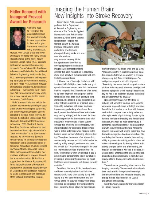
To address this issue, Hidler developed upper<br />
and lower extremity test devices that allow<br />
researchers to study brain activity during fMRI<br />
testing in a well-controlled manner. <strong>The</strong> arm<br />
device allows for accurate measures <strong>of</strong> forces<br />
generated by subjects at <strong>the</strong>ir wrist while <strong>the</strong><br />
lower extremity device allows for <strong>the</strong> measurement<br />
<strong>of</strong> forces at <strong>the</strong> ankle, knee and hip joints.<br />
“This was definitely a challenging project since<br />
<strong>the</strong> magnetic fields we are working in are very<br />
strong — up to 3 Tesla or 30,000 gauss.” A<br />
refrigerator magnet is about 5-10 gauss!<br />
“Essentially this means that all magnetic materials<br />
have to be replaced; o<strong>the</strong>rwise <strong>the</strong> object will<br />
become a projectile or will heat up. Needless to<br />
say we had to be extremely cautious throughout<br />
<strong>the</strong> development <strong>of</strong> <strong>the</strong> devices.”<br />
Now, researchers can run fMRI tests in<br />
patients with very little function, such as in <strong>the</strong><br />
very acute stages <strong>of</strong> stroke, with high precision.<br />
One <strong>of</strong> <strong>the</strong> first studies to be done with <strong>the</strong> new<br />
devices is to compare brain activity before and<br />
after eight weeks <strong>of</strong> gait training. Funded by <strong>the</strong><br />
National Institute on Disability and Rehabilitation<br />
Research, this NRH-led multi-center study is<br />
comparing <strong>the</strong> effectiveness <strong>of</strong> robotic gait training<br />
with conventional gait training. Adding <strong>the</strong><br />
imaging component will provide insight into how<br />
<strong>the</strong> brain re-organizes to enhance function. “We<br />
are finding that in some subjects, <strong>the</strong>ir walking<br />
ability improves substantially, while in o<strong>the</strong>rs we<br />
notice only small gains. By looking at how brain<br />
activity changes before and after training, we<br />
may get a better idea <strong>of</strong> why this is <strong>the</strong> case. If<br />
we can better understand <strong>the</strong> process <strong>of</strong> neural<br />
plasticity and neural recovery, in <strong>the</strong> future we<br />
may be able to develop more effective interventions.”<br />
<strong>The</strong> devices are generating a buzz around <strong>the</strong><br />
imaging community. <strong>The</strong> arm device has already<br />
been replicated for Georgetown <strong>University</strong>’s<br />
Center for Functional and Molecular Imaging and<br />
<strong>the</strong> leg module has been built for UCLA’s<br />
Department <strong>of</strong> Neurology.<br />
See http://cabrr.cua.edu for more information<br />
on Hidler’s research.<br />
fall2006 | 5